. 2013 Jul 15;305(2):C182-9.
doi: 10.1152/ajpcell.00086.2013. Epub 2013 May 1.

NLRP3 deletion protects from hyperoxia-induced acute lung injury

Affiliations

NLRP3 deletion protects from hyperoxia-induced acute lung injury

Jutaro Fukumoto et al. Am J Physiol Cell Physiol. .

Abstract

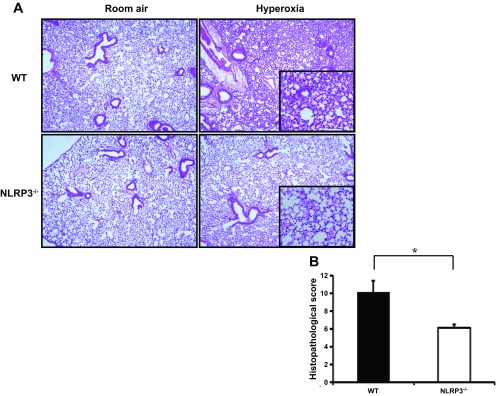
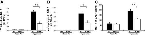
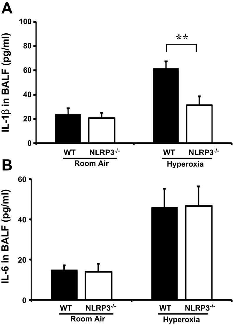
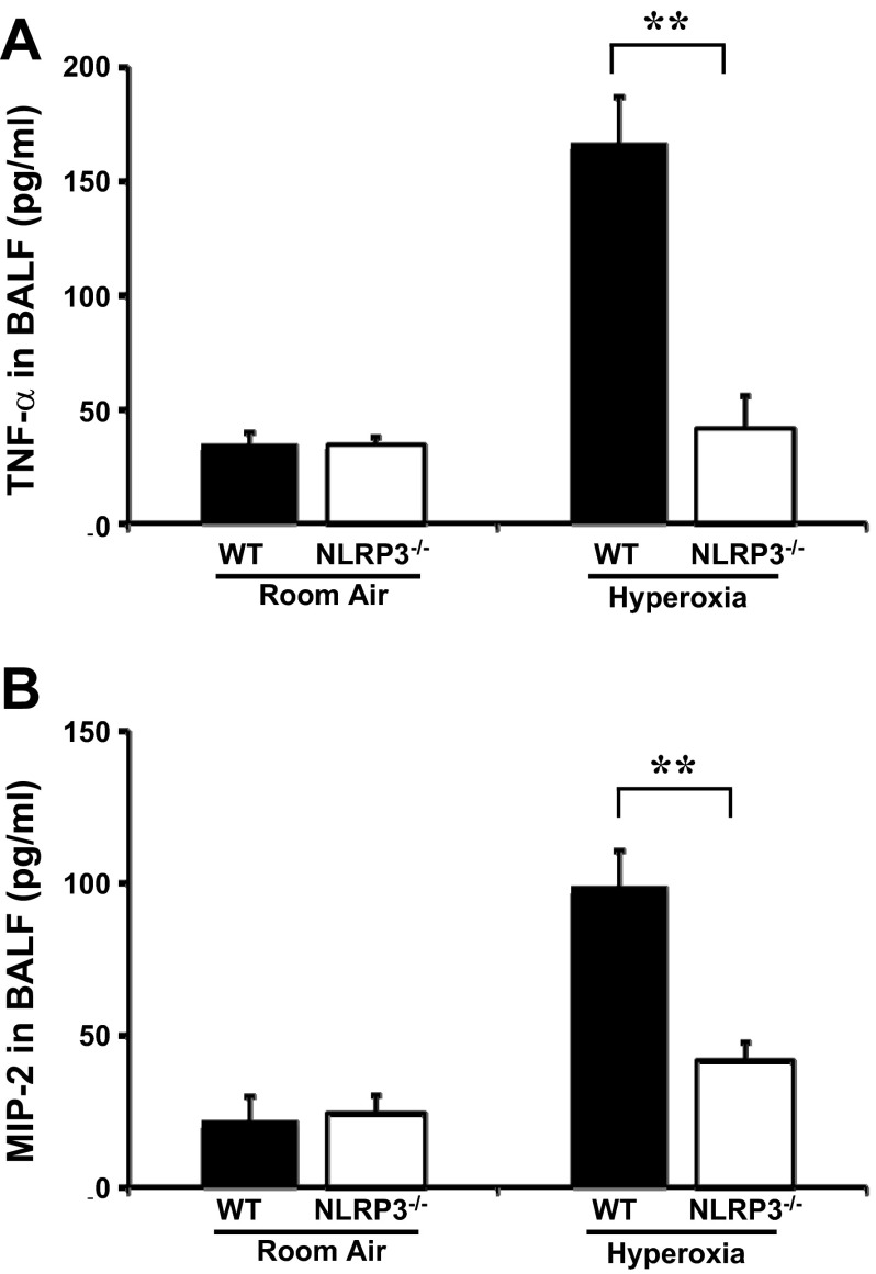
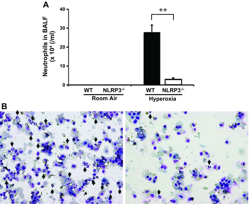
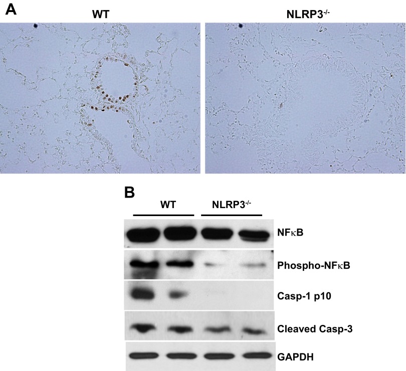
Inspiration of a high concentration of oxygen, a therapy for acute lung injury (ALI), could unexpectedly lead to reactive oxygen species (ROS) production and hyperoxia-induced acute lung injury (HALI). Nucleotide-binding domain and leucine-rich repeat PYD-containing protein 3 (NLRP3) senses the ROS, triggering inflammasome activation and interleukin-1β (IL-1β) production and secretion. However, the role of NLRP3 inflammasome in HALI is unclear. The main aim of this study is to determine the effect of NLRP3 gene deletion on inflammatory response and lung epithelial cell death. Wild-type (WT) and NLRP3(-/-) mice were exposed to 100% O2 for 48-72 h. Bronchoalveolar lavage fluid and lung tissues were examined for proinflammatory cytokine production and lung inflammation. Hyperoxia-induced lung pathological score was suppressed in NLRP3(-/-) mice compared with WT mice. Hyperoxia-induced recruitment of inflammatory cells and elevation of IL-1β, TNFα, macrophage inflammatory protein-2, and monocyte chemoattractant protein-1 were attenuated in NLRP3(-/-) mice. NLRP3 deletion decreased lung epithelial cell death and caspase-3 levels and a suppressed NF-κB levels compared with WT controls. Taken together, this research demonstrates for the first time that NLRP3-deficient mice have suppressed inflammatory response and blunted lung epithelial cell apoptosis to HALI.

Keywords: hyperoxia; inflammation; injury; lung; reactive oxygen species.


Figures

Fig. 1.
Fig. 1.
Hyperoxia-exposed Nucleotide-binding domain and leucine-rich repeat PYD-containing protein 3-deficient (NLRP3−/−) mice show attenuated lung injury. wild-type (WT) and NLRP3−/− mice were exposed to room air or 100% O2 for 48 h. Mice were euthanized and lung tissues were collected. A: representative hematoxylin-eosin (H&E)-stained lung tissue sections. Original magnifications: ×40 (insets, ×100). B: semiquantitative lung injury analysis by evaluation of H&E-stained lung tissue sections. Total histopathological score was expressed as summed scores for 4 independent parameters: alveolar congestion, hemorrhage, aggregation of neutrophil or leukocyte infiltration, and thickness of the alveolar wall. Results (means ± SE; n = 8 in each group) are cumulative data of 2 independent experiments. *P < 0.05.
Fig. 2.
Fig. 2.
Hyperoxia-induced recruitment of inflammatory cells and elevation of monocyte chemoattractant protein-1 (MCP-1) were attenuated in NLRP3−/− mice. WT and NLRP3−/− mice were exposed to room air or 100% O2 for 72 h and euthanized, and bronchoalveolar lavage fluid (BALF) was collected. A: total cell counts. B: total number of macrophages. Results of A and B (means ± SE; n = 4 in each group) are representative of 2 independent experiments. C: levels of MCP-1 were measured by ELISA. Results (means ± SE; n = 4 in each group for normoxia; n = 8 in each group for hyperoxia) are cumulative data of 2 independent experiments. *P < 0.05 **P < 0.01.
Fig. 3.
Fig. 3.
Hyperoxia-induced elevation of IL1-β is NLRP3 dependent. WT and NLRP3−/− mice exposed to room air or 100% O2 for 72 h were euthanized and BALF was collected. IL-1β (A) and IL-6 (B) concentrations in BALF were measured by ELISA. Results (means ± SE; n = 4 in each group for normoxia; n = 7–8 in each group for hyperoxia) are cumulative data of 2 independent experiments. **P < 0.01.
Fig. 4.
Fig. 4.
Hyperoxia-induced elevation of TNF-α and macrophage inflammatory protein-2 (MIP-2) is suppressed in NLRP3−/− mice. WT and NLRP3−/− mice exposed to room air or 100% O2 for 72 h were euthanized and BALF was collected. TNF-α (A) and MIP-2 (B) concentrations were measured by ELISA. Results (means ± SE; n = 4 in each group for normoxia, n = 8 in each group for hyperoxia) are cumulative data of 2 independent experiments. **P < 0.01.
Fig. 5.
Fig. 5.
Hyperoxia-induced neutrophil recruitment was decreased in NLRP3−/− mice. WT and NLRP3−/− mice exposed to room air or 100% O2 were euthanized and BALF was collected. A: means ± SE of total number of neutrophils (n = 4 in each group) from 1 of the 2 independent experiments is shown. B: mixture of equal amount of BALF from 4 different mice per each group was cytospined and stained with Diff-Quik. Infiltrating neutrophils are indicated by arrows. Magnification: ×200. **P < 0.01.
Fig. 6.
Fig. 6.
Hyperoxia-induced lung epithelial cell death is attenuated in NLRP3−/− mice. A: WT and NLRP3−/− mice (n = 8 in each group) exposed to 100% O2 for 48 h were euthanized and lung tissues were collected. Lung tissue sections were stained using terminal deoxynucleotidyl transferase dUTP-mediated nick-end labeling (TUNEL) assay. Shown are representative images of 2 independent experiments. TUNEL-positive apoptotic cells are observed in lung epithelium in WT mice, but they are hardly detectable in NLRP3−/− mice. Magnification: ×200. B: Western blot analysis of NF-κB, phosphor-NF-κB, caspase-1 p10, and cleaved caspase-3 in whole lung lysates after 48-h hyperoxic exposure. Results are representative of 2 independent experiments.

Similar articles

Cited by

References

    1. Abraham E, Carmody A, Shenkar R, Arcaroli J. Neutrophils as early immunologic effectors in hemorrhage- or endotoxemia-induced acute lung injury. Am J Physiol Lung Cell Mol Physiol 279: L1137–L1145, 2000 - PubMed
    1. Agouridakis P, Kyriakou D, Alexandrakis MG, Prekates A, Perisinakis K, Karkavitsas N, Bouros D. The predictive role of serum and bronchoalveolar lavage cytokines and adhesion molecules for acute respiratory distress syndrome development and outcome. Respir Res 3: 25, 2002 - PMC - PubMed
    1. Ayala A, Chung CS, Lomas JL, Song GY, Doughty LA, Gregory SH, Cioffi WG, LeBlanc BW, Reichner J, Simms HH, Grutkoski PS. Shock-induced neutrophil mediated priming for acute lung injury in mice: divergent effects of TLR-4 and TLR-4/FasL deficiency. Am J Pathol 161: 2283–2294, 2002 - PMC - PubMed
    1. Bethea JR, Chung IY, Sparacio SM, Gillespie GY, Benveniste EN. Interleukin-1 beta induction of tumor necrosis factor-alpha gene expression in human astroglioma cells. J Neuroimmunol 36: 179–191, 1992 - PubMed
    1. Byers DE, Holtzman MJ. Alternatively activated macrophages and airway disease. Chest 140: 768–774, 2011 - PMC - PubMed

Publication types

MeSH terms

LinkOut - more resources