. 2013 Jun 15;19(12):3276-84.
doi: 10.1158/1078-0432.CCR-12-3768. Epub 2013 May 1.

Noninvasive detection of HER2 amplification with plasma DNA digital PCR

Affiliations

Noninvasive detection of HER2 amplification with plasma DNA digital PCR

Heidrun Gevensleben et al. Clin Cancer Res. .

Abstract

Purpose: Digital PCR is a highly accurate method of determining DNA concentration. We adapted digital PCR to determine the presence of oncogenic amplification through noninvasive analysis of circulating free plasma DNA and exemplify this approach by developing a plasma DNA digital PCR assay for HER2 copy number.

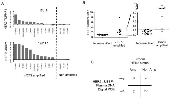
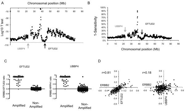
Experimental design: The reference gene for copy number assessment was assessed experimentally and bioinformatically. Chromosome 17 pericentromeric probes were shown to be suboptimal, and EFTUD2 at chromosome position 17q21.31 was selected for analysis. Digital PCR assay parameters were determined on plasma samples from a development cohort of 65 patients and assessed in an independent validation cohort of plasma samples from 58 patients with metastatic breast cancer. The sequential probability ratio test was used to assign the plasma DNA digital PCR test as being HER2-positive or -negative in the validation cohort.

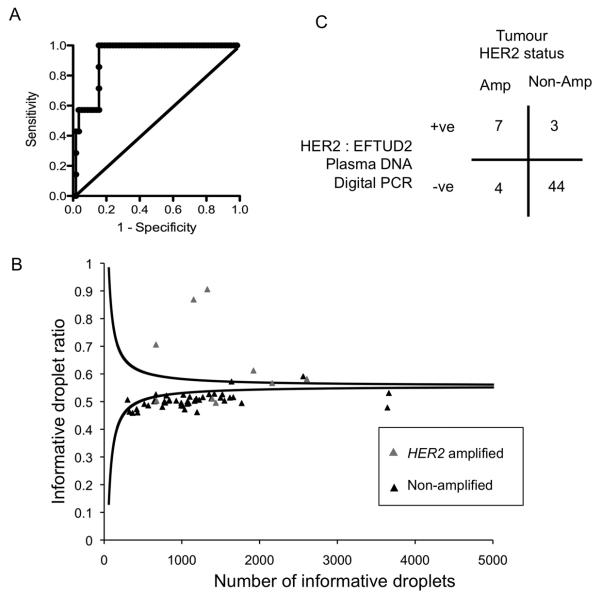
Results: In the development cohort, the HER2:EFTUD2 plasma DNA copy number ratio had a receiver operator area under the curve (AUC) = 0.92 [95% confidence interval (CI), 0.86-0.99, P = 0.0003]. In the independent validation cohort, 64% (7 of 11) of patients with HER2-amplified cancers were classified as plasma digital PCR HER2-positive and 94% (44 of 47) of patients with HER2-nonamplified cancers were classified as digital PCR HER2-negative, with a positive and negative predictive value of 70% and 92%, respectively.

Conclusion: Analysis of plasma DNA with digital PCR has the potential to screen for the acquisition of HER2 amplification in metastatic breast cancer. This approach could potentially be adapted to the analysis of any locus amplified in cancer.


Figures

Figure 1
Figure 1. Digital PCR assessment with chromosome 17 peri-centromeric probes
A. Assessment of reference probes for digital PCR. HER2:reference copy number ratio from a single 384 well digital PCR assessment of DNA from 11 HER2 amplified cell lines and 3 non-amplified cell lines. The HER2:UBBP4 (17p11.1) ratio is substantially elevated in all amplified cancer cell lines, but the HER2:TUFMP1 is not raised in 3 HER2 amplified cell lines due to co-amplification of 17q peri-centromeric DNA (Supplementary Figure 1). B. Development cohort for UBBP4 reference probe. Plasma DNA digital PCR HER2:UBBP4 ratio from 9 patients with HER2 positive and 35 patients with HER2 negative cancers. Left full data-set and Right expanded y axis for ratios 1.0-2.0, dashed line indicates a ratio of 1.25. C. Assessment of HER2:UBBP4 in an independent validation cohort. Tabulated results of plasma DNA digital PCR analysed by SPRT on plasma samples from 10 patients with HER2 amplified cancer and 36 patients with HER2 non-amplified cancer. p=0.003 Fishers Exact test.
Figure 2
Figure 2. Identification of an optimal chromosome 17 reference probe for digital PCR assay
A. Analysis of microarray CGH data from 311 primary breast cancers (16). For each cancer the ERBB2 (HER2) to reference copy number ratio was calculated for every genomic position along chromosome 17. The ERBB2 to reference copy number ratio of HER2 amplified cancers was compared with HER2 negative cancers using Student’s T test. Displayed is the log 10 p value for each genomic position, with arrow indicating the locus of EFTUD2 with the most significant difference between amplified and non-amplified cancers. B. The corresponding sensitivity for each reference genomic position was assessed for each genomic position. C. Comparison of ERBB2 : EFTUD2 and ERBB2 : UBBP4 copy number ratios in HER2 amplified and non-amplified cancers from the same micro-array CGH series(16). D. Correlation of ERBB2 and EFTUD2, along with ERBB2 and UBBP4, copy number in the 246 HER2 non-amplified cancers. The copy number of ERBB2 and EFTUD2 are highly correlated as low level gain or loss of ERBB2 extends to EFTUD2. EFTUD2 therefore is predicted to generate stable copy number ratios in the analysis of non-amplified cancers.
Figure 3
Figure 3. Digital PCR assay for HER2 copy number assessment by droplet digital PCR
A. Plasma is separated within 2 hours of venepuncture and stored at −80°C before extraction of free circulating DNA. B. Droplet Digital PCR with a FAM labeled HER2 probe and VIC labeled EFTUD2 (reference) probe. DNA is partitioned into ~14,000 droplets per reaction. After single molecule PCR droplets are assessed by a fluorescent reader. The concentration of DNA in each sample can be quantified from the number of wells positive using the Poisson distribution. C. Validation cohort: Analysis of digital PCR with Sequential Probability Ratio Test (SPRT) using informative droplets, those droplets positive for HER2 or EFTUD2 alone, and not those positive for both or neither. The SPRT assesses whether the proportion of informative wells positive for HER2, informative wells ratio, is elevated as data accumulates. SPRT defines two boundaries, with a ratio above the upper boundary being considered HER2 positive and below the lower boundary considered HER2 negative. A ratio between the two boundaries is considered as unassigned, and the sample is subjected to further rounds of digital PCR until the result is above or below the boundaries.
Figure 4
Figure 4. Plasma DNA digital PCR with the EFTUD2 reference probe has high diagnostic accuracy in an independent cohort
A. Development cohort for EFTUD2 reference probe. Plasma DNA digital PCR HER2:EFTUD2 ratio was assessed in plasma samples from 65 patients with metastatic breast cancer, consisting of 7 with HER2 amplified cancer and 58 with HER2 non-amplified cancer. ROC analysis with an AUC 0.92 (95% CI 0.86 to 0.99, P=0.0003). A cut-off of 1.25 was selected to define HER2 amplification in the validation series. B. Assessment of HER2:EFTUD2 in an independent validation cohort. Plasma DNA digital PCR results analysed by SPRT on plasma samples from 11 patients with HER2 amplified cancers and 47 patients with HER2 non-amplified cancers. Grey triangle indicates patients with HER2 amplified tumours and black triangle HER2 non-amplified tumours. The displayed SPRT decision boundaries are for illustrative purposes only, as the exact level varies according to the EFTUD2 control probe concentration (MEFTUD2), with the displayed boundaries calculated with MEFTUD2=0.025. Cases with a number of informative droplets > 5000 are not displayed. C. Tabulated results of Plasma DNA digital PCR analysed by SPRT on independent validation cohort. p=0.0001 Fishers Exact test.

References

    1. Slamon DJ, Leyland-Jones B, Shak S, Fuchs H, Paton V, Bajamonde A, et al. Use of chemotherapy plus a monoclonal antibody against HER2 for metastatic breast cancer that overexpresses HER2. N Engl J Med. 2001;344:783–92. - PubMed
    1. Bang YJ, Van Cutsem E, Feyereislova A, Chung HC, Shen L, Sawaki A, et al. Trastuzumab in combination with chemotherapy versus chemotherapy alone for treatment of HER2-positive advanced gastric or gastro-oesophageal junction cancer (ToGA): a phase 3, open-label, randomised controlled trial. Lancet. 2010;376:687–97. - PubMed
    1. Thompson AM, Jordan LB, Quinlan P, Anderson E, Skene A, Dewar JA, et al. Prospective comparison of switches in biomarker status between primary and recurrent breast cancer: the Breast Recurrence In Tissues Study (BRITS) Breast Cancer Res. 2010;12:R92. - PMC - PubMed
    1. Houssami N, Macaskill P, Balleine RL, Bilous M, Pegram MD. HER2 discordance between primary breast cancer and its paired metastasis: tumor biology or test artefact? Insights through meta-analysis. Breast Cancer Res Treat. 2011;129:659–74. - PubMed
    1. Turke AB, Zejnullahu K, Wu YL, Song Y, Dias-Santagata D, Lifshits E, et al. Preexistence and clonal selection of MET amplification in EGFR mutant NSCLC. Cancer Cell. 2010;17:77–88. - PMC - PubMed

Publication types

MeSH terms