. 2013 May 1;33(18):8009-21.
doi: 10.1523/JNEUROSCI.4505-12.2013.

A role for an Hsp70 nucleotide exchange factor in the regulation of synaptic vesicle endocytosis

Affiliations

A role for an Hsp70 nucleotide exchange factor in the regulation of synaptic vesicle endocytosis

Jennifer R Morgan et al. J Neurosci. .

Abstract

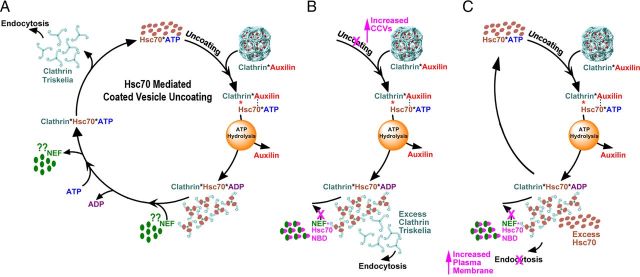
Neurotransmission requires a continuously available pool of synaptic vesicles (SVs) that can fuse with the plasma membrane and release their neurotransmitter contents upon stimulation. After fusion, SV membranes and membrane proteins are retrieved from the presynaptic plasma membrane by clathrin-mediated endocytosis. After the internalization of a clathrin-coated vesicle, the vesicle must uncoat to replenish the pool of SVs. Clathrin-coated vesicle uncoating requires ATP and is mediated by the ubiquitous molecular chaperone Hsc70. In vitro, depolymerized clathrin forms a stable complex with Hsc70*ADP. This complex can be dissociated by nucleotide exchange factors (NEFs) that release ADP from Hsc70, allowing ATP to bind and induce disruption of the clathrin:Hsc70 association. Whether NEFs generally play similar roles in vesicle trafficking in vivo and whether they play such roles in SV endocytosis in particular is unknown. To address this question, we used information from recent structural and mechanistic studies of Hsp70:NEF and Hsp70:co-chaperone interactions to design a NEF inhibitor. Using acute perturbations at giant reticulospinal synapses of the sea lamprey (Petromyzon marinus), we found that this NEF inhibitor inhibited SV endocytosis. When this inhibitor was mutated so that it could no longer bind and inhibit Hsp110 (a NEF that we find to be highly abundant in brain cytosol), its ability to inhibit SV endocytosis was eliminated. These observations indicate that the action of a NEF, most likely Hsp110, is normally required during SV trafficking to release clathrin from Hsc70 and make it available for additional rounds of endocytosis.


Figures

Figure 1.
Figure 1.
Hsc70-mediated coated vesicle uncoating. A, In all panels, the color of the text matches the color of the corresponding molecule. Clathrin coat disassembly begins when auxilin binds clathrin and recruits Hsc70:ATP to the clathrin coat. After ATP hydrolysis, the clathrin coat disassembles, auxilin is released, and clathrin forms a long-lived complex with Hsc70:ADP. Interaction of clathrin:Hsc70:ADP with an NEF results in the release of ADP, and subsequent binding of ATP to Hsc70 results in release of the NEF and clathrin. The role of auxilin and Hsc70 in this process has been demonstrated in vivo, but although NEFs have been shown to stimulate uncoating in vitro (Schuermann et al., 2008), there is as yet no data on the role of Hsp70 NEFs in vesicle trafficking in vivo. B, Expected effects of an NEF inhibitor if the amount of clathrin in neuronal cytosol is greater than the amount of Hsc70. With free clathrin available, plasma membrane endocytosis can occur, but with most Hsc70 sequestered in clathrin*Hsc70*ADP complexes, uncoating would be inhibited and an increased number of CCVs would be observed. C, Expected effects of an NEF inhibitor if the amount of Hsc70 is greater than the amount of clathrin. With free Hsc70 available, uncoating can occur, but with most clathrin sequestered in clathrin*Hsc70*ADP complexes, plasma membrane endocytosis would be inhibited and increased plasma membrane area would be observed.
Figure 2.
Figure 2.
An ATP-binding-deficient Hsc70 NBD inhibits Hsp110 stimulation of Hsc70 ATPase activity. A, Rationale for construction of an Hsp70 NEF inhibitor: (1) NEFs such as Hsp110 (shown in space-filling representation in yellow) bind the Hsp70 NBD (cyan) when the latter is nucleotide free or bound to ADP. (2) The interaction between Hsp110 and the Hsp70 NBD induces the latter to release its bound ADP, and the resulting complex with nucleotide-free Hsp70 NBD is stable. (3) Binding of ATP to the Hsp70 NBD induces complex dissociation; an Hsp70 NBD that cannot bind ATP would therefore form a stable complex that could not be induced to dissociate by ATP and such an NBD would sequester and inhibit Hsp70 NEFs. B, Backbone model of the bovine Hsc70 NBD (1kax.pdb) (Flaherty et al., 1994) is shown with bound ATP (red) and residues G201/G202 (green) in space-filling representation. C, Steady-state rates of ATP hydrolysis for the indicated reaction mixes: (1) Hsc70 D152K (Hsc70D/K) + auxilin (J); (2) Hsc70 D152K + auxilin + yeast Hsp110; (3) Hsc70 D152K + auxilin + yeast Hsp110 + WT NBD; (4) Hsc70 D152K + auxilin + yeast Hsp110 + GG/ED NBD; (5) yeast Hsp110 + WT NBD; and (6) yeast Hsp110 + GG/ED NBD. In these experiments, yeast Hsp110 was used instead of mammalian Hsp110 because it has a lower ATPase rate. Human Hsp110 was used for all subsequent experiments.
Figure 3.
Figure 3.
An ATP-binding-deficient Hsc70 NBD inhibits NEF stimulation of clathrin basket disassembly. A, Clathrin coat disassembly as monitored by dynamic light scattering. Disassembly reactions contained clathrin baskets and auxilin only (black squares); clathrin baskets with ATP, auxilin, and Hsc70 (red circles); clathrin + ATP + auxilin + Hsc70 + Hsp110 (green circles); or clathrin + ATP + auxilin + Hsc70 + Hsp110 and varying amounts of GG/ED NBD (0.04 μm: blue circles; 0.05 μm: cyan circles; 0.1 μm: magenta circles; 0.25 μm: yellow circles; 0.5 μm: purple circles; 1.0 μm: olive circles). Data are globally fit to y = y0 + A*(exp(−t1*x) − 1) − k*x, where y0 is the photons scattered at x = 0 s (∼1,275,000 for 1 μm clathrin), A is the amplitude of first (rapid) exponential phase, t1 is the time constant of first exponential phase, k is the rate of second (slow) linear phase (the decrease/second in the number of scattered photons). Data were globally fit with the y0 value held constant because this value is determined by the (constant) amount of clathrin baskets added to each reaction and with A and t1 shared since the kinetic scheme has A and t1 determined by the constant amount of Hsc70 present in all reactions. B, Percentage inhibition of Hsp110 stimulation of uncoating in A plotted as a function of the GG/ED NBD concentration. C, As in A, but using Bag1 at 2.0 μm instead of Hsp110 at 0.1 μm, and with the GG/ED NBD at 0.2 μm (blue circles), 0.4 μm (cyan circles), 0.6 μm (magenta circles), 0.8 μm (yellow circles), 1.0 μm (purple circles), 2.0 μm (olive circles), or 4.0 μm (gray circles). D, As in B, but with data from C plotted. E, NEFs cannot promote coat disassembly in the absence of Hsc70. Disassembly reactions contained clathrin baskets and auxilin only (black squares), or clathrin baskets with auxilin and ATP (red circles), or clathrin + auxilin + ATP + varying amounts of NEFs or control proteins: 0.1 μm Hsp110 (green), 2 μm Hsp110 (purple), 2 μm Bag1 (blue), 0.1 μm BSA (magenta), or 2 μm BSA (yellow). Although a very slight decrease in scattering intensity was observed when ATP was added to clathrin baskets and auxilin, no further decrease in scattering intensity was observed when any of the NEFs or control proteins was added in the absence of Hsc70. F, The GG/ED NBD has no effect on clathrin assembly. Clathrin was induced to assemble by AP2 (squares) or AP180 (circles) at pH 6.5 in the absence (solid symbols) or presence (open symbols) of 50 μm GG/ED NBD. The addition of GG/ED NBD had no effect on either AP2- or AP180-mediated coat assembly.
Figure 4.
Figure 4.
Hsc70 is in large excess of clathrin in brain cytosol. A, Quantitative Western blots were performed to determine the content of clathrin, Hsc70, Hsp110, or Bag1 in bovine brain cytosol in the presence of calibrated standards of each protein, as well as a purified CCV control enriched in clathrin (Pearse, 1975; Blondeau et al., 2004). Note that there are two isoforms of Hsp110 (Ishihara et al., 1999) and three isoforms of Bag1 (Cutress et al., 2003). Bands were quantified separately by densitometry and summed for each protein in the analysis shown in B. B, Quantitative analysis of clathrin, Hsc70, Hsp110, and Bag 1 in brain cytosol. The bars represent the mean of the respective protein amounts in micrograms/milligram of total protein ± SEM from 5 to 13 independent experiments. Asterisks indicate statistical significance using ANOVA.
Figure 5.
Figure 5.
Mutations in the Hsc70 region that interacts specifically with Hsp110 specifically disrupt binding to Hsp110. A, Surface representations of the Hsc70 NBD with different regions of the surface colored according to whether they interact with Bag1/Bag5 (orange; Sondermann et al., 2001; Arakawa et al., 2010), Bag2/BNB (yellow; Xu et al., 2008), HspBp1 (red; Shomura et al., 2005), auxilin J domain (green; Jiang et al., 2007), or Hsp110 (brown; Polier et al., 2008; Schuermann et al., 2008). There is some overlap between the regions contacted by the different co-chaperones, but the region encompassing residues R299-R311 is not known to interact with any co-chaperone except Hsp110. B, Native gel electrophoresis of the indicated proteins. Lane 1: Hsc70 GG/ED NBD; lane 2: GG/ED RRR/EEE NBD; lane 3: Bag1; lane 4: Hsp110; lane 5: Bag1 + GG/ED NBD; lane 6: Bag1 + GG/ED RRR/EEE NBD; lane 7: Hsp110 + GG/ED NBD; lane 8: Hsp110 + GG/ED RRR/EEE NBD.
Figure 6.
Figure 6.
An ATP- and Hsp110-binding deficient Hsc70 NBD inhibits Bag1 but not Hsp110. A, Clathrin coat disassembly as monitored by dynamic light scattering. Disassembly reactions contained clathrin baskets and auxilin only (black squares), or clathrin baskets with ATP, auxilin, and Hsc70 (red circles); clathrin + ATP + auxilin + Hsc70 + Hsp110 (green circles); or clathrin + ATP + auxilin + Hsc70 + Hsp110 and varying amounts of GG/ED,RRR/EEE NBD (25 μm: blue circles; 50 μm: cyan circles; 100 μm: magenta circles; 200 μm: yellow circles; 280 μm: purple circles. B, Percentage inhibition of Hsp110 stimulation of uncoating in A plotted as a function of the GG/ED,RRR/EEE NBD concentration. C, As in A, but using Bag1 at 2.0 μm instead of Hsp110 at 0.1 μm, and with the GG/ED,RRR/EEE NBD at 0.2 μm (blue circles), 0.4 μm (cyan circles), 0.6 μm (magenta circles), 0.8 μm (yellow circles), 1.0 μm (purple circles), 2.0 μm (olive circles), or 4.0 μm (gray circles). D, As in B, but with data from C plotted. Data were fit as described in the legend to Figure 3.
Figure 7.
Figure 7.
GG/ED NBD inhibits the ability of Hsp110 to disrupt Hsp70:clathrin interactions. A, Quantitative analysis (top; filled circles: clathrin; empty circles: Hsc70) of SDS PAGE (bottom) of basket disassembly reactions (1 μm clathrin heavy and light chains, 0.6 μm Hsc70, 0.1 μm auxilin, 1 mm ATP) resolved by gel exclusion chromatography. Under these conditions, most of the Hsc70 co-elutes with clathrin. B, As in A, but with 0.1 μm Hsp110 added. Under these conditions, most of the Hsc70 elutes at a position later than clathrin because Hsp110 promotes its dissociation from the clathrin. C, As in B, but with 4 μm GG/ED NBD added. Now, most of the Hsc70 co-elutes with clathrin, again, because Hsp110 is being sequestered by the GG/ED NBD. D, As in B but with 4 μm GG/ED,RRR/EEE NBD added. Now, most of the Hsc70 no longer co-elutes with clathrin because the GG/ED,RRR/EEE NBD does not bind to Hsp110.
Figure 8.
Figure 8.
Hsc70 GG/ED NBD inhibits SV endocytosis from the plasma membrane. A, A control synapse after buffer injection and stimulation (20 Hz, 5 min). The plasma membrane (highlighted in red) is only moderately ruffled in appearance because SV endocytosis is able to keep up with exocytosis. B, In contrast, after treatment with Hsc70 GG/ED NBD and stimulation, synapses exhibit enlarged plasma membrane evaginations due to inhibition of vesicle retrieval from the plasma membrane. C, The endocytic defect is not observed after injection of the Hsc70 GG/ED,RRR/EEE NBD, indicating that disruption of the NBD interaction with Hsp110 disrupts its ability to inhibit endocytosis. Scale in A applies to AC. Asterisks mark postsynaptic dendrites. D, Quantification of the size of plasma membrane evaginations from 20 to 34 synapses per experimental condition. Bars represent mean ± SEM. Asterisks indicate statistical significance using ANOVA. E, Quantification of the number of CCVs from 20 to 34 synapses per experimental condition. Bars represent mean ± SEM. F, Quantification of the number of CCPs from 20 to 34 synapses per experimental condition. Bars represent mean ± SEM. G, Quantification of the number of SVs from 20 to 34 synapses per experimental condition. Bars represent mean ± SEM.

Similar articles

Cited by

References

    1. Ahle S, Ungewickell E. Purification and properties of a new clathrin assembly protein. EMBO J. 1986;5:3143–3149. - PMC - PubMed
    1. Arakawa A, Handa N, Ohsawa N, Shida M, Kigawa T, Hayashi F, Shirouzu M, Yokoyama S. The C-terminal BAG domain of BAG5 induces conformational changes of the Hsp70 nucleotide-binding domain for ADP-ATP exchange. Structure. 2010;18:309–319. doi: 10.1016/j.str.2010.01.004. - DOI - PubMed
    1. Augustine GJ, Morgan JR, Villalba-Galea CA, Jin S, Prasad K, Lafer EM. Clathrin and synaptic vesicle endocytosis: studies at the squid giant synapse. Biochem Soc Trans. 2006;34:68–72. doi: 10.1042/BST0340068. - DOI - PMC - PubMed
    1. Black MM, Chestnut MH, Pleasure IT, Keen JH. Stable clathrin: uncoating protein (hsc70) complexes in intact neurons and their axonal transport. J Neurosci. 1991;11:1163–1172. - PMC - PubMed
    1. Blondeau F, Ritter B, Allaire PD, Wasiak S, Girard M, Hussain NK, Angers A, Legendre-Guillemin V, Roy L, Boismenu D, Kearney RE, Bell AW, Bergeron JJ, McPherson PS. Tandem MS analysis of brain clathrin-coated vesicles reveals their critical involvement in synaptic vesicle recycling. Proc Natl Acad Sci U S A. 2004;101:3833–3838. doi: 10.1073/pnas.0308186101. - DOI - PMC - PubMed

Publication types

MeSH terms

LinkOut - more resources