Review
. 2013 May 1;5(5):a008698.
doi: 10.1101/cshperspect.a008698.

Death receptor-ligand systems in cancer, cell death, and inflammation

Affiliations
Review

Death receptor-ligand systems in cancer, cell death, and inflammation

Henning Walczak. Cold Spring Harb Perspect Biol. .

Abstract

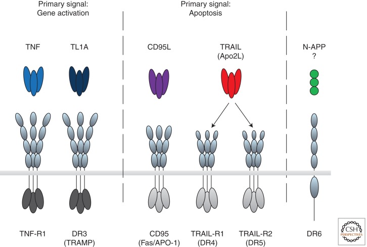
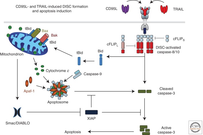
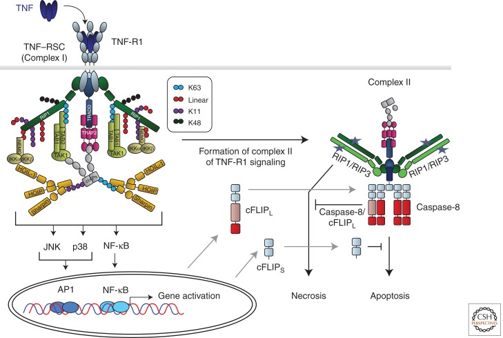
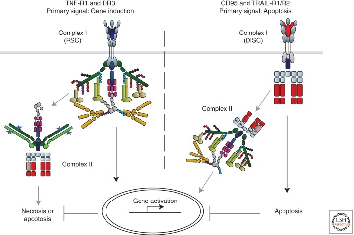
The discovery of tumor necrosis factor (TNF) marked the beginning of one of the most fascinating journeys in modern biomedical research. For the moment, this journey has culminated in the development of drugs that inhibit TNF. TNF blockers have revolutionized the treatment of many chronic inflammatory diseases. Yet, the journey seems far from over. TNF is the founding member of a family of cytokines with crucial functions in cell death, inflammation, and cancer. Some of these factors, most prominently TNF, CD95L, and TRAIL, can induce cell death. The receptors that mediate this signal are therefore referred to as death receptors, even though they also activate other signals. Here I will take you on a journey into the discovery and study of death receptor-ligand systems and how this inspired new concepts in cancer therapy and our current understanding of the interplay between cell death and inflammation.


Figures

Figure 1.
Figure 1.
Death receptors and their ligands. The death domain-containing receptors are transmembrane proteins that contain 2–4 cysteine-rich repeats in their extracellular domain, required for ligand binding, and an intracellular death domain capable of recruiting specific adaptors that define their downstream interactors and signals (light gray for CD95/TRAIL and dark gray for TNF-R1/DR3 systems). Death receptors trigger two main signals. TNF-R1 and DR3 induce gene activation as their primary signaling output, whereas CD95, TRAIL-R1, and TRAIL-R2 induce apoptosis as their primary signal. DR6 has been proposed to be ligated by N-APP but this is unconfirmed, and DR6 signaling is altogether less well understood.
Figure 2.
Figure 2.
CD95L- and TRAIL-induced DISC formation and apoptosis induction. Binding of CD95L or TRAIL to their cognate receptors leads to receptor trimerization and formation of the death-inducing signaling complex (DISC). The DD of one FADD molecule interacts with the DDs of the three cross-linked receptors. Subsequently, procaspases 8 and 10 are recruited by interaction of their DED with that of FADD. Heterodimers between cFLIPL and caspase-8 or -10 are not inactive as proteolytic enzymes but their proteolytic activity is edited as compared with caspase-8 or -10 homodimers. cFLIPS in turn inhibits caspase activity at the DISC by preventing dimerization of caspase-8/10/cFLIPL. Interestingly, one FADD molecule recruits six to 10 DED-containing caspase-8, -10, and cFLIP proteins. DISC-activated caspase-8/-10 cleaves caspase-3, enabling autoactivation of caspase-3, which renders the enzyme fully active. This latter step can, however, be blocked by X-linked inhibitor of apoptosis protein (XIAP). DISC-activated caspase-8/-10 also cleaves Bid (tBID). Interaction of tBid with Bak and Bax in the mitochondrial outer membrane induces Bax/Bak oligomerization, resulting in mitochondrial outer-membrane permeabilization (MOMP) so that cytochrome c and Smac/DIABLO are released from the mitochondrial intermembrane space to the cytosol. Cytochrome c, together with Apaf-1 and caspase-9, forms the apoptosome, the activation platform for caspase-9, which can, however, also be inhibited by XIAP. Smac/DIABLO binds to XIAP, which releases caspase-3 and -9 from XIAP-imposed inhibition. Activation of these caspases enables execution of apoptotic cell death.
Figure 3.
Figure 3.
TNF-R1-induced gene activation and cell death signaling. Cross-linking of TNF-R1 by TNF results in formation of the TNF-R1 signaling complex (TNF–RSC). Cross-linked TNF-R1 recruits TRADD and RIP1 to the DD of the receptor. Subsequently, TRADD recruits TRAF2, which in turn provides the platform for cIAP1/2. cIAPs then place ubiquitin chains, linked via different interubiquitin linkages, on various TNF–RSC components. cIAP-Mediated ubiquitination is required to recruit LUBAC. Once recruited, LUBAC places linearly linked ubiquitin linkages on RIP1 and NEMO. Together, the different cIAP- and LUBAC-generated ubiquitin chains, placed in defined positions and sequences on specific components of the TNF–RSC, enable the physiologically required gene-activatory capacity of this complex by mediating the exact positioning of both the IKK and TAB/TAK complexes in the TNF–RSC. The different ubiquitin linkages are indicated in different colors. The depicted chain lengths, the sequence of the different linkages in them, and their exact positioning on different TNF–RSC components are only shown as examples in this model of the TNF–RSC as they are currently mostly unknown. Most likely involving the action of deubiquitinases (DUBs; not depicted here), the TNF–RSC releases TRADD, together with RIP1 and other cytoplasmic constituents of the complex, into the cytosol. This secondary complex, complex II, recruits FADD, caspase-8/10, and, when expressed, the different isoforms of cFLIP and RIP3. RIP1/3-induced necrosis from complex II is counteracted by the activity of the caspase-8/cFLIPL heteromer, and FADD/caspase-8-mediated apoptosis by cFLIPS and possibly also by cFLIPL. Depending on the relative presence of the components in complex II, it can therefore either initiate FADD/caspase-8-dependent apoptosis or RIP1/RIP3-kinase-activity-dependent necrosis, or, when cFLIP and perhaps other, currently unknown inhibitory factors are present in the complex at sufficiently high levels, its cell-death-inducing capacity may be entirely inhibited.
Figure 4.
Figure 4.
Comparison of CD95/TRAIL-R1/R2 and TNF-R1/DR3 signaling. For both the proapoptotic CD95 and TRAIL systems as well as the proinflammatory TNF and DR3 systems, the complex defined as complex I is the protein complex that forms at the plasma membrane and exerts the primary function of the respective receptor (i.e., apoptosis for CD95 and TRAIL-R1/R2 and gene activation via NF-κB and MAPK by TNF-R1 and DR3). The two primary complexes dissociate from the DD of the respective receptor and recruit additional proteins from the cytosol to form complex II, which triggers the respective secondary signal. In the case of CD95 and TRAIL-R1/R2 the second signal is gene activation via the NF-κB and MAPK pathways; in the case of TNF-R1/DR3 it is induction of necrosis or apoptosis. The signaling outputs of the respective secondary complexes are prevented or attenuated in case the respective primary complexes reach theirs.

Similar articles

Cited by

References

    1. Ackery A, Robins S, Fehlings MG 2006. Inhibition of Fas-mediated apoptosis through administration of soluble Fas receptor improves functional outcome and reduces posttraumatic axonal degeneration after acute spinal cord injury. J Neurotrauma 23: 604–616 - PubMed
    1. Alderson MR, Armitage RJ, Maraskovsky E, Tough TW, Roux E, Schooley K, Ramsdell F, Lynch DH 1993. Fas transduces activation signals in normal human T lymphocytes. J Exp Med 178: 2231–2235 - PMC - PubMed
    1. Alderson MR, Tough TW, Braddy S, Davis-Smith T, Roux E, Schooley K, Miller RE, Lynch DH 1994. Regulation of apoptosis and T cell activation by Fas-specific mAb. Int Immunol 6: 1799–1806 - PubMed
    1. Alderson MR, Tough TW, Davis-Smith T, Braddy S, Falk B, Schooley KA, Goodwin RG, Smith CA, Ramsdell F, Lynch DH 1995. Fas ligand mediates activation-induced cell death in human T lymphocytes. J Exp Med 181: 71–77 - PMC - PubMed
    1. Ashkenazi A, Pai RC, Fong S, Leung S, Lawrence DA, Marsters SA, Blackie C, Chang L, McMurtrey AE, Hebert A, et al. 1999. Safety and antitumor activity of recombinant soluble Apo2 ligand. J Clin Invest 104: 155–162 - PMC - PubMed

MeSH terms

Substances