Review
. 2013 May 1;5(5):a012757.
doi: 10.1101/cshperspect.a012757.

Repair of double-strand breaks by end joining

Affiliations
Review

Repair of double-strand breaks by end joining

Kishore K Chiruvella et al. Cold Spring Harb Perspect Biol. .

Abstract

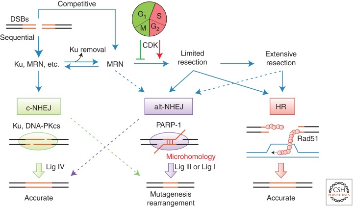
Nonhomologous end joining (NHEJ) refers to a set of genome maintenance pathways in which two DNA double-strand break (DSB) ends are (re)joined by apposition, processing, and ligation without the use of extended homology to guide repair. Canonical NHEJ (c-NHEJ) is a well-defined pathway with clear roles in protecting the integrity of chromosomes when DSBs arise. Recent advances have revealed much about the identity, structure, and function of c-NHEJ proteins, but many questions exist regarding their concerted action in the context of chromatin. Alternative NHEJ (alt-NHEJ) refers to more recently described mechanism(s) that repair DSBs in less-efficient backup reactions. There is great interest in defining alt-NHEJ more precisely, including its regulation relative to c-NHEJ, in light of evidence that alt-NHEJ can execute chromosome rearrangements. Progress toward these goals is reviewed.


Figures

Figure 1.
Figure 1.
Ku and DNA-PKcs. (A) Structural representation of the CTD of human Ku70 (PDB 1JJR) (Zhang et al. 2001). (B) The human Ku70–Ku80 heterodimer bound to DNA (PDB 1JEY) (Walker et al. 2001). (C) The CTD of human Ku80 (PDB 1RW2) (Zhang et al. 2004). (Dashed black lines) Connections between structures. (Light blue) Ku70; (blue) Ku80; (orange) DNA. (D) Low-resolution structure of human DNA-PKcs plus the Ku80 CTD (PDB 3KGV) (Sibanda et al. 2010). (Yellow) Kinase domain, (green) HEAT repeats, (white) “brow,” (blue) putative DBD, and (red) remainder.
Figure 2.
Figure 2.
DNA ligase IV assembly. (A) The adenylation domain (AdD) of Lig4 (blue, PDB 3VNN) (Ochi et al. 2012) is superimposed on a structural representation of Lig1 bound to a DNA nick (light gray, PDB 1X9N) (Pascal et al. 2004) as a surrogate model of how Lig4 might bind DNA. (OBD) Oligonucleotide/oligosaccharide binding domain; (green) 5′ AMP; (orange) DNA. (B) The human XRCC4 homodimer bound to the Lig4 tandem BRCT repeat region (PDB 3II6) (Wu et al. 2009). (C) Human XLF homodimer (PDB 2QM4) (Li et al. 2008b). (D) Surface representation of the XRCC4–XLF axial filament with a bound Lig4 BRCT region, created by superimposing PDB 3II6 onto PDB 3RWR (Andres and Junop 2011). (Blue) Lig4; (shades of green) XRCC4; (shades of red) XLF. (E) Idealized models of DNA engagement and end bridging by XRCC4–XLF multimers, colored the same as in D. “Axial” and “parallel” refer to the orientation of XRCC4–XLF interactions that drive the assembly.
Figure 3.
Figure 3.
Mre11–Rad50–Nbs1 (MRN) complex. (A) Fission yeast Nbs1 FHA domain bound to a Ctp1 phosphopeptide (PDB 3HUF) (Williams et al. 2009). (B) Fission yeast Mre11 globular domain bound to an Nbs1 internal peptide (PDB 4FBW) (Schiller et al. 2012). (C) Pyrococcus furiosus Mre11 globular domain bound to DNA (PDB 3DSD) (Williams et al. 2008). (D) Superimposed structures of the Thermotoga maritima Mre11 globular domain in the open (PDB 3QG5) (Lammens et al. 2011) and closed (PDB 3THO) (Mockel et al. 2012) conformations showing the large Rad50 domain movement induced by adenosine nucleotide binding. (E) P. furiosus Rad50 Zn hook motif (PDB 1L8D) (Hopfner et al. 2002). (Red) Ctp1 peptide; (green) Nbs1; (shades of blue) Mre11; (shades of red) Rad50; (green) ADP; (purple) Zn and Mn ions; (orange) DNA. (Dashed black lines) Connections between structures.
Figure 4.
Figure 4.
Pol X polymerases. (A) Human Pol λ catalytic domain bound to a 1-base gap and incoming nucleotide (light gray; PDB 1XSN) (Garcia-Diaz et al. 2005). (Dark blue) Loop 1; (green) ddTTP; (orange) DNA. (B) Human Pol μ BRCT domain (PDB 2DUN, RIKEN Structural Genomics/Proteomics Initiative, similar to PDB 2HTF) (DeRose et al. 2007). (Dashed black line) Connection between structures. (C) Line diagrams depicting the different requirements imposed on a DNA polymerase (blue) filling a DSB gap (blue arrow) at 3′ (left panel) versus 5′ (right panel) overhangs with respect to placement of the template strand break. (D) dRP lyase activity as an example of end processing, illustrating different requirements for handling terminal versus internal base damage. AP, Abasic site. (E) The Tdp1 fidelity control mechanism that prevents Pol X-dependent insertional mutagenesis by transiently cleaving and blocking termini with a 3′ phosphate.
Figure 5.
Figure 5.
Disposition of DSBs between repair pathways. Diagram illustrating the relationships between c-NHEJ, alt-NHEJ, and HR and the factors that influence the disposition of DSBs between these repair pathways. See text for further discussion.

References

    1. Ahnesorg P, Smith P, Jackson SP 2006. XLF interacts with the XRCC4-DNA ligase IV complex to promote DNA nonhomologous end-joining. Cell 124: 301–313 - PubMed
    1. Andres SN, Junop MS 2011. Crystallization and preliminary X-ray diffraction analysis of the human XRCC4–XLF complex. Acta Crystallogr Sect F Struct Biol Cryst Commun 67: 1399–1402 - PMC - PubMed
    1. Andres SN, Modesti M, Tsai CJ, Chu G, Junop MS 2007. Crystal structure of human XLF: A twist in nonhomologous DNA end-joining. Mol Cell 28: 1093–1101 - PubMed
    1. Andres SN, Vergnes A, Ristic D, Wyman C, Modesti M, Junop M 2012. A human XRCC4–XLF complex bridges DNA. Nucleic Acids Res 40: 1868–1878 - PMC - PubMed
    1. Arlt MF, Rajendran S, Birkeland SR, Wilson TE, Glover TW 2012. De novo CNV formation in mouse embryonic stem cells occurs in the absence of Xrcc4-dependent nonhomologous end joining. PLoS Genet 8: e1002981. - PMC - PubMed

MeSH terms