

Review
doi: 10.1101/cshperspect.a013201.
Protein folding in the endoplasmic reticulum
Affiliations
- PMID: 23637286
- PMCID: PMC3632058
- DOI: 10.1101/cshperspect.a013201
Item in Clipboard
Review
Protein folding in the endoplasmic reticulum
Cold Spring Harb Perspect Biol.
.
Abstract
In this article, we will cover the folding of proteins in the lumen of the endoplasmic reticulum (ER), including the role of three types of covalent modifications: signal peptide removal, N-linked glycosylation, and disulfide bond formation, as well as the function and importance of resident ER folding factors. These folding factors consist of classical chaperones and their cochaperones, the carbohydrate-binding chaperones, and the folding catalysts of the PDI and proline cis-trans isomerase families. We will conclude with the perspective of the folding protein: a comparison of characteristics and folding and exit rates for proteins that travel through the ER as clients of the ER machinery.
Figures
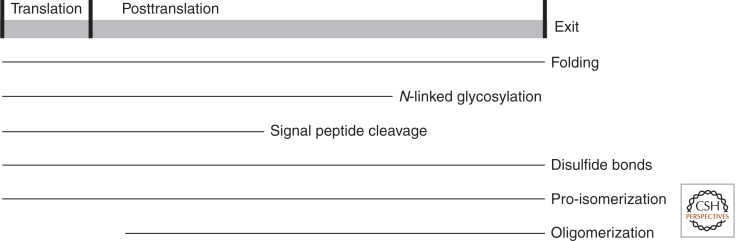
Parallel events during protein folding. The gray bar represents a time course, where folding starts during translation and continues until the protein has reached its native conformation and leaves the ER. If not properly folded or assembled it may exit as misfolded protein.
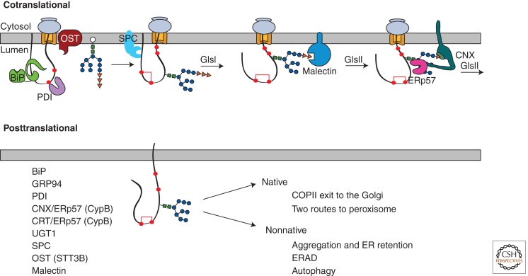
Co- and posttranslational protein folding in the ER lumen. Top panel, the ribosome (grey) sits on the Sec61 translocon (orange) to support cotranslational translocation of the nascent chain into the ER lumen. The oligosaccharyltransferase (OST) attaches preassembled glycans (tree structure) to Asn on the nascent chain. BiP (green) and PDI (purple) are positioned for early assistance. Disulfide bonds start to form. The amino-terminal signal sequence is cleaved by the signal sequence peptidase complex (SPC, light blue). Glucosidase I (GlsI) removes the terminal glucose residue (orange triangle) from the N-linked glycan. The diglucosylated glycan can bind to the membrane-associated lectin, malectin (dark blue). Glucosidase II (GlsII) removes a second glucose to generate a monoglucosylated glycan structure that is bound to calnexin (CNX, green), a lectin chaperone associated with the oxidoreductase ERp57 (pink). Lectin chaperone binding continues until GlsII removes the final glucose residue. Bottom panel, the listed factors interact co- and posttranslationally, after the translation of the nascent chain has been completed. These factors help with maturation and the sorting of the native or nonnative protein for its various fates. Calreticulin (CRT) is a soluble paralogue of calnexin.
References
-
- Allen S, Naim HY, Bulleid NJ 1995. Intracellular folding of tissue-type plasminogen activator. Effects of disulfide bond formation on N-linked glycosylation and secretion. J Biol Chem 270: 4797–4804 - PubMed
-
- Anttonen AK, Mahjneh I, Hamalainen RH, Lagier-Tourenne C, Kopra O, Waris L, Anttonen M, Joensuu T, Kalimo H, Paetau A, et al. 2005. The gene disrupted in Marinesco-Sjogren syndrome encodes SIL1, an HSPA5 cochaperone. Nat Genet 37: 1309–1311 - PubMed
-
- Arnold SM, Kaufman RJ 2003. The noncatalytic portion of human UDP-glucose: Glycoprotein glucosyltransferase I confers UDP-glucose binding and transferase function to the catalytic domain. J Biol Chem 278: 43320–43328 - PubMed
Publication types
MeSH terms
Substances
Grants and funding
LinkOut - more resources
Full Text Sources
Other Literature Sources