. 2013 Jul;87(13):7608-21.
doi: 10.1128/JVI.03186-12. Epub 2013 May 1.

Cellular chaperonin CCTγ contributes to rabies virus replication during infection

Affiliations

Cellular chaperonin CCTγ contributes to rabies virus replication during infection

Jinyang Zhang et al. J Virol. 2013 Jul.

Abstract

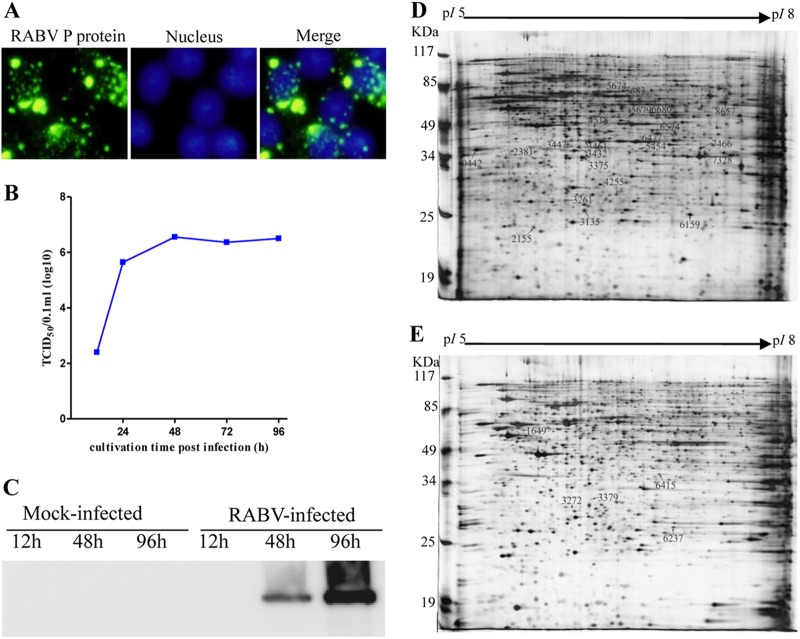
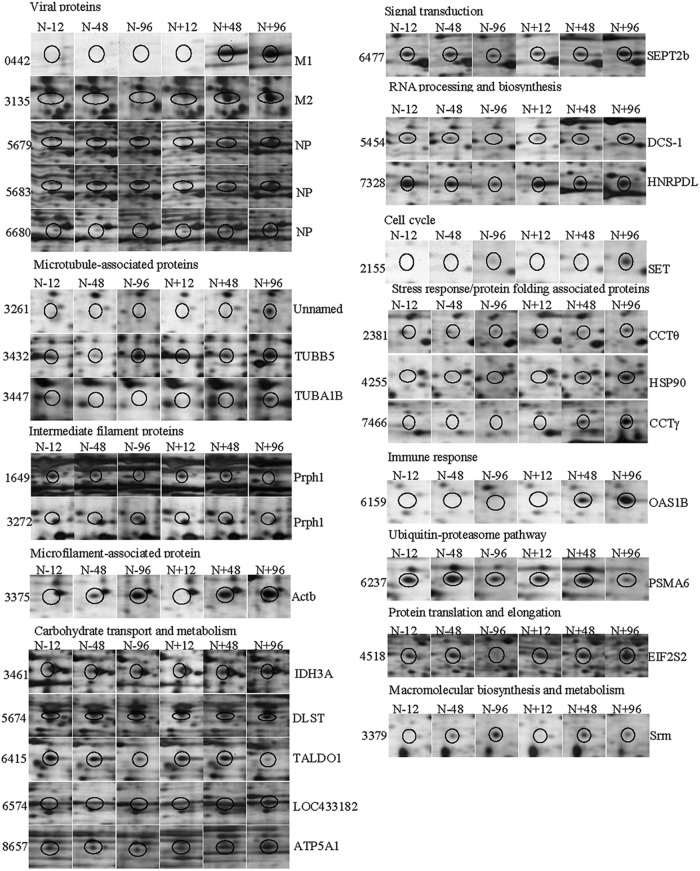
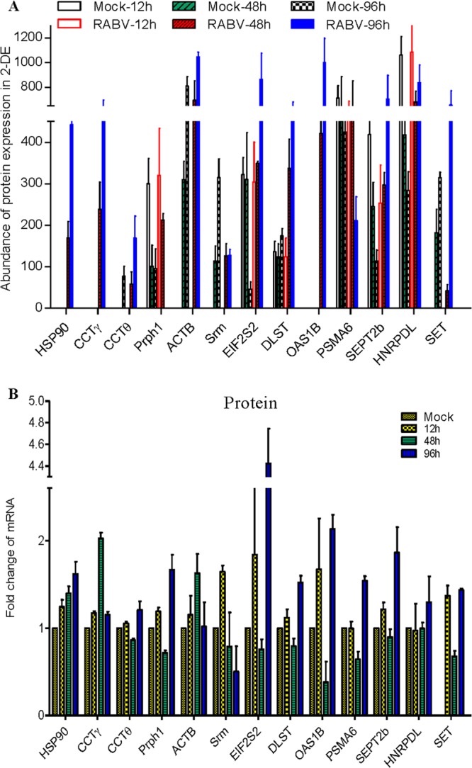
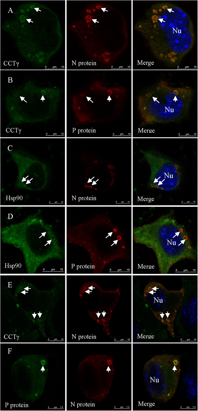
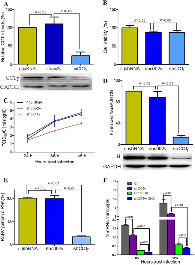
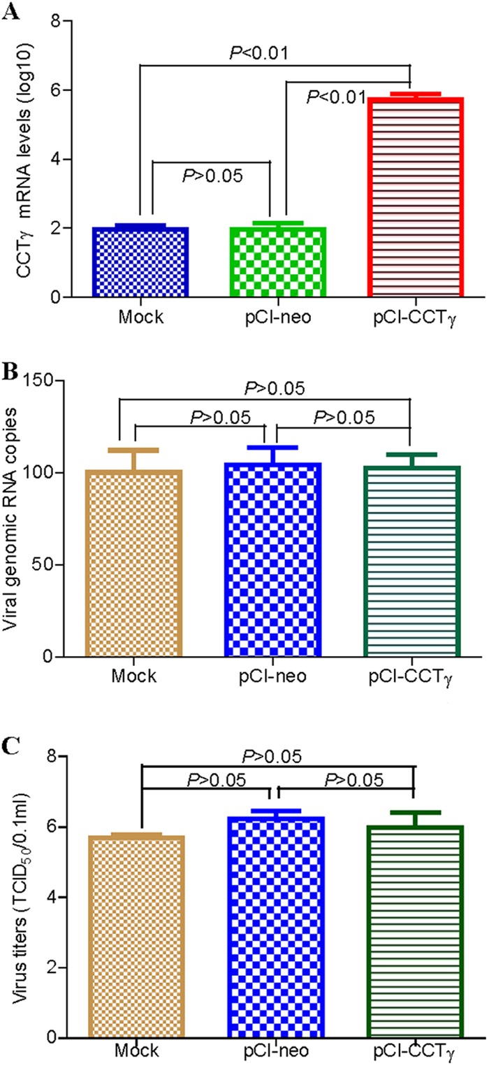
Rabies, as the oldest known infectious disease, remains a serious threat to public health worldwide. The eukaryotic cytosolic chaperonin TRiC/CCT complex facilitates the folding of proteins through ATP hydrolysis. Here, we investigated the expression, cellular localization, and function of neuronal CCTγ during neurotropic rabies virus (RABV) infection using mouse N2a cells as a model. Following RABV infection, 24 altered proteins were identified by using two-dimensional electrophoresis and mass spectrometry, including 20 upregulated proteins and 4 downregulated proteins. In mouse N2a cells infected with RABV or cotransfected with RABV genes encoding nucleoprotein (N) and phosphoprotein (P), confocal microscopy demonstrated that upregulated cellular CCTγ was colocalized with viral proteins N and P, which formed a hollow cricoid inclusion within the region around the nucleus. These inclusions, which correspond to Negri bodies (NBs), did not form in mouse N2a cells only expressing the viral protein N or P. Knockdown of CCTγ by lentivirus-mediated RNA interference led to significant inhibition of RABV replication. These results demonstrate that the complex consisting of viral proteins N and P recruits CCTγ to NBs and identify the chaperonin CCTγ as a host factor that facilitates intracellular RABV replication. This work illustrates how viruses can utilize cellular chaperonins and compartmentalization for their own benefit.


Figures

Fig 1
Fig 1
Identification of RABV-infected mouse N2a cells and representative 2-DE proteomic profiles from mock- and RABV-infected N2a cells. (A) Identification of RABV-infected mouse N2a cells by IFA using the MAb to RABV P. (B) RABV infection kinetics in N2a cells. (C) Detection of the viral N protein in RABV-infected and mock-infected N2a cells by Western blotting using a specific MAb. (D) 2-DE gel of RABV-infected N2a cells with upregulated protein spots labeled. (E) 2-DE gel of mock-infected N2a cells with downregulated protein spots marked.
Fig 2
Fig 2
Dynamic 2-DE profiles of the differentially expressed proteins in RABV-infected N2a cells. Differentially expressed protein spots are circled. N+ and N- indicate the RABV-infected and mock-infected mouse N2a cells, respectively, and numbers indicate time (h) postransfection.
Fig 3
Fig 3
Translational and transcriptional profiling of altered proteins in RABV-infected mouse N2a cells. (A) Translational profiling of the differentially expressed proteins in 2-DE. The spot densities (total pixel intensity within spot boundaries) in 2-DE maps were carried out with PDQuest 2-D analysis software 8.0.1. (B) Total cellular mRNA of mouse N2a cells with or without RABV infection was analyzed by quantitative real-time PCR. All samples were normalized with the results for the GAPDH gene as an internal control and with the results for mock-infected N2a cells at each time point as the reference. The values are quantitation ratios of fold increase or decrease relative to the results for mock-infected cells, and mock-infected-cell transcripts were normalized to 1. Error bars represent standard deviations.
Fig 4
Fig 4
Subcellular distribution of CCTγ and Hsp90 in N2a cells by confocal microscopy. Immunostaining was conducted using MAbs to RABV N and P proteins followed by TRITC-conjugated IgG (red) or FITC-conjugated IgG (green) and counterstaining using rabbit antibodies to CCTγ and Hsp90 followed by FITC-conjugated IgG (green). Nuclei (Nu) were stained with DAPI (blue). The triple-stained cells were observed by confocal microscopy. (A and B) Colocalization of CCTγ with viral proteins N and P, revealing a hollow ring-like structure in RABV-infected N2a cells. (C and D) Hsp90 has some colocalization with the hollow ring-like structure containing viral N and P proteins in RABV-infected N2a cells. (E) CCTγ colocalized with viral protein N, revealing a hollow ring-like structure in mouse N2a cells cotransfected with RABV N and P genes. (F) Colocalization of viral proteins P (green) and N (red) formed a hollow cricoid-like structure (Merge) in N2a cells cotransfected with RABV N and P genes. Scale bars = 10 μm.
Fig 5
Fig 5
CCTγ knockdown inhibits RABV replication and transcription. (A) Western blotting of CCTγ-silenced N2a cells. (-)shRNA, no shRNA. (B) Viability assay of CCTγ-silenced N2a cells. Cell viability is expressed as the percentage of (-)shRNA N2a cells. (C) Growth curve of RABV in CCTγ-silenced N2a cells. N2a cells only transfected with nontargeting shRNA shcoo2v or CCTγ-specific shRNA, as well as no shRNA, used as positive control [(-)shRNA], were infected at an MOI of 0.1. At 48 h.p.i., the virus titer in cells transfected with CCTγ-specific shRNA decreased nearly 20-fold in comparison with that in cells transfected with nontargeting shRNA shcoo2v. (D) Expression of RABV N protein in N2a cells as determined by Western blotting. (E) Quantitation of RABV genomic RNA by quantitative real-time PCR. The genomic RNA of RABV in shRNA-silenced cells was decreased ∼80% (P < 0.01) in comparison with that in cells transfected with nontargeting shRNA shcoo2v. (F) The mRNA transcripts of RABV N gene as determined by real-time comparative quantitative PCR. The transcription of RABV N was decreased significantly after silencing of CCTγ, although CHX interfered significantly in the N2a cells transfected with and without CCTγ-specific shRNA. Values are means ± standard deviations of three independent experiments.
Fig 6
Fig 6
RABV replication in CCTγ-overexpressing N2a cells. (A) CCTγ mRNA transcript. CCTγ mRNA was detected from the transfected N2a cells for 48 h by comparative quantitative PCR (qPCR). GAPDH gene was used as a control. (B) Detection of cellular RABV genomic RNA. The cellular RABV genomic RNA was quantified by absolute qPCR from the RABV-infected N2a cells for 48 h. The RABV genomic copies in the mock N2a cells were normalized to be 100%. (C) Virus titer of RABV. The virus titer in cell supernatants is represented as TCID50.

Similar articles

Cited by

References

    1. Lafon M. 2011. Evasive strategies in rabies virus infection. Adv. Virus Res. 79:33–53 - PubMed
    1. Jamin M, Leyrat C, Ribeiro EA, Gerard FCA, Ivanov I, Ruigrok RWH. 2011. Structure, interactions with host cell and functions of rhabdovirus phosphoprotein. Future Virol. 6:465–481
    1. WHO September 2012. Rabies: fact sheet no. 99. World Health Organization, Geneva, Switzerland: http://www.who.int/mediacentre/factsheets/fs099/en/ Accessed 26 December 2012
    1. Negri A. 1903. Contributo allo studio dell'eziologia della rabbia. Boll. Soc. Med. Chir. Pavia 2:88–115
    1. Suchy A, Bauder B, Gelbmann W, Lohr CV, Teifke JP, Weissenbock H. 2000. Diagnosis of feline herpesvirus infection by immunohistochemistry, polymerase chain reaction, and in situ hybridization. J. Vet. Diagn. Invest. 12:186–191 - PubMed

Publication types

MeSH terms