. 2013 Jul;87(13):7637-45.
doi: 10.1128/JVI.03519-12. Epub 2013 May 1.

Structures of the procapsid and mature virion of enterovirus 71 strain 1095

Affiliations

Structures of the procapsid and mature virion of enterovirus 71 strain 1095

Javier O Cifuente et al. J Virol. 2013 Jul.

Abstract

Enterovirus 71 (EV71) is an important emerging human pathogen with a global distribution and presents a disease pattern resembling poliomyelitis with seasonal epidemics that include cases of severe neurological complications, such as acute flaccid paralysis. EV71 is a member of the Picornaviridae family, which consists of icosahedral, nonenveloped, single-stranded RNA viruses. Here we report structures derived from X-ray crystallography and cryoelectron microscopy (cryo-EM) for the 1095 strain of EV71, including a putative precursor in virus assembly, the procapsid, and the mature virus capsid. The cryo-EM map of the procapsid provides new structural information on portions of the capsid proteins VP0 and VP1 that are disordered in the higher-resolution crystal structures. Our structures solved from virus particles in solution are largely in agreement with those from prior X-ray crystallographic studies; however, we observe small but significant structural differences for the 1095 procapsid compared to a structure solved in a previous study (X. Wang, W. Peng, J. Ren, Z. Hu, J. Xu, Z. Lou, X. Li, W. Yin, X. Shen, C. Porta, T. S. Walter, G. Evans, D. Axford, R. Owen, D. J. Rowlands, J. Wang, D. I. Stuart, E. E. Fry, and Z. Rao, Nat. Struct. Mol. Biol. 19:424-429, 2012) for a different strain of EV71. For both EV71 strains, the procapsid is significantly larger in diameter than the mature capsid, unlike in any other picornavirus. Nonetheless, our results demonstrate that picornavirus capsid expansion is possible without RNA encapsidation and that picornavirus assembly may involve an inward radial collapse of the procapsid to yield the native virion.


Figures
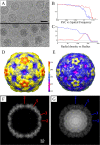
Fig 1
Fig 1
Cryo-EM reconstructions of EV71-9105 procapsid and capsid. (A) Representative areas of cryomicrographs used for the reconstructions with procapsid (top) and mature virion (bottom). Bar = 300 Å. (B) Fourier shell correlation versus spatial frequency. Resolution of the reconstructions is assessed where the FSC curve crosses below a correlation value of 0.5. (C) Radial density profiles revealing that the radius of the procapsid is greater than that of the virion capsid. (D and E) Surface renditions colored by radius of the procapsid and mature capsid density maps, respectively, illustrate the differences in size and angularity between the procapsid and capsid forms. The regions at the 5-fold vertices are distinctly raised in the procapsid by ∼11 Å compared to the mature capsid. (F and G) Corresponding central sections from the cryo-EM maps of the procapsid and the virion, respectively, show the arrangement of the capsid density without and with a packaged RNA genome and before and after cleavage of VP0. Locations of the 2-, 3-, and 5-fold axes of symmetry are indicated.
Fig 2
Fig 2
Central surface projections and radial density projections. (A) Radial plot of a central section in the standard orientation for the capsid surfaces, displayed centered to a circle with an r of 135 Å and labeled positions for 2-, 3-, and 5-fold axes of symmetry that cross the section. Red surfaces for the procapsid are rendered at 1 σ. For the virion (blue), the external surface is rendered at 1 σ and the inner surface is depicted at 2.35 σ, which is the level at which the RNA core can be distinguished from the protein capsid shell. The figure shows the rearrangement of both capsid surfaces between procapsid and virion, with notable differences in the width of the capsid wall, which is more regular and has more-densely packed protein in the virion. (B) The radial density projection is a 2D representation of the distribution of density of a spherical shell of the map at a given radius, r, of 135 Å. Density is white, and black represents the absence of density. The asymmetric unit is marked by a yellow line, with the icosahedral axes indicated. Comparison of the densities at the same radii shows intricate rearrangement of the capsid structural proteins in the virion relative to the procapsid. Specifically, at the 3-fold axis, rearrangement introduces directionality and fills the virion capsid density; at the 2-fold axis of the procapsid, there is notably less density in the procapsid; differences around the 5-fold axis are also noted due to the rearrangement of the density from movement of the protomers in the virion compared to the procapsid.
Fig 3
Fig 3
(A) EV71-1095 procapsid crystal structure. Peptide chains are colored using the canonical picornavirus coloring scheme, with VP1 in blue, VP0 in green, and VP3 in red. The first 81 N-terminal residues of VP0 are disordered; hence, displayed here is the structure of VP0 residues 82 to 318, which correspond to VP2 residues 13 to 249, after cleavage. (B) Sites where EV71-1095 superimposed over the crystal structure of 3VBU (shown in gold) with an RMSD greater than 2 Å: VP1 residues 218 to 219 (GH loop, including disordered residues 211 to 217), VP0 residues 43 to 51 (η1-η2 loop or AB loop) and 138 to 139 (βE-α2 loop or EF loop), and VP3 residues 180 to 183 (βG-η3 loop or GH loop) (specific nomenclature of loops from Wang et al. [9]). (C) The zoomed view shows the EV71-1095 structure (blue) superimposed with the Fuyang structure, 3VBU (gold), to show the VP1 BC, DE, and HI loops of the 5-fold vertex. Side chains of the specific residues were depicted by sticks and labeled to show the slight but significant differences in residues known to affect virulence and PSGL-1 receptor binding.
Fig 4
Fig 4
Cryo-EM reconstructions of the procapsid (A) (red) and the virion (B) (blue), displayed at 1 σ, showing the fitting of crystal structures depicted as ribbons and colored according the canonical picornavirus coloring: VP1 in blue, VP0 in green, and VP3 in red for the procapsid and VP1 in blue, VP2 in green, VP3 in red, and VP4 in yellow for the virion. Full map and cutaway views are shown for each, with closeup views of the docked crystal structures adjacent. (A) Procapsid reconstruction fitted with the crystal structure of the empty particle (procapsid) 4GMP and VP4 from 3VBS (9). The front hemisphere of the reconstruction is displayed in transparent red, and the back hemisphere of the reconstruction shows the inner and outer surfaces. (B) Same views of the virion reconstruction (less RNA) fitted with the crystal structure 3VBS (9), with the reconstruction density displayed in transparent blue. The outer surface was rendered at 1 σ and the inner surface was rendered at 2.35 σ, as described in Fig. 2.
Fig 5
Fig 5
Detail of the difference map in which the density corresponding to an 8-Å map simulated from the EV71-1095 procapsid X-ray crystal structure was subtracted from the cryo-EM map of the procapsid, resulting in an internal 5-fold plateau of density. The EV71-1095 procapsid structure was fitted with additional VP1 and VP4 portions of mature virus (3VBS) spliced in to provide the structure of those parts that are disordered in the procapsid crystal structure. The first 81 N-terminal residues of VP0 disordered in the procapsid correspond to all of VP4 (yellow ribbon) and five residues of VP2 (data not shown). The N-terminal 72 residues of VP1, which are also disordered in the crystal structure, are shown with a blue ribbon. The view is centered on a 5-fold axis of symmetry and shows the inner portion of the internal difference density map surface in translucent gray (radii, 0 to 140 Å). Noncolored portions of the 3VBS structure appear with a white ribbon. Scale bar = 30 Å.
Fig 6
Fig 6
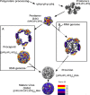
Picornavirus assembly flowchart incorporating the new EV71 structural information and displaying the structural proteins, intermediates of assembly, and protein stoichiometry. The single RNA transcript is translated into a polypeptide that is proteolysed to produce the structural proteins VP0, VP1, and VP3. These proteins assemble into protomers, five of which self-assemble to form a pentamer. Pentamers (14S) assemble into the provirion either by procapsid self-assembly followed by progeny genome packaging (A) or by capsid condensation around a progeny genome (B). The latter model suggests that the empty procapsids are an off-pathway structure. The final step of maturation involves the cleavage of VP0 to generate VP2 and VP4, a step which is induced by the packaged RNA. In the flowchart, the observed and hypothetical paths are depicted by solid and dashed arrows, respectively.

Similar articles

Cited by

References

    1. Schmidt NJ, Lennette EH, Ho HH. 1974. An apparently new enterovirus isolated from patients with disease of the central nervous system. J. Infect. Dis. 129:304–309 - PubMed
    1. Reference deleted.
    1. Solomon T, Lewthwaite P, Perera D, Cardosa MJ, McMinn P, Ooi MH. 2010. Virology, epidemiology, pathogenesis, and control of enterovirus 71. Lancet Infect. Dis. 10:778–790 - PubMed
    1. Huang SW, Hsu YW, Smith DJ, Kiang D, Tsai HP, Lin KH, Wang SM, Liu CC, Su IJ, Wang JR. 2009. Reemergence of enterovirus 71 in 2008 in Taiwan: dynamics of genetic and antigenic evolution from 1998 to 2008. J. Clin. Microbiol. 47:3653–3662 - PMC - PubMed
    1. Tyler KL. 2009. Emerging viral infections of the central nervous system: part 1. Arch. Neurol. 66:939–948 - PMC - PubMed

Publication types

LinkOut - more resources