. 2013 Jun;24(12):2034-44.
doi: 10.1091/mbc.E12-12-0893. Epub 2013 May 1.

Phosphorylation of centromeric histone H3 variant regulates chromosome segregation in Saccharomyces cerevisiae

Affiliations

Phosphorylation of centromeric histone H3 variant regulates chromosome segregation in Saccharomyces cerevisiae

Lars Boeckmann et al. Mol Biol Cell. 2013 Jun.

Abstract

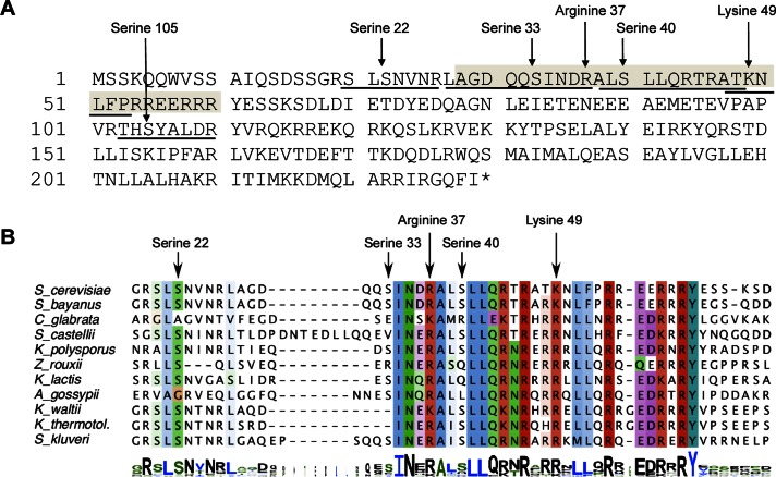
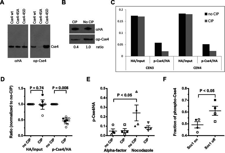
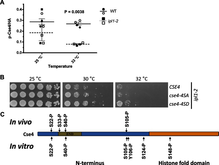
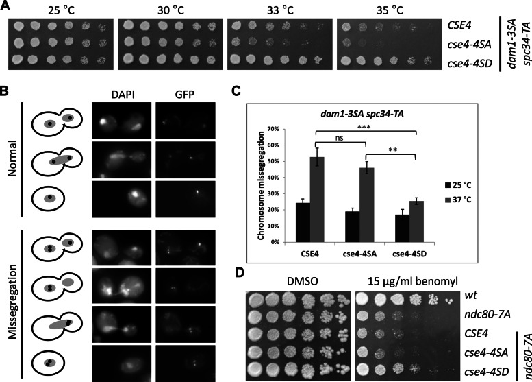
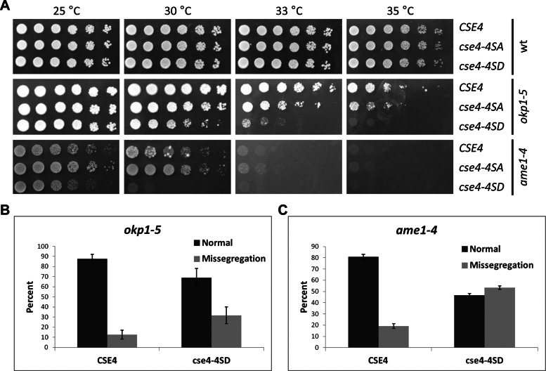
The centromeric histone H3 variant (CenH3) is essential for chromosome segregation in eukaryotes. We identify posttranslational modifications of Saccharomyces cerevisiae CenH3, Cse4. Functional characterization of cse4 phosphorylation mutants shows growth and chromosome segregation defects when combined with kinetochore mutants okp1 and ame1. Using a phosphoserine-specific antibody, we show that the association of phosphorylated Cse4 with centromeres increases in response to defective microtubule attachment or reduced cohesion. We determine that evolutionarily conserved Ipl1/Aurora B contributes to phosphorylation of Cse4, as levels of phosphorylated Cse4 are reduced at centromeres in ipl1 strains in vivo, and in vitro assays show phosphorylation of Cse4 by Ipl1. Consistent with these results, we observe that a phosphomimetic cse4-4SD mutant suppresses the temperature-sensitive growth of ipl1-2 and Ipl1 substrate mutants dam1 spc34 and ndc80, which are defective for chromosome biorientation. Furthermore, cell biology approaches using a green fluorescent protein-labeled chromosome show that cse4-4SD suppresses chromosome segregation defects in dam1 spc34 strains. On the basis of these results, we propose that phosphorylation of Cse4 destabilizes defective kinetochores to promote biorientation and ensure faithful chromosome segregation. Taken together, our results provide a detailed analysis, in vivo and in vitro, of Cse4 phosphorylation and its role in promoting faithful chromosome segregation.


Figures

FIGURE 1:
FIGURE 1:
Amino acids in Cse4 that are posttranslationally modified are evolutionarily conserved. (A) Cse4 is phosphorylated, methylated, and acetylated. The peptides identified by mass spectrometry are underlined, and the modified amino acid residues indicated (also see Table 1). The essential N-terminal domain (END) is shaded (amino acids 28–60). (B) The region containing the PTM sites within the Cse4 N-terminus is conserved. ClustalW alignment of the Cse4 END regions of point centromere yeasts. Alignment in logo format is shown at the bottom (Schneider and Stephens, 1990).
FIGURE 2:
FIGURE 2:
Centromeric association of phosphorylated Cse4. (A) Specificity of Cse4 phospho antibody (αp-Cse4). Western blots of Cse4 proteins purified from strains (BY4741) expressing wild-type Cse4 (pMB1601), cse4-4SA (pMB1566), or cse4-4SD (pMB1563) were probed with αHA (loading control) or αp-Cse4. (B) Phosphatase treatment of Cse4 reduces affinity of αp-Cse4 to Cse4. Cse4 protein purified from BY4741 expressing wild-type Cse4 (pMB1601) was treated with or without CIP and analyzed by Western blots probed with αHA (loading control) or αp-Cse4. (C) Phosphorylated Cse4 is associated with centromeres. ChIP of extracts treated with (CIP) or without CIP (no CIP) was performed with αHA or αp-Cse4 (YMB8378) and assayed by PCR for CEN3 and CEN4 sequences. The graph shows an example of a single experiment. Analysis of four independent experiments and statistical significance are shown in D. (D) Ratios of αHA/input DNA and αp-Cse4/αHA signals normalized to the no-CIP condition. Four independent biological replicates were performed. Error bars show the mean ± 95% confidence intervals. Statistical significance was determined by paired t test. Open symbols, CEN3; filled symbols, CEN4. (E) Centromere-associated phosphorylated Cse4 is increased in nocodazole-treated cells. ChIP samples from wild-type strain (YMB8378) treated with α-factor or nocodazole were analyzed as described in C. The αp-Cse4 signals (normalized to αHA) before and after CIP treatment were calculated. Results from two independent experiments were pooled. Lines on the scatter plots show the mean ± SE of the mean. Statistical significance was determined by one-way analysis of variance (ANOVA). Open symbols, CEN3; filled symbols, CEN4. (F) Phosphorylation of Cse4 is increased in cells depleted of Scc1. ChIP was performed on strain YMB8674 arrested in metaphase (Cdc20 OFF) with defective cohesion (Scc1 OFF). The control strain (Scc1 ON) was grown in galactose medium. Fraction of phospho-Cse4 is the fraction of the total αp-Cse4 signal removed by CIP treatment. Results from two independent experiments were pooled and plotted as in D. Significance was determined by t test. Open symbols, CEN3; filled symbols, CEN4.
FIGURE 3:
FIGURE 3:
Ipl1 phosphorylates Cse4 in vivo and in vitro and phosphomimetic cse4-4SD suppresses temperature sensitivity of an ipl1-2 strain. (A) Ipl1 contributes to the phosphorylation of centromere-associated Cse4. ChIP experiments were performed with WT (YMB8525) and ipl1-2 (YMB8528) strains grown at 25°C and shifted to 33°C for 3 h. The p-Cse4 signal is plotted, with lines showing the mean ± SE of the mean. Results from two independent experiments were pooled. Statistical significance was analyzed by two-way ANOVA (strain, temperature), which indicated that observed variation was significant and predominately due to differences between the wild-type and ipl1-2 strains (p = 0.0038). Results of pairwise tests were corrected for multiple comparisons (Bonferroni). Open symbols, CEN3; filled symbols, CEN4. (B) Phosphomimetic cse4-4SD suppresses the temperature sensitivity of an ipl1-2 strain. Serial dilutions of ipl1-2 (KT1963) with CSE4 (YMB8528), cse4-4SA (YMB8529), or cse4-4SD (YMB8530) were plated on YPD medium and incubated at the indicated temperatures for 2–3 d. (C) Ipl1 phosphorylates Cse4 in vitro. Products of in vitro kinase assays using purified Cse4, ATP, and Ipl1-Sli15 at 30°C for 90 min were analyzed by mass spectrometry. Cse4 modification sites are shown, along with those identified by in vivo analysis (Figure 1A; also see Tables 1 and 2 and Supplemental Figure 3B).
FIGURE 4:
FIGURE 4:
Phosphomimetic cse4-4SD suppresses phenotypes of nonphosphorylatable dam1 spc34 and ndc80 strains. (A) Suppression of temperature sensitivity of dam1 spc34 strains by cse4-4SD. Serial dilutions of dam1-3SA scp34-TA (DDY2503) with CSE4 (YMB8390), cse4-4SA (YMB8458), or cse4-4SD (YMB8393) were assayed for growth at the indicated temperatures on YPD medium for 2–3 d. (B, C) Suppression of chromosome segregation of dam1 spc34 by cse4-4SD. Segregation of a GFP-labeled chromosome (GFP) in strains from A grown at 25 or 37°C was assayed as described in Materials and Methods. Fluorescence images in B show examples of normal or missegregation events scored and quantified in (C). DAPI staining shows the position of the nucleus in each of the cells. Statistical significance was determined by two-way ANOVA (strain, temperature), correcting for multiple comparisons (Bonferroni). ***p < 0.001, **p < 0.01; ns, not significant. Error bars represent SE of the mean. In each of three independent experiments at least 100 cells were counted for each strain. (D) Suppression of benomyl sensitivity of ndc80-7SA by cse4-4SD. Serial dilutions of ndc80-7A (SBY7123) with CSE4 (YMB8395), cse4-4SA (YMB8087), or cse4-4SD (YMB8086) were assayed for growth on YPD containing 15 μg/ml benomyl (or DMSO as control) at 30°C for 4 d.
FIGURE 5:
FIGURE 5:
cse4 phosphorylation mutations cause growth defects and increased chromosome segregation defects in okp1 and ame1 strains. (A) Enhanced temperature sensitivity of cse4 phosphorylation mutants in okp1 and ame1 strains. Serial dilutions of wild-type strain (DDY1925), okp1-5 (YV167), or ame1-4 (YV171) with CSE4 (YMB8378, YMB8384, YMB8654), cse4-4SA (YMB8382, YMB8388, YMB8655), or cse4-4SD (YMB8379, YMB8386, YMB8656) were assayed at the indicated temperatures on YPD medium for 2–3 d. (B, C) cse4-4SD enhances the chromosome segregation defects of okp1-5 (B) and ame1-4 (C) strains. Strains expressing either CSE4 (YMB8384, YMB8672) or cse4-4SD (YMB8386, YMB8673) were arrested with α-factor and shifted to 33 and 30°C, respectively, for 3 h. Segregation of a GFP-labeled chromosome was scored in large-budded cells undergoing mitosis (n > 35), and missegregation of the GFP-labeled chromosome was quantified as described in Figure 4B and Materials and Methods. Error bars represent mean deviations for two independent experiments.

References

    1. Akiyoshi B, Nelson CR, Ranish JA, Biggins S. Analysis of Ipl1-mediated phosphorylation of the Ndc80 kinetochore protein in Saccharomyces cerevisiae. Genetics. 2009;183:1591–1595. - PMC - PubMed
    1. Bharucha JP, Larson JR, Gao L, Daves LK, Tatchell K. Ypi1, a positive regulator of nuclear protein phosphatase type 1 activity in Saccharomyces cerevisiae. Mol Biol Cell. 2008;19:1032–1045. - PMC - PubMed
    1. Biggins S, Severin FF, Bhalla N, Sassoon I, Hyman AA, Murray AW. The conserved protein kinase Ipl1 regulates microtubule binding to kinetochores in budding yeast. Genes Dev. 1999;13:532–544. - PMC - PubMed
    1. Black BE, Jansen LE, Maddox PS, Foltz DR, Desai AB, Shah JV, Cleveland DW. Centromere identity maintained by nucleosomes assembled with histone H3 containing the CENP-A targeting domain. Mol Cell. 2007;25:309–322. - PubMed
    1. Bloom K, Joglekar A. Towards building a chromosome segregation machine. Nature. 2010;463:446–456. - PMC - PubMed

Publication types

MeSH terms