. 2013;9(4):e1003297.
doi: 10.1371/journal.ppat.1003297. Epub 2013 Apr 18.

A mechanistic paradigm for broad-spectrum antivirals that target virus-cell fusion

Affiliations

A mechanistic paradigm for broad-spectrum antivirals that target virus-cell fusion

Frederic Vigant et al. PLoS Pathog. 2013.

Abstract

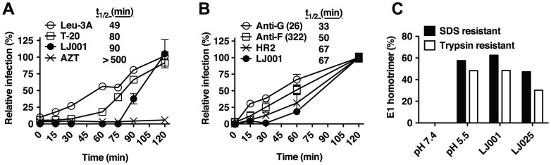
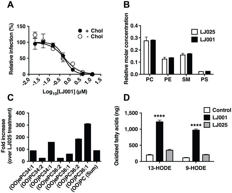
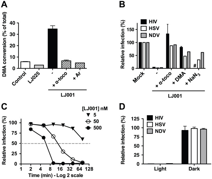
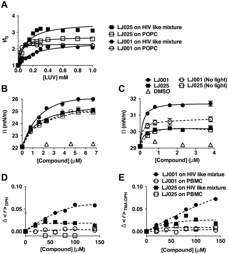
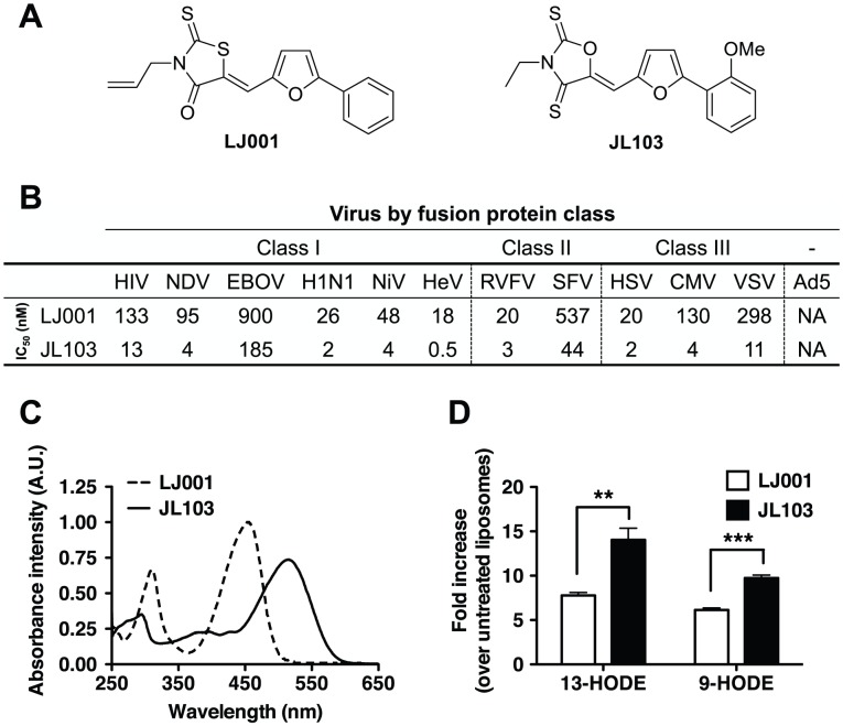
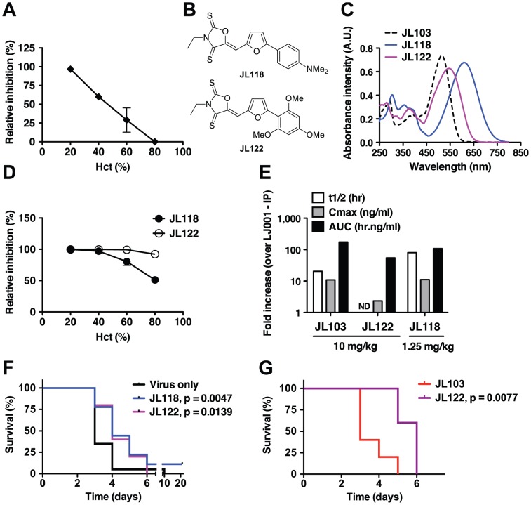
LJ001 is a lipophilic thiazolidine derivative that inhibits the entry of numerous enveloped viruses at non-cytotoxic concentrations (IC50 ≤ 0.5 µM), and was posited to exploit the physiological difference between static viral membranes and biogenic cellular membranes. We now report on the molecular mechanism that results in LJ001's specific inhibition of virus-cell fusion. The antiviral activity of LJ001 was light-dependent, required the presence of molecular oxygen, and was reversed by singlet oxygen ((1)O2) quenchers, qualifying LJ001 as a type II photosensitizer. Unsaturated phospholipids were the main target modified by LJ001-generated (1)O2. Hydroxylated fatty acid species were detected in model and viral membranes treated with LJ001, but not its inactive molecular analog, LJ025. (1)O2-mediated allylic hydroxylation of unsaturated phospholipids leads to a trans-isomerization of the double bond and concurrent formation of a hydroxyl group in the middle of the hydrophobic lipid bilayer. LJ001-induced (1)O2-mediated lipid oxidation negatively impacts on the biophysical properties of viral membranes (membrane curvature and fluidity) critical for productive virus-cell membrane fusion. LJ001 did not mediate any apparent damage on biogenic cellular membranes, likely due to multiple endogenous cytoprotection mechanisms against phospholipid hydroperoxides. Based on our understanding of LJ001's mechanism of action, we designed a new class of membrane-intercalating photosensitizers to overcome LJ001's limitations for use as an in vivo antiviral agent. Structure activity relationship (SAR) studies led to a novel class of compounds (oxazolidine-2,4-dithiones) with (1) 100-fold improved in vitro potency (IC50<10 nM), (2) red-shifted absorption spectra (for better tissue penetration), (3) increased quantum yield (efficiency of (1)O2 generation), and (4) 10-100-fold improved bioavailability. Candidate compounds in our new series moderately but significantly (p≤0.01) delayed the time to death in a murine lethal challenge model of Rift Valley Fever Virus (RVFV). The viral membrane may be a viable target for broad-spectrum antivirals that target virus-cell fusion.


Conflict of interest statement

The authors have declared that no competing interests exist.

Figures

Figure 1
Figure 1. LJ001 inhibits a late stage of viral fusion.
(A) Time-of-addition experiment (see Figure S1). HIV-1JRCSF infection of TZM-bl cells was synchronized by spinoculation for 2 h at 4°C. The plates were subsequently incubated at room temperature (t = 0) for the first 60 min, then to 37°C. LJ001 (20 µM) or HIV entry inhibitors specifically blocking CD4-attachment (Leu-3A, 10 µg/ml), or 6-HB formation (T-20 or enfuvirtide, 5 µM) were added at different times. AZT (10 µM) blocks reverse transcription, a post-entry step. Luciferase expression in cell lysates 48 h post-infection was expressed relative to untreated control (100%). Data representing the mean ± SD of triplicate experiments were graphed, and t 1/2 values calculated using GraphPad PRISM. (B) VSV-ΔG-rluc pseudotyped with NiV envelope glycoproteins, F and G, was spinoculated for 2 h at 4°C onto VERO cells to synchronize the infection. The plates were subsequently shifted to room temperature (t = 0) for 1 h before incubating at 37°C. Inhibitors of NiV entry specifically blocking: attachment (Anti-G, Mab26, 1 µg/ml), fusion triggering (Anti-F, Mab322, 1 µg/ml), or 6-HB formation (HR2, peptide equivalent of T-20 in the HIV system, 1 µM) , , and LJ001 (10 µM) were added at different times. Luciferase expression in cell lysates was analyzed 24 h post-infection and expressed relative to untreated control (100%). Data representing the mean ± SD of duplicate experiments were graphed, and t 1/2 values calculated using GraphPad PRISM. (C) Radiolabeled SFV treated with 6.15 µM of LJ001, or the inactive control LJ025, was allowed to adsorb to BHK cells on ice. After washing, membrane fusion was triggered by low pH, 1 min at 37°C. Controls included non-treated cell-bound virus incubated at low or neutral pH. After fusion triggering, cell lysates were collected and the trypsin- and SDS-resistant E1 homotrimer in each sample was quantified by SDS-PAGE and phosphorimaging. Results, representative of two independent experiments, are expressed as a percent of the total E1 present.
Figure 2
Figure 2. LJ001 oxidizes unsaturated fatty acids in viral membranes.
(A) Equivalent titers of Semliki forest virus (SFV) grown in cholesterol-depleted or control C6/36 mosquito cells were treated with increasing concentrations of LJ001 and their infectivity on target BHK cells determined by immunofluorescence (as in Figure S2). Results are presented as % of infection (mean ± SD, n = 3) relative to that obtained in the absence of LJ001 treatment. The IC50 for LJ001's antiviral activity was determined by non-linear regression using GraphPad PRISM (Top = 100%, Bottom = 0%). (B–C) Purified influenza A virus (A/PR/8/34 H1N1) was treated with 5 µM of LJ001, or control LJ025, and exposed to light for 1 h. The total lipid content was extracted and the viral lipidome analyzed by high-resolution LC-MS (see Figure S3). (B) Relative molar concentration of the major phospholipid species present in the viral lipidome. (C) The amount of peroxidized phosphatidylcholine (PC) species, presented as fold-increase in LJ001- over LJ025-treated samples. Similar results were obtained in two independent experiments with two technical replicates each. PE: Phosphatidylethanolamine, PS: Phosphatidylserine, SM: Sphingomyelin, (OO)ePC: oxidized (hydroperoxide) ether PC, (OO)PC oxidized (hydroperoxide) PC. (D) Liposomes (150 µg in 1 ml) were treated with LJ001 (10 µM), or control LJ025, and exposed to light for 1 h. After de-esterification, fatty acids were extracted, and the amount of 9-hydroxy-10E,12Z-octadecadienoic acid (9-HODE) and 13-hydroxy-9Z,11E-octadecadienoic acid (13-HODE) was determined by LC-MS/MS. Data represents the mean ± SD of triplicates. ****: p<0.0001, LJ001 vs LJ025, Two-way ANOVA, Bonferroni post-test using GraphPad PRISM.
Figure 3
Figure 3. The antiviral activity of LJ001 is dependent on its ability to generate singlet oxygen (1O2).
(A) LJ001, or control LJ025, was added to a solution of DMA and kept under light. After 6 h, DMA conversion was detected by 1H-NMR (DMA∶oxiDMA = 3.1 ppm:2.1 ppm (methyl peak)). Reactions were performed in CDCl3 using 1 equivalent of each reagent. CDCl3 was saturated with oxygen by bubbling O2 through the solvent for 30 min and the reaction was kept under O2 gas atmosphere, except for Ar where oxygen was exchanged with argon by freeze/thaw method. Data represents the mean ± SD of duplicate experiments. (B) HIV-1IIIB, Herpes Simplex Virus-1 (HSV) or Newcastle disease virus (NDV) were incubated with 0.25 µM of LJ001 in the presence of 50 µM α-tocopherol or DMA, or 100 mM NaN3. Infectivity was determined as described in Materials and Methods, and results presented as infection relative to untreated virus (100%). HIV: mean ± SD of duplicate measurements, representative of three independent experiments. HSV and NDV: results representative of three independent experiments. #: NaN3 was toxic to TZM-Bl cells used to assay HIV entry. (C) HSV was incubated with 5, 50 or 500 nM of LJ001 and exposed to white light for 2, 5, 10, 20, 40 or 80 min. Infectivity was determined as described in Materials and Methods, and results presented as infection relative to untreated virus (100%) at a given time, to account for loss of infectivity over time, and as a function of time of light exposure. Data are representative of two independent experiments. (D) HIV-1IIIB, HSV or NDV were treated in the dark with 1 µM of LJ001, and subsequently either exposed to a white light source or left in the dark, for 10 min, before infection of cells in the dark. Relative infectivity was determined as in (B). LJ001-treated viruses exposed to light had >99% reduction in infectivity. Data represents the mean ± SD of two independent experiments.
Figure 4
Figure 4. The effect of LJ001 on the biophysical properties of model versus cellular membranes.
(A) Relative fluorescence intensity increase, of the sample compounds in the presence (I) or absence (I0) of the indicated amounts of membrane, due to partition of LJ001 and LJ025 into large unilamellar vesicles (LUV), performed by successive additions of a concentrated LUV suspension of pure POPC (1-palmitoyl-2-oleyl-sn-glycero-3-phosphocholine, a lipid with packing density and fluidity properties similar to mammalian cell membranes) or HIV membrane-like mixture (POPC 5.3%, DPPC 3.5%, cholesterol 45.3%, SM 18.2%, POPE 19.3% and POPS 8.4%; mol %[15]). Data are representative of three independent experiments. The partition coefficients (Kp) and the fluorescence intensity ratios (ILipids/IWater) resulting from the curve fitting shown here can be found in Table S1. (B–C) Surface pressure measurements on a lipid monolayer comprised of (B) pure POPC or (C) HIV membrane-like mixture with increasing addition of LJ001, LJ025, or DMSO (vehicle control), in the presence or absence of light. Data represent the mean ± SD of duplicate measurements and are representative of three independent experiments. (D–E) Changes in fluorescence anisotropy (<r>) as a function of LJ001 or LJ025 addition to LUV with HIV membrane-like mixture or peripheral blood mononuclear cells (PMBC) using the fluorescent probes (D) DPH or (E) TMA-DPH. Control measurements of <r> vs temperature, using LUV of a reference lipid, showed that the probes were able to correctly detect the membrane phase transition, demonstrating that the compounds did not interfere with the correct assessment of membrane fluidity. Each point is the average of at least triplicates of independent samples.
Figure 5
Figure 5. Improved antiviral and photophysical properties of the oxazolidine-2,4-dithione JL103.
(A) Structures of LJ001 and JL103. (B) IC50 of LJ001 and JL103 against representative viruses that use different classes of fusion proteins (see Figure S8). (C) Absorption spectra of LJ001 and JL103 (100 µM in DMSO). (D) Liposomes (150 µg in 1 ml) were treated with JL103 or LJ001 (10 µM) and exposed to light for 1 h. Fatty acids were extracted as in Figure 2D, and the amount of 9- and 13-HODE was determined by LC-MS/MS. Results are shown as the fold-increase (mean ± S.D., n = 3) in oxidized lipids over untreated samples. Student's t test: **, p = 0.0097; ***, p = 0.0009.
Figure 6
Figure 6. Evaluation of candidate oxazolidine-2,4-dithiones for antiviral activity in vivo.
(A, D) Antiviral efficacy of (A) JL103 or (D) JL118 and JL122 at varying hematocrits (Hct). RBCs in PBS were spiked with HIV-1JR-CSF and brought to the indicated Hct. Normal human Hct is 45±5%. Thin layers of spiked RBCs were treated with 20 µM of the indicated compound under light, for 1 h under constant agitation. Remaining infectivity in the supernatant of treated RBCs was evaluated by inoculating reporter TZM-bl cells. Data represent the relative infectivity (mean ± SD, n = 2) measured 48 h post-infection (untreated control = 100%) from one of two representative experiments. (B) Structures of JL118 and JL122. (C) Absorption spectra of JL103, JL118 and JL122 (100 µM in DMSO). (E) Pharmacokinetics of JL103, JL122 and JL118 in mice. ND: Not determined. (F) Mice lethally challenged IP with 20 pfu of RVFV ZH501 were treated IP once a day for 7 days, starting 1 h post-challenge, with JL118 (1.25 mg/kg) or JL122 (10 mg/kg). n = 20 per group. (G) Mice lethally challenged IP with 50 pfu of RVFV ZH501 were treated IP at 1, 12, 24 and 48 h post-challenge with JL103 (10 mg/kg) or JL122 (10 mg/kg). n = 5 per group. For both (F) and (G), mice were monitored daily and survival as a Kaplan-Meier plot was compared with the Log-rank (Mantel-Cox) test using GraphPad PRISM. Respective p values are indicated on the graphs. (F) JL118 or JL122 treatment moderately, but significantly, increased median survival times compared to the untreated group. (G) Median survival significantly increased from 3 to 6 days for JL103- vs JL122-treated mice, respectively.

References

    1. De Clercq E (2004) Antivirals and antiviral strategies. Nat Rev Microbiol 2: 704–720. - PMC - PubMed
    1. Zasloff M, Adams AP, Beckerman B, Campbell A, Han Z, et al. (2011) Squalamine as a broad-spectrum systemic antiviral agent with therapeutic potential. Proc Natl Acad Sci U S A 108: 15978–15983. - PMC - PubMed
    1. Kesel AJ (2011) Broad-spectrum antiviral activity including human immunodeficiency and hepatitis C viruses mediated by a novel retinoid thiosemicarbazone derivative. Eur J Med Chem 46: 1656–1664. - PubMed
    1. Wolf MC, Freiberg AN, Zhang T, Akyol-Ataman Z, Grock A, et al. (2010) A broad-spectrum antiviral targeting entry of enveloped viruses. Proc Natl Acad Sci U S A 107: 3157–3162. - PMC - PubMed
    1. St Vincent MR, Colpitts CC, Ustinov AV, Muqadas M, Joyce MA, et al. (2010) Rigid amphipathic fusion inhibitors, small molecule antiviral compounds against enveloped viruses. Proc Natl Acad Sci U S A 107: 17339–17344. - PMC - PubMed

Publication types

MeSH terms