. 2013;9(4):e1003312.
doi: 10.1371/journal.ppat.1003312. Epub 2013 Apr 18.

Development of a highly protective combination monoclonal antibody therapy against Chikungunya virus

Affiliations

Development of a highly protective combination monoclonal antibody therapy against Chikungunya virus

Pankaj Pal et al. PLoS Pathog. 2013.

Abstract

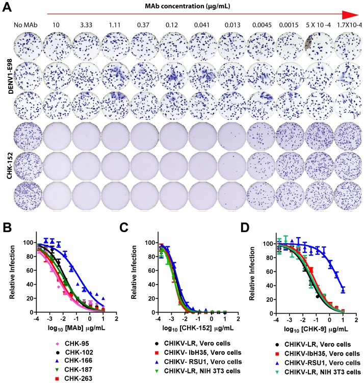
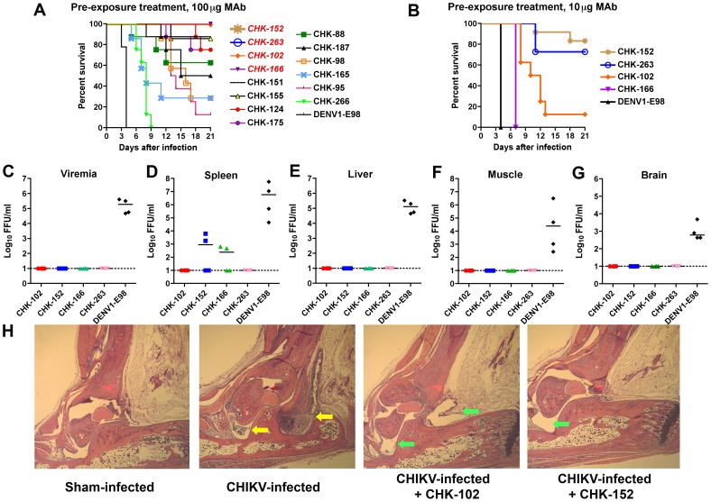
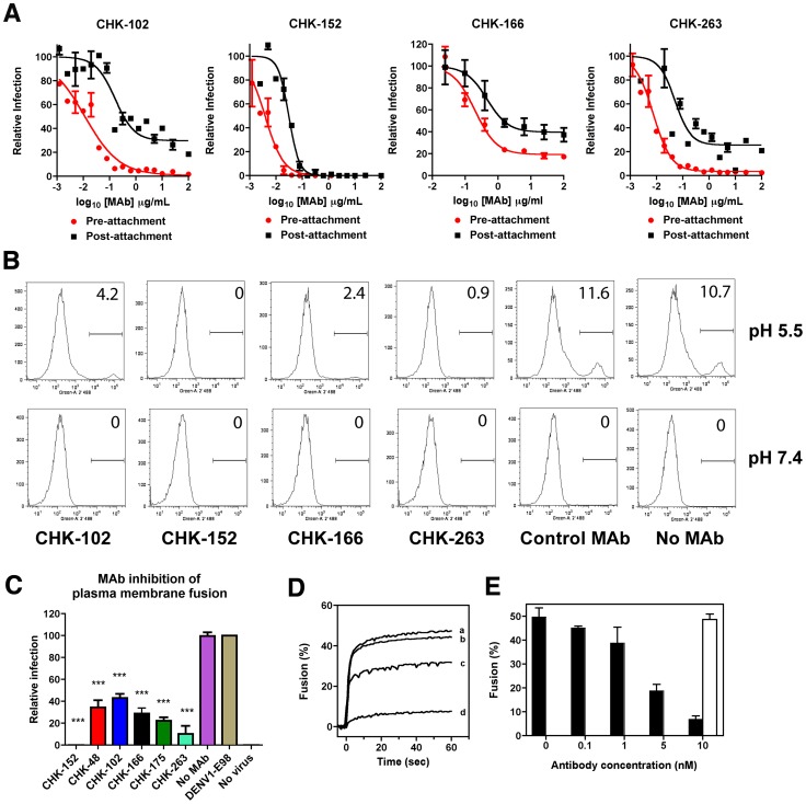
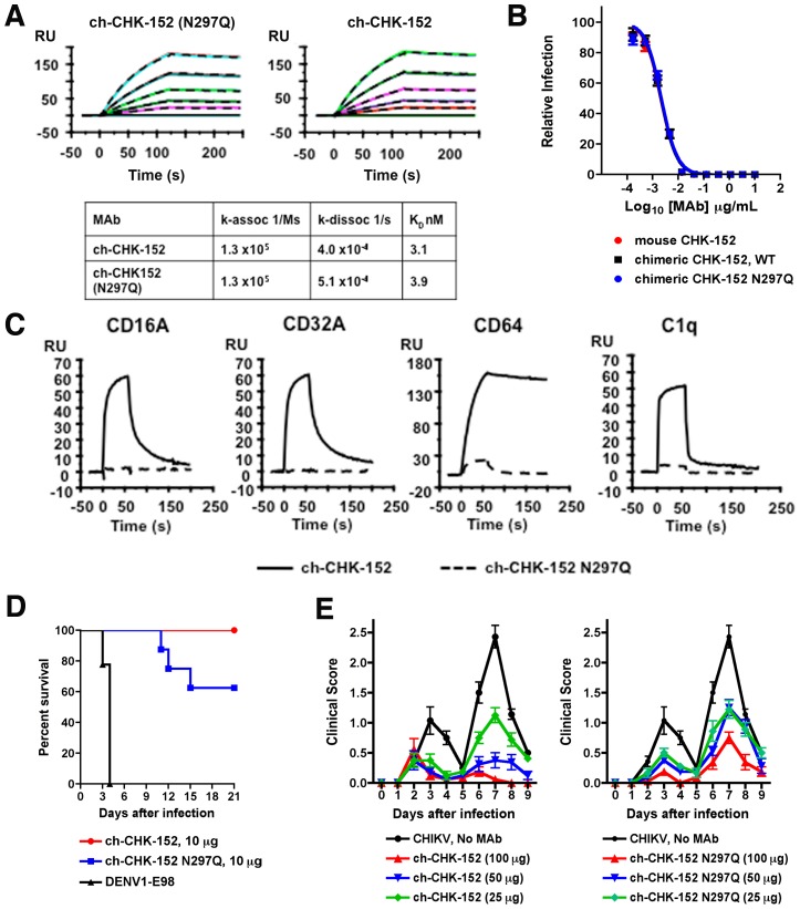
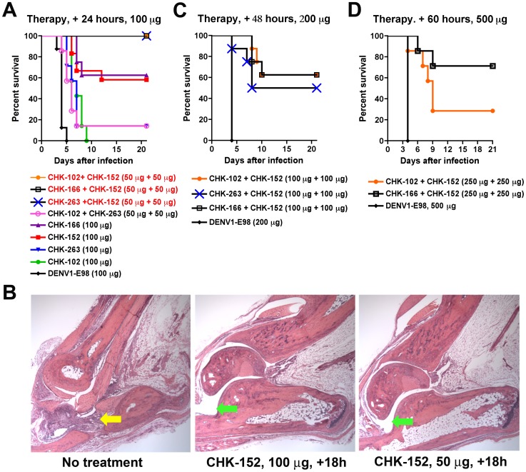
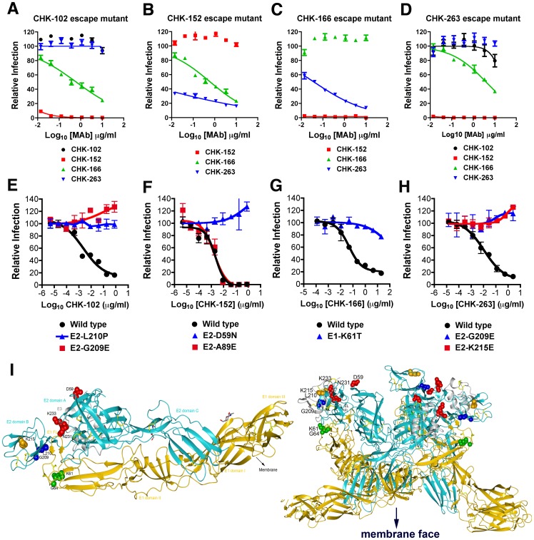
Chikungunya virus (CHIKV) is a mosquito-transmitted alphavirus that causes global epidemics of a debilitating polyarthritis in humans. As there is a pressing need for the development of therapeutic agents, we screened 230 new mouse anti-CHIKV monoclonal antibodies (MAbs) for their ability to inhibit infection of all three CHIKV genotypes. Four of 36 neutralizing MAbs (CHK-102, CHK-152, CHK-166, and CHK-263) provided complete protection against lethality as prophylaxis in highly susceptible immunocompromised mice lacking the type I IFN receptor (Ifnar(-/-) ) and mapped to distinct epitopes on the E1 and E2 structural proteins. CHK-152, the most protective MAb, was humanized, shown to block viral fusion, and require Fc effector function for optimal activity in vivo. In post-exposure therapeutic trials, administration of a single dose of a combination of two neutralizing MAbs (CHK-102+CHK-152 or CHK-166+CHK-152) limited the development of resistance and protected immunocompromised mice against disease when given 24 to 36 hours before CHIKV-induced death. Selected pairs of highly neutralizing MAbs may be a promising treatment option for CHIKV in humans.


Conflict of interest statement

I have read the journal's policy and have the following conflicts: S. Johnson and S. Gorlatov are employees of MacroGenics, which has an option for licensure of the anti-CHKV MAbs for commercial development. M. Diamond is a paid consultant for MacroGenics. This does not alter our adherence to all PLoS Pathogens policies on sharing data and materials.

Figures

Figure 1
Figure 1. Profile of neutralizing MAbs against CHIKV.
A. Examples of MAb neutralization as judged by a reduction in the number of FFU using the Biospot Macroanalyzer. Rows 2 to 12 going across represent decreasing (3-fold) concentrations of CHK-152 or the negative control DENV1-E98 MAb. Column 1 shows infection in the absence of MAb. B. Increasing concentrations of CHK-95, CHK-102, CHK-166, CHK-187, or CHK-263 were mixed with 100 to 150 FFU of CHIKV-LR for one hour at 37°C and Vero cells were infected. Neutralization was determined by FFU assay. C–D. CHK-152 (C) or CHK-9 (D) was mixed with CHIKV-LR (East, Central and South African genotype), CHIKV-RSUI (Asian genotype), or CHIKV IbH35 (West African genotype) for one hour at 37°C and Vero or NIH 3T3 cells were infected as indicated. Neutralization was determined by FFU assay. Data in this Figure is pooled from three independent experiments performed in duplicate or triplicate. All error bars represent the standard deviations.
Figure 2
Figure 2. Efficacy of anti-CHIKV MAb prophylaxis.
A. Six to eight week-old Ifnar −/− C57BL/6 mice were passively transferred 100 µg of the indicated MAbs via an i.p. injection one day before infection with 10 FFU of CHIKV-LR via a s.c. route. The percentage and number of surviving mice were as follows: DENV1-E98 (0%, 0 of 9), CHK-88 (62.5%; 5 of 8), CHK-95 (12.5%; 1 of 8), CHK-98 (28.6%; 2 of 7), CHK-102 (100%; 8 of 8), CHK-124 (75%; 6 of 8), CHK-151 (87.5%; 7 of 8), CHK-152 (100%; 8 of 8), CHK-155 (85.7%; 6 of 7), CHK-165 (28.6%; 2 of 7), CHK-166 (100%; 8 of 8), CHK-175 (75%; 6 of 8), CHK-187 (50%; 4 of 8), CHK-263 (100%; 8 of 8), or CHK-266 (0%; 0 of 8). MAbs italicized in red in the Figure provided 100% protection. B. Ifnar −/− mice were passively transferred 10 µg of MAb via an i.p. injection one day before infection with 10 FFU of CHIKV-LR via a s.c. route. The percentage and number of surviving mice were as follows: DENV1-E98 (0%; 0 of 7), CHK-102 (12.5%; 1 of 8), CHK-152 (83%; 10 of 12), CHK-166 (0%; 0 of 12), or CHK-263 (73%; 8 of 11). For (A) and (B) the survival curves were constructed from data of at least two independent experiments. All anti-CHK MAbs provided statistically significant protection in the percentage of surviving animals or mean survival time compared to the control DENV1-E98 MAb (P<0.05). C–G. Viral burden in MAb-treated Ifnar −/− mice. Animals were passively transferred 100 µg of the indicated MAbs (CHK-102, CHK-152, CHK-166, CHK-263, or isotype control DENV1-E98) via an i.p. injection one day before infection with 10 FFU of CHIKV-LR via a s.c. route. Two days later, viremia (C) and tissues (D, spleen; E, liver; F, muscle; and G, brain) were harvested and infectious virus was titrated by focus-forming assay. Results are pooled from two independent experiments (n = 4 mice per group). The dashed line indicates the limit of detection of the assay and the solid bar indicates the median values. All viral burden results with CHK-102, CHK-152, CHK-166, and CHK-263 were statistically different (P<0.02) from those obtained with DENV1-E98, as analyzed by the Mann-Whitney test. H. Four week-old female WT C57BL/6 mice were sham-treated or administered 100 µg of CHK-102 or CHK-152 via an i.p. route. 24 hours later, mice were infected with 100 PFU of CHIKV-SL 15649 and at day 10, virus-induced pathology in the foot and ankle joint was assessed. (Outer left) Sham-infected, (middle left) CHIKV infected and sham-treated, (middle right) CHIKV-infected and CHK-102 treated, and (outer right) CHIKV infected and CHK-152 treated. Shown are representative images after hematoxylin and eosin staining from at least 3 mice per group at 100× magnification. Yellow and green arrows indicate regions of inflammation or normal joints, respectively.
Figure 3
Figure 3. Mechanism of neutralization by CHIKV MAbs.
A. Pre- and post-attachment inhibition assays. Vero cells were pre-chilled to 4°C and 100 FFU of CHIKV-LR was added to each well for one hour. After extensive washing at 4°C, the indicated MAbs were added for one hour at 4°C, and then the FRNT protocol was completed (black lines, Post). In comparison, a standard pre-incubation FRNT with all steps performed at 4°C is shown for reference. Virus and MAb are incubated together for one hour at 4°C, prior to addition to cells (red lines, Pre). Data shown are representative of three experiments performed in duplicate with error bars representing standard deviation. B–C. FFWO assay. CHIKV was incubated with Vero cells at 4°C to allow virus attachment. Free virus was removed after washing and 50 µg/ml of the indicated MAbs (including DENV1-E98, a negative control MAb) were added at 4°C. Viral fusion at the plasma membrane was induced after a brief exposure to a low pH buffer. After pH normalization, cells were cultured for 14 hours in the presence of NH4Cl to inhibit infection through the endosomal pathway. Cells were analyzed for infection by staining with an anti-E2 MAb. Representative histograms are shown (B) and the data was pooled from four independent experiments for statistical analysis (C). For simplicity of display, not all of the MAbs included in the summary graph are shown by flow cytometry analysis. Asterisks indicate values that are statistically different (P<0.05) from the control MAb. Error bars represent standard deviations. Note low pH-triggered viral fusion at the plasma membrane is an inefficient process with only 10 to 20% of cells becoming infected even when a high MOI was used. D–E. Viral membrane fusion with liposomes. Fusion of pyrene-labeled CHIKV was measured at pH 4.7 (37°C) using liposomes consisting of phosphatidylcholine, phosphatidylethanolamine, sphingomyelin, and cholesterol in a molar ratio of (1/1/1/1.5), as described in the Methods. (D) Curve a, no MAb; curve b, 0.1 nM CHK-152; curve c, 1 nM CHK-152; curve d, 10 nM CHK-152. (E) Extent of fusion (average value between 50 to 60 seconds post acidification) at increasing concentrations of MAb. Black bars, CHK-152; white bar, isotype control (MAb 0031, only included at 10 nM concentration). All fusion measurements were performed at least three independent times.
Figure 4
Figure 4. The effector functions of CHK-152 contribute to protection in vivo.
A. Comparison of binding of ch-CHK-152 and agylocsyl ch-CHK-152 N297Q to pE2-E1, as measured by surface plasmon resonance. A single representative sensogram is shown for each MAb. The experimental curves (colored lines) were fit using a 1∶1 Langmuir analysis (dashed lines), after double referencing, to determine the kinetic parameters presented in the Table immediately below. B. Comparison of neutralizing activity of murine CHK-152, ch-CHK-152, and ch-CHK-152 N297Q, as measured by FRNT on Vero cells. C. Comparison of binding of ch-CHK-152 and ch-CHK-152 N297Q to FcγR (CD16A, 500 nM; CD32A, 100 nM; and CD64, 100 nM) or C1q (50 nM), as measured by surface plasmon resonance. D. Comparison of pre-exposure protective activity of ch-CHK-152 and ch-CHK-152 N297Q. Ifnar −/− mice were administered via an i.p. injection 10 µg of ch-CHK-152 and ch-CHK-152 N297Q one day before infection with 10 FFU of CHIKV-LR via a s.c. route. Mice were monitored for survival for 21 days after infection. The survival curves were constructed from data of at least two independent experiments and the number of animals for each antibody ranged from 8 to 10 per group. ch-CHK-152 provided statistically greater protection than ch-CHK-152 N297Q (P<0.05). E. Five week-old WT C57BL/6 mice were infected with 100 PFU of CHIKV in the left rear footpad and either sham-treated, or treated with 100, 50, or 25 µg of ch-CHK-152 (left panel) or ch-CHK-152 N297Q (right panel) at 18 hours post infection. Mice were scored daily for virus-induced footpad swelling, where a score of 0 = no swelling, 1 = mild swelling where the top of the foot is slightly raised, 2 = moderate swelling with the entire top of foot raised, and 3 = severe swelling involving both the top and bottom of the foot. Scores are the mean values for 7 to 8 mice per treatment group and are representative of three independent experiments. Ch-CHK-152 mediated protection was significantly greater than ch-CHK-152 N297Q on days 7, 8, and 9 post infection for the 100 µg antibody dose, and at day 7 post infection for the 50 µg dose, as determined by the Kruskal-Wallace test with Bonferroni correction (P<0.05). No statistically significant differences between ch-CHK-152 and ch-CHK-152 N297Q were observed with the 25 µg dose. Of note, we observed a reproducible decrease in clinical score on day 5 in many animals. This reflects the biphasic pattern of swelling: during the first 3 to 4 days, swelling is due to edema, whereas after day 5, it is due to inflammatory cell infiltration into the foot and ankle.
Figure 5
Figure 5. Therapeutic efficacy of anti-CHIKV MAbs.
A. Ifnar −/− mice were passively transferred via an i.p. injection 100 µg of DENV1-E98, CHK-102, CHK-152, CHK-166, or CHK-263 or 50 µg each of CHK-102+CHK-152, CHK-166+CHK-152, CHK-263+CHK-152, or CHK-102+CHK-263 at 24 hours after CHIKV infection. B. Five week-old WT C57BL/6 mice were infected with 100 PFU of CHIKV in the footpad and either sham-treated, or treated with 100 or 50 µg of CHK-152 at 18 hours post infection. Virus induced pathology in the foot and ankle joint was assessed by histopathological analysis at day 10 post-infection. (Left) CHIKV-infected, sham-treated, (middle) CHIKV-infected, CHK-152 (100 µg) treated at +18 hours, and (right) CHIKV-infected, CHK-152 (50 µg) treated at +18 hours. Shown are representative images after hematoxylin and eosin staining from 3 mice per group at 100× magnification. Yellow and green arrows indicate regions of inflammation or normal joints, respectively. C. Ifnar −/− mice were passively transferred via an i.p. injection 200 µg of DENV1-E98 or 100 µg each of CHK-102+CHK-152, CHK-166+CHK-152, or CHK-263+CHK-152 at 48 hours after CHIKV infection. D. Ifnar −/− mice were passively transferred via an i.p. injection 500 µg of DENV1-E98 or 250 µg each of CHK-102+CHK-152 or CHK-166+CHK-152 at 60 hours after CHIKV infection. For A, C, and D the survival curves were constructed from data of at least two independent experiments. The number of animals for each antibody ranged from 8 to 10 per group, with the exception of CHK-102+CHK-263, which was performed with 7 mice only. Statistically significant differences in protection compared to DENV1-E98 are described in the text.
Figure 6
Figure 6. Characterization and mapping of neutralization escape mutants.
A–D. FRNT assay with bulk virus obtained after three to six passages under selection of (A) CHK-102, (B) CHK-152, (C) CHK-166, or (D) CHK-263 on Vero cells. Bulk virus also was tested for infectivity in the presence of the non-selecting MAbs. Results are representative of two to three independent experiments performed in triplicate. E–H. Confirmation of resistant phenotype with SFV-CHIKV-GFP containing the indicated single engineered point mutations. Serial dilutions of MAb were incubated with chimeric SFV-CHIKV virus (WT or mutant stocks) for one hour at room temperature. MAb-virus complexes were added to Vero cells plated in 96-well plates and incubated at 37°C. After 8 hours cells were trypsinized, fixed, and the number of GFP-positive, infected cells was assessed by flow cytometry. Curves are representative of 2 to 3 independent experiments. I. Epitope mapping of anti-CHIKV MAbs on the crystal structure of the mature envelope glycoprotein complex (PDB code 3N44). (Left) The domains on E2 (cyan) and E1 (gold) are indicated, and the fusion loop on E1 (E1 FL) is delineated. Amino acid residues of neutralizing MAbs were determined by escape selection, sequencing, and reverse genetic confirmation. CHK-102 and CHK-263 recognize the B domain on E2, CHK-152 recognizes a residue on the wings of the A domain on E2, and CHK-166 recognizes an amino acid in domain II of E1 proximal to the conserved fusion loop. (Right) The mature envelope glycoprotein docked onto the trimer conformation (PDB code 2XFB) that is present on the virion. E3, E2, and E1 and the escape residues are colored as in the left panel. Neutralization escape residues are readily accessible on the top of the trimer, distal to the viral membrane.

References

    1. Staples JE, Breiman RF, Powers AM (2009) Chikungunya fever: an epidemiological review of a re-emerging infectious disease. Clin Infect Dis 49: 942–948. - PubMed
    1. Burt FJ, Rolph MS, Rulli NE, Mahalingam S, Heise MT (2012) Chikungunya: a re-emerging virus. Lancet 379: 662–671. - PubMed
    1. Schuffenecker I, Iteman I, Michault A, Murri S, Frangeul L, et al. (2006) Genome microevolution of chikungunya viruses causing the Indian Ocean outbreak. PLoS Med 3: e263. - PMC - PubMed
    1. Arankalle VA, Shrivastava S, Cherian S, Gunjikar RS, Walimbe AM, et al. (2007) Genetic divergence of Chikungunya viruses in India (1963–2006) with special reference to the 2005–2006 explosive epidemic. J Gen Virol 88: 1967–1976. - PubMed
    1. Powers AM, Brault AC, Tesh RB, Weaver SC (2000) Re-emergence of Chikungunya and O'nyong-nyong viruses: evidence for distinct geographical lineages and distant evolutionary relationships. J Gen Virol 81: 471–479. - PubMed

Publication types

MeSH terms