. 2013 Apr;9(4):e1003425.
doi: 10.1371/journal.pgen.1003425. Epub 2013 Apr 25.

Six homeoproteins directly activate Myod expression in the gene regulatory networks that control early myogenesis

Affiliations

Six homeoproteins directly activate Myod expression in the gene regulatory networks that control early myogenesis

Frédéric Relaix et al. PLoS Genet. 2013 Apr.

Abstract

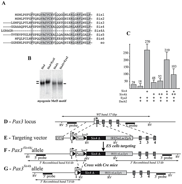
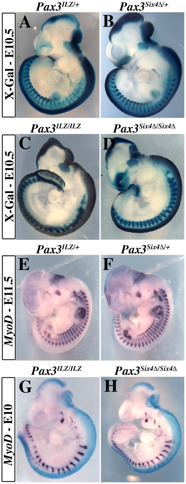
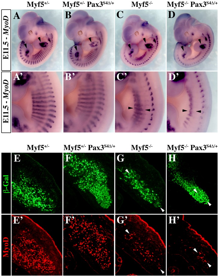
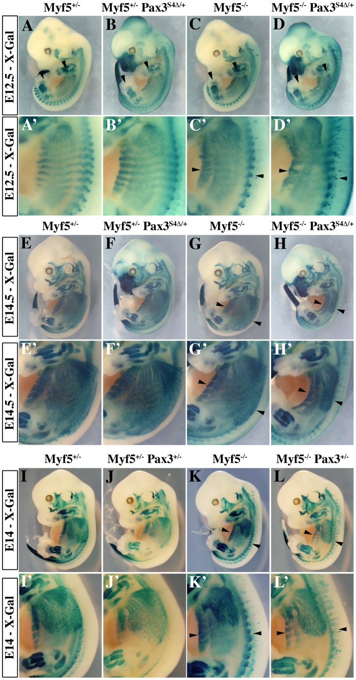
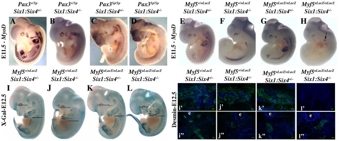
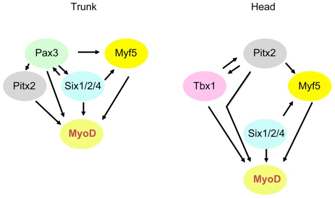
In mammals, several genetic pathways have been characterized that govern engagement of multipotent embryonic progenitors into the myogenic program through the control of the key myogenic regulatory gene Myod. Here we demonstrate the involvement of Six homeoproteins. We first targeted into a Pax3 allele a sequence encoding a negative form of Six4 that binds DNA but cannot interact with essential Eya co-factors. The resulting embryos present hypoplasic skeletal muscles and impaired Myod activation in the trunk in the absence of Myf5/Mrf4. At the axial level, we further show that Myod is still expressed in compound Six1/Six4:Pax3 but not in Six1/Six4:Myf5 triple mutant embryos, demonstrating that Six1/4 participates in the Pax3-Myod genetic pathway. Myod expression and head myogenesis is preserved in Six1/Six4:Myf5 triple mutant embryos, illustrating that upstream regulators of Myod in different embryonic territories are distinct. We show that Myod regulatory regions are directly controlled by Six proteins and that, in the absence of Six1 and Six4, Six2 can compensate.


Conflict of interest statement

The authors have declared that no competing interests exist.

Figures

Figure 1
Figure 1. Genetic relationships between Six1 and Pax3.
X-Gal staining of Six1nLacZ/+ heterozygous embryos on a wild type background (A–C) and on a Pax3 mutant background (Pax3Sp/Sp) (G–I) at E11.5 (A–B, G–H) and E13.5 (C, I) shows that Six1 expression, followed by the nLacZ reporter, is maintained in the absence of Pax3. Comparison of Pax3Sp/Sp : Six1nLacZ/+ embryos (G–I) with Six1nLacZ/nLacZ mutants (D–F) shows a reduction of the extent of the somite where SixnLacZ is expressed, particularly hypaxially at E11.5 (E, H). Disorganisation and loss of hypaxial muscle fibers is observed at E13.5 (F, I) in the interlimb level. These phenotypes are more severe in Pax3Sp/Sp : Six1−/− double mutants (J,K), notably at E13.5 (L). B,E,H,K and C,F,I,L show enlargements in the interlimb region. M-N, Whole mount in situ hybridization using an Eya2 probe on Pax3nLacZ/+ (M) and Pax3nLacZ/nLacZ (N) embryos at E10.5 shows that Eya2 expression is independent of Pax3. O-V, co-immunohistochemistry with Eya2 (Q,R,U,V) and Myod (P,T,R,V) antibodies on interlimb sections of Pax3nLacZ/+ and Pax3nLacZ/nLacZ embryos confirms continuing expression of Eya2 in the absence of Pax3. Reduction of Eya2 expression, notably in hypaxial lips of the somites, is due to dermomyotome reduction in the Pax3 mutant. O,S, DAPI staining.
Figure 2
Figure 2. Targeting of a sequence encoding dominant negative Six4 into the Pax3 locus.
A, Alignment of Six protein sequences shows conservation of the N-terminal-most regions of the Six-domain. This region is absent in the Six4Δ mRNA splicing variant. B, Bandshift assays show that Six4 and Six4Δ bind the MEF3 site, but that only Six4 can interact with Eya2 protein to form a larger complex. C, Transfection experiments performed in primary cultures of chick myoblasts show that Six4 and Eya2 synergistically activate transcription of a luciferase reporter driven by the multimerized MEF3 sequence. In contrast, Six4Δ and Eya2 display no functional synergy, and increasing amounts of Six4Δ compete for Six4-Eya2 transcriptional activation. Y axis, ratio between Luciferase and Renilla activities in arbitrary units. D-G, Strategy for targeting the Six4Δ coding sequence into an allele of Pax3. The probes and restriction enzymes (EcoRV: RV) are indicated, with the size of the resulting wild-type and recombined restriction fragments. The targeting construct (E) contains 2.4 kb and 4 kb of 5′ and 3′ genomic flanking sequences of the mouse Pax3 gene. A floxed puromycin-pA selection marker (Puro), replaces the coding sequence in exon 1 of Pax3 (D), followed by a di-cistronic cassette containing the murine Six4Δ cDNA comprising the whole coding region, followed by an IRESnLacZ cassette and by a final pA signal. The IRESnLacZ allows easy detection of Six4Δ expression . A counter-selection cassette encoding the A subunit of Diptheria Toxin (DTA) was inserted at the 5′end of the vector. After homologous recombination in embryonic stem (ES) cells, Six4Δ-IRESnLacZ expression from the Pax3(Six4 Δ-IRESnLacZ) allele is blocked by the floxed puromycin-pA cassette (F) and is therefore conditional to removal by crossing with a PGK-Cre mouse . This generates the Pax3Six4Δ-IRESnLacZ allele (abbreviated Pax3Six4Δ) (G).
Figure 3
Figure 3. Expression of Six4Δ does not perturb normal embryonic development nor rescue Pax3 mutant deficiencies.
A–B, X-Gal staining of Pax3IRESnLacZ/+ (Pax3ILZ/+, A) and Pax3Six4Δ/+ (B) embryos at E10.5 demonstrates correct expression of the Six4Δ transgene. C–D, X-Gal staining of homozygotes Pax3ILZ/ILZ (C) and Pax3Six4Δ/Six4Δ (D) embryos at E10.5 demonstrates that the Six4Δ sequence does not rescue deficiencies due to the absence of Pax3 (Exencephaly, spina bifida, lack of limb muscles, somitic defects and neural crest cell deficiencies). E–F, Whole mount in situ hybridization using a Myod probe on Pax3ILZ/+ (E) and Pax3Six4Δ/+ (F) embryos at E11.5 shows that the Six4Δ sequence does not overtly perturb Myod expression. G–H, Whole mount in situ hybridization using a Myod probe on homozygote Pax3ILZ/ILZ (G) and Pax3Six4Δ/Six4Δ (H) embryos at E10 shows that the onset of Myod expression is similar to that of a Pax3 mutant, in the absence of Pax3 but in the presence of a dominant negative Six4Δ (H).
Figure 4
Figure 4. Six4Δ affects Myod expression and myogenesis in the absence of Myf5.
A–D′, Whole mount in situ hybridization experiments using a Myod probe on Myf5+/− (A, A′), Myf5+/− : Pax3Six4Δ/+ (B, B′), Myf5−/− (C, C′) and Myf5−/− : Pax3Six4Δ/+ (D, D′) embryos at E11.5. At this stage, in Myf5−/− embryos (C, C′), Myod is activated and begins to rescue the formation of the myotome (arrowheads in C′). However, in Myf5 deficient embryos which express Six4Δ under the control of Pax3 regulatory elements, Myf5−/− : Pax3Six4Δ/+ (D, D′), Myod expression is reduced, affecting the rescue of myotome formation (D′, arrowheads). In contrast, in thoracic somites of Myf5+/− : Pax3Six4Δ/+ (B, B′) Myod expression is not altered compared to Myf5+/− embryos (A,A′). A′–D′, show enlargements in the interlimb region of A–D. E–H′, co-immunohistochemistry on transverse sections of hypaxial somites from Myf5+/− (E, E′), Myf5+/− : Pax3Six4Δ/+ (F, F′), Myf5−/− (G, G′) and Myf5−/− : Pax3Six4Δ/+ (H, H′) embryos at E11.5 using anti-β-Galactosidase (β-Gal) (green, E–H) and anti-Myod (red, E′–H′) antibodies confirms the severe reduction of Myod expression in Myf5−/− : Pax3Six4Δ/+ (H, H′) embryos. Arrowheads indicate examples of cells in which the β-Gal reporter from the Myf5nLacZ allele is expressed and which co-express Myod.
Figure 5
Figure 5. Impaired myogenesis in the presence of Six4Δ, in the absence of Myf5.
A–L′; X-Gal staining of E12.5 (A–D′), E14.5 (E–H′) or E14 (I–L′) Myf5+/− (A, A′, E, E′, I, I′), Myf5+/− : Pax3Six4Δ/+ (B, B′, F, F′), Myf5−/− (C, C′, G, G′,K, K′) and Myf5−/− : Pax3Six4Δ/+ (D, D′, H, H′), Myf5+/− : Pax3+/− (J, J′), Myf5−/− : Pax3+/− (L, L′) embryos demonstrates that in Myf5 deficient embryos which express Six4Δ under the control of Pax3 regulatory elements, the localisation of myogenic cells, marked by the Myf5nLacZ reporter is impaired, notably in trunk muscles (H′ compared with L′). A′–D′, E′–H′, and I′–L′ are enlargements in the interlimb region of A–D, E–H and I–L respectively.
Figure 6
Figure 6. Axial Myod expression is lost in Myf5−/−:Six1−/−/Six4−/− embryos.
A–H, Whole mount in situ hybridization using a Myod probe, I–L, X-Gal staining, and i′–l′, i″–l″ immunohistochemistry on sagittal sections of I–L embryos at the interlimb somites or head level using Desmin antibodies, at E11.5 (A–H) or E12.5 (I–L) with Pax3+/SpSix1+/−Six4+/− (A), Pax3+/SpSix1−/−Six4−/− (B) Pax3Sp/SpSix1+/−Six4+/− (C), Pax3Sp/SpSix1−/−Six4−/− (D), Myf5+/−Six1+/−Six4+/− (E, I), Myf5−/−Six1+/−Six4+/− (F, J) Myf5+/−Six1−/−Six4−/− (G, K), Myf5−/−Six1−/−Six4−/− (H, L) embryos, showing the role of Pax3/Six proteins and Myf5 acting upstream of Myod during trunk myogenesis. Desmin expression in E12.5 compound embryos at the axial level (i′–l′) and at the head level (i″–l″) is not detected in Myf5−/−Six1−/−Six4−/− embryos at the axial level (l′) but at the head level (l″), showing that craniofacial myogenesis can take place in this compound mutant. e: eye. White arrow in L shows the presence of craniofacial muscles.
Figure 7
Figure 7. Six proteins are required for Myod expression in the mouse embryo.
A- Chromatin Immunoprecipitation (ChIP) experiments performed with Eya antibodies or control IgG, on chromatin prepared from Pax3-GFP cells separated by flow cytometry from the trunk region of Pax3GFP/+ embryos at E11.5. ChIP experiments reveal association of Eya proteins with the core enhancer (CE) and distal regulatory region (DRR) 5′ of the Myod gene. B- Sequence of mouse Myod core enhancer (CE) and DRR. MEF3 sites are in red, E boxes in blue, Pitx sites in purple and Pax3 site in green. Underlined sequences correspond to the LS4 and LS15 linker-scanner mutagenesis performed on the human core enhancer . C- Electromobility shift assays showing the interaction of Six1 and Six4 proteins with three distinct MEF3 DNA elements present in the regulatory regions of Myod. Radioactively labelled oligonucleotides with the Myogenin MEF3 site (Myog) were incubated with in vitro translated Six1 and Six4 proteins as a control (1). A 60 or 300 fold excess of unlabelled oligonucleotides containing the MEF3 Myogenin site (2,3), the MEF3 DRR site (4,5), the MEF3 CE1 site (6,7), the MEF3 CE2 site (8,9) or a 300 fold excess of unrelated Myogenin NFI oligonucleotides (10) were added in the reaction mix. D- Wild type and MEF3 mutant Myod transgenes used in the study, (not to scale). PRR, proximal regulatory region corresponding to the Myod promoter. E-Transient transgenic embryos with wild type or mutant Myod sequences at E12-E12.5. X-Gal staining of transgenic embryos with wt CE-MD6.0-nLacZ (a–c) or mut3MEF3-CE-MD6.0-nLacZ (d–f) transgenes. Six out of ten wild type transgenes expressed the LacZ reporter with the same expression pattern, three of them are shown. The number of transgenes inserted varied between 3 and 34 for X-Gal-positive (X-Gal+) embryos, and from 1 to 14 for X-Gal-negative (X-Gal−) embryos. Three out of eight mutant transgenic embryos expressed the LacZ reporter, all three are shown. The number of transgenes inserted was 23 (f), 39 (d) and 40 (e) for X-Gal+ embryos, and from 1 to 51 for X-Gal− embryos. F- Sections for one wild type (c) and for the three mutant transgenic embryos expressing the LacZ transgene were analysed for Myod protein by immunohistochemistry at the thoracic (Th) (c–f), and eye (c″–f″) levels to detect myogenic cells, thus revealing the % of transgene expression (X-Gal+ cells, c′–f′ and c′″–f′″) in the myogenic cell population (Myod-positive cells). While most Myod+ cells express the wt Myod transgene (c′, c′″), very few are marked by expression of the mutant Myod transgene (d′–f′, d′″–f′″).
Figure 8
Figure 8. Six2 proteins bind Myod regulatory elements.
Whole-mount in situ hybridization using a Six2 probe on an E9.5 embryo. Note Six2 expression in dorsal aspects of newly formed somites (black arrow), and in the center of the first branchial arch (white arrow). B- Electromobility shift assays showing the interaction of Six2 and Six5 proteins with three distinct MEF3 DNA elements present in the regulatory regions of Myod. Radioactively labelled oligonucleotides with the Myogenin MEF3 site (Myog) were incubated with in vitro translated Six2 and Six5 proteins as a control (1). A 60 or 300 fold excess of unlabelled oligonucleotides containing the MEF3 Myogenin site (2,3), the MEF3 DRR site (4,5), the MEF3 CE1 site (6,7), the MEF3 CE2 site (8,9) or a 300 fold excess of unrelated Myogenin NFI oligonucleotides (10) were added in the reaction mix. C- Chromatin Immunoprecipitation (ChIP) experiments performed with Six2 antibodies or control IgG, on chromatin prepared from E12 Six1−/−Six4−/− embryos, and showing binding of Six2 in vivo on the regulatory elements of Myod. D- Immunocytochemistry performed with Six2 and Myod antibodies on E12.5 Myf5+/−Six1+/−Six4+/− (a), Myf5+/−Six1−/−Six4−/− (b) Myf5−/−Six1+/−Six4+/− (c), Myf5−/−Six1−/−Six4−/− embryos (d) at the masseter level, demonstrating Six2 (red) accumulation in Myod-positive (green) cells (white arrowheads).
Figure 9
Figure 9. Schematic representation of genetic networks that activate Myod during myogenesis in the trunk and head.
Variations in the interactions of factors in sub-domains at different developmental stages are not included here.

Similar articles

Cited by

References

    1. Gehring WJ, Ikeo K (1999) Pax 6: mastering eye morphogenesis and eye evolution. Trends in genetics 15: 371–377. - PubMed
    1. Kumar JP (2010) Retinal determination the beginning of eye development. Current topics in developmental biology 93: 1–28. - PMC - PubMed
    1. van Heyningen V, Williamson KA (2002) PAX6 in sensory development. Human molecular genetics 11: 1161–1167. - PubMed
    1. Jemc J, Rebay I (2007) The eyes absent family of phosphotyrosine phosphatases: properties and roles in developmental regulation of transcription. Annual review of biochemistry 76: 513–538. - PubMed
    1. Buckingham M, Relaix F (2007) The role of Pax genes in the development of tissues and organs: Pax3 and Pax7 regulate muscle progenitor cell functions. Annual review of cell and developmental biology 23: 645–673. - PubMed

Publication types

LinkOut - more resources