. 2013 Apr;9(4):e1003461.
doi: 10.1371/journal.pgen.1003461. Epub 2013 Apr 18.

The histone demethylase Jarid1b ensures faithful mouse development by protecting developmental genes from aberrant H3K4me3

Affiliations

The histone demethylase Jarid1b ensures faithful mouse development by protecting developmental genes from aberrant H3K4me3

Mareike Albert et al. PLoS Genet. 2013 Apr.

Abstract

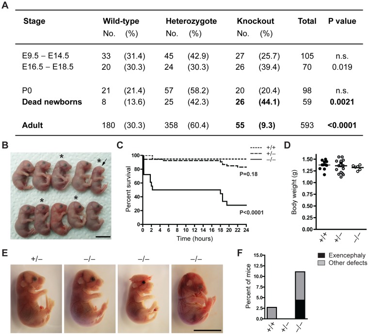
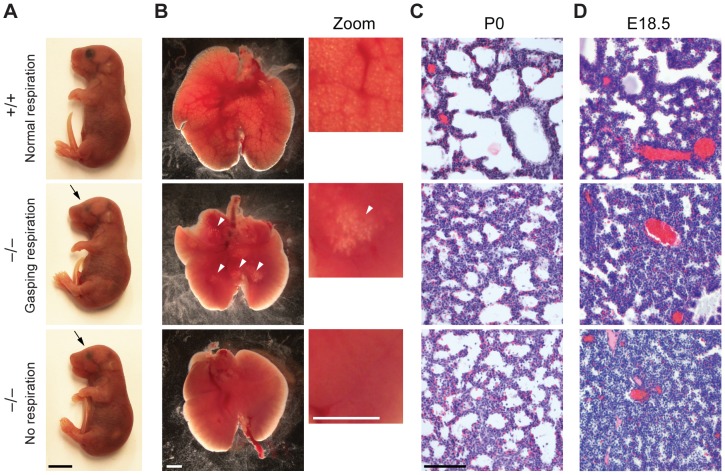
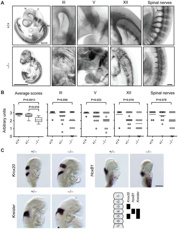
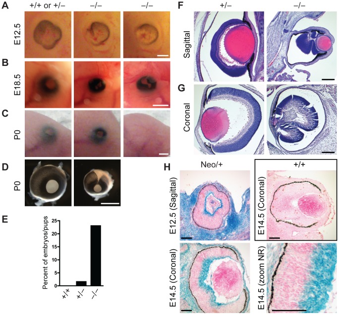
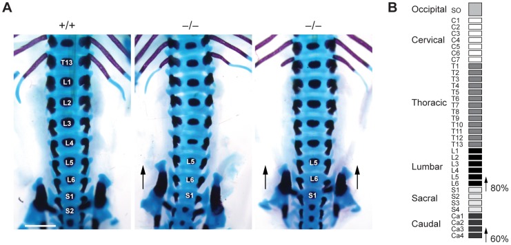
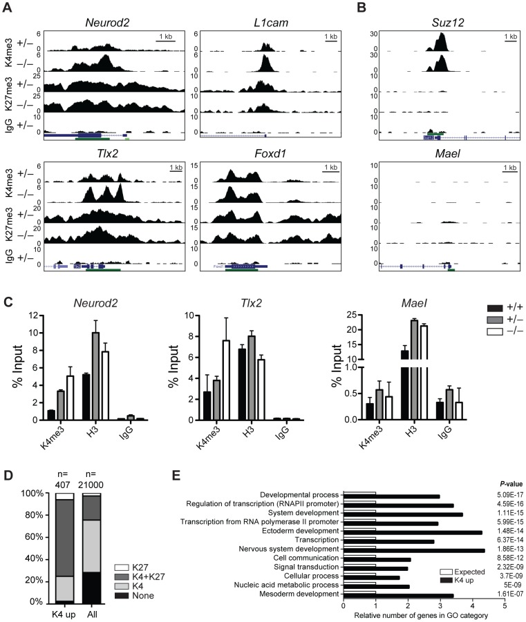
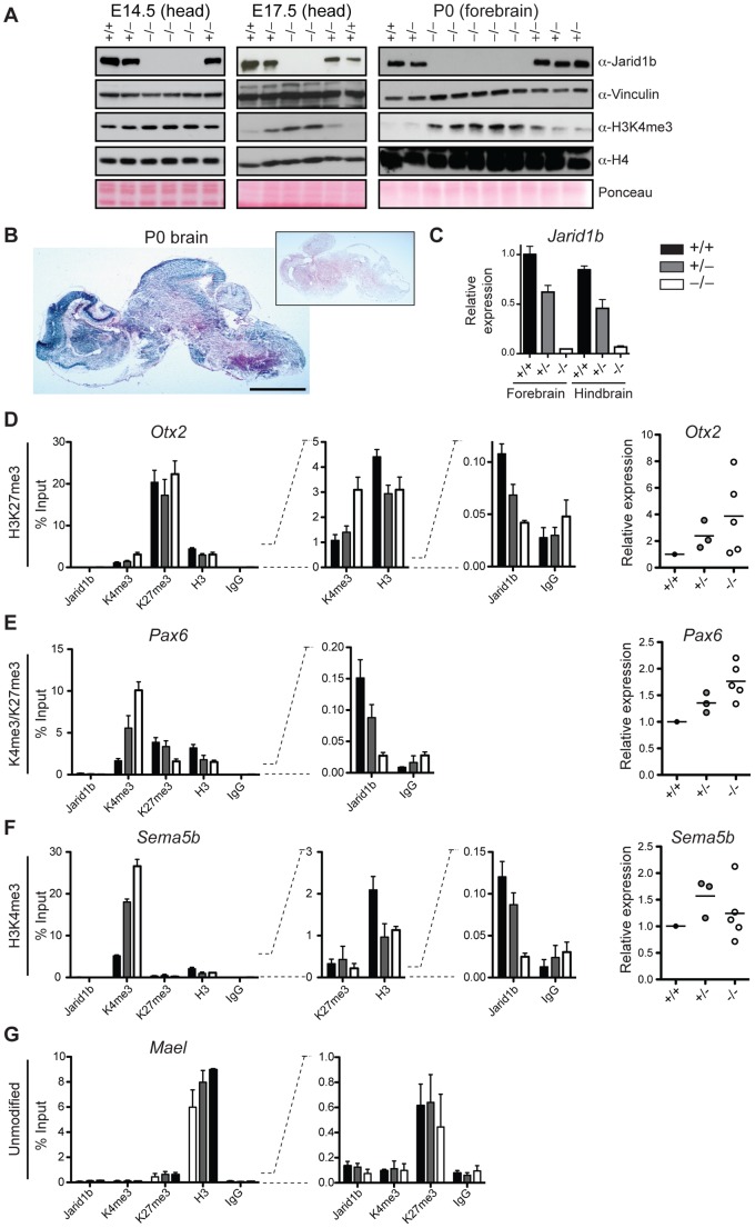
Embryonic development is tightly regulated by transcription factors and chromatin-associated proteins. H3K4me3 is associated with active transcription and H3K27me3 with gene repression, while the combination of both keeps genes required for development in a plastic state. Here we show that deletion of the H3K4me2/3 histone demethylase Jarid1b (Kdm5b/Plu1) results in major neonatal lethality due to respiratory failure. Jarid1b knockout embryos have several neural defects including disorganized cranial nerves, defects in eye development, and increased incidences of exencephaly. Moreover, in line with an overlap of Jarid1b and Polycomb target genes, Jarid1b knockout embryos display homeotic skeletal transformations typical for Polycomb mutants, supporting a functional interplay between Polycomb proteins and Jarid1b. To understand how Jarid1b regulates mouse development, we performed a genome-wide analysis of histone modifications, which demonstrated that normally inactive genes encoding developmental regulators acquire aberrant H3K4me3 during early embryogenesis in Jarid1b knockout embryos. H3K4me3 accumulates as embryonic development proceeds, leading to increased expression of neural master regulators like Pax6 and Otx2 in Jarid1b knockout brains. Taken together, these results suggest that Jarid1b regulates mouse development by protecting developmental genes from inappropriate acquisition of active histone modifications.


Conflict of interest statement

KH is a cofounder of EpiTherapeutics and has shares and warrants in the company. All other authors have declared that no competing financial interests exist.

Figures

Figure 1
Figure 1. The majority of Jarid1b knockouts die within the first day after birth.
(A) The number of Jarid1b wild-type, heterozygous and knockout mice is presented for the indicated stages. P0 refers to pups immediately after caesarian delivery. Dead newborns are pups that were found dead after natural birth (total number of pups examined n = 649; Note that dead pups are often cannibalized by the mothers, and thus the numbers present an underestimation). P values were calculated using a Chi-square test. (B) Litter obtained from Jarid1b heterozygotes immediately after caesarean delivery. Asterics indicate Jarid1b knockout pups. Note that the Jarid1b knockout pup marked by an arrow lacks eyes. (C) Survival curve of Jarid1b wild-type (n = 19), heterozygous (n = 54) and knockout (n = 18) pups during the first day after caesarean delivery. (D) Body weight of newborns. (E) Examples of E18.5 embryos with indicated genotypes. While the left knockout embryo appears normal, the second shows exencephaly while the third one has heterogeneous defects. (F) Frequency of Jarid1b wild-type (n = 37), heterozygous (n = 83) and knockout (n = 50) pups and late embryos that develop exencephaly or other heterogeneous defects. Scale bars, 1 cm.
Figure 2
Figure 2. Reduced lung inflation of Jarid1b knockout pups.
(A) Jarid1b wild-type and knockout littermates immediately after caesarian delivery. Both knockout pups died within 2 hours. Note that both knockout pups have smaller eyes (arrows). Scale bar, 0.5 cm. (B) Lungs isolated 1–4 hours after delivery. Lung inflation is visible in zoomed images on the right. Note that some regions of the lung are inflated (arrowheads) in the knockout pup that shows gasping respiration, while no inflation is visible in the knockout pup that shows no respiration. Scale bars, 1 mm. (C, D) Hematoxylin and eosin staining of paraffin-embedded left lungs at P0 (C) and E18.5 (D). Scale bar, 100 µm.
Figure 3
Figure 3. Cranial and spinal nerves are disorganized in Jarid1b knockout embryos.
(A) Embryos at E10.5 were stained with anti-neurofilament antibody. Shown are examples of a Jarid1b wild-type and knockout embryo (scale bar, 1 mm) and zoomed images (scale bar, 100 µm) of cranial nerves III, V and XII and spinal nerves. Note that the tail has been removed from the knockout embryo to facilitate imaging. (B) Scoring of nerve integrity: 3 = well defined; 2 = not very distinct, 1 = dysmorphic, 0 = absent. P values were determined by Mann-Whitney test. (C) Whole-mount in situ hybridization for Krox20, HoxB1 and Kreisler in E8.75 (5–12 somite pairs) Jarid1b heterozygous and knockout embryos. Scale bar, 200 µm. A scheme illustrating normal gene expression in the rhombomeric hindbrain is shown at the bottom right.
Figure 4
Figure 4. Defects in eye development in Jarid1b knockout embryos.
(A) External morphology of E12.5 eyes. Ventral is to the left. Note that the optic fissure is not completely closed in the Jarid1b knockouts while it is closed in controls (pooled +/+ and +/−). Scale bar, 200 µm. (B) Eyes of E18.5 embryos. Scale bar, 1 mm. (C) Eyes of newborn mice. Note that one of the knockouts has an open eyelid (left) while the other knockout completely lacks the eye (right). Scale bar, 1 mm. (D) Isolated eyes of newborn mice. Scale bar, 1 mm. (E) Frequency of the externally visible defects described in (B) and (C) for wild-types (n = 47), heterozygotes (n = 123) and knockouts (n = 78). (F, G) Hematoxylin and eosin staining of paraffin-embedded embryos at E18.5 showing the left eye cut sagittally (F) and the right eye cut coronally (G). Note that both knockout eyes are microphthalmic and the neural retina is misfolded. Scale bar, 100 µm. (H) Staining for β-galactosidase on sections of E12.5 and E14.5 eyes representing Jarid1b expression. For E14.5, a zoom on the neural retina (NR) is shown at the bottom right. Staining of a wild-type embryo is shown as negative control.
Figure 5
Figure 5. Skeletal transformations in Jarid1b knockout embryos.
(A) Skeletal preparations of E17.5 Jarid1b embryos stained with Alcian blue (cartilage) and Alizarin red (bone). Shown is a ventral view of the lumbar-sacral region. Note that the sacroiliac joints are attached asymmetrically in the knockout shown on the left (n = 5/10) while the joints are symmetrically attached to L6 in the knockout shown on the right (n = 3/10). Scale bar, 1 mm. (B) Schematic representation of the frequency of posterior skeletal transformations in Jarid1b knockout embryos.
Figure 6
Figure 6. H3K4me3 is increased at bivalent and repressed genes in early Jarid1b embryos.
(A, B) Genome wide profiles of H3K4me3 and H3K27me3 were determined by ChIP-seq in the head region of Jarid1b heterozygous and knockout embryos at E8.5. Representative loci with elevated (A) and unchanged (B) H3K4me3 levels in the knockout are shown. y-axis denotes number of sequence tag reads/million. Schematic presentation of Refseq transcripts is in dark blue, and dark green bars represent CpG islands. (C) ChIP-qPCR for H3K4me3, H3 and IgG in E8.5 embryos. Error bars represent S.D. of three PCR amplifications. (D) Percentage of H3K27me3, H3K4me3/H3K27me3, H3K4me3 and unmodified genes among genes with elevated H3K4me3 levels compared to all genes. (E) Gene ontology analysis of genes with elevated H3K4me3 levels generated using PANTHER. The 12 top scoring categories are shown.
Figure 7
Figure 7. Loss of Jarid1b leads to increased global levels of H3K4me3 and higher expression of key developmental regulators during embryogenesis.
(A) Immunoblots for Jarid1b and H3K4me3 of Jarid1b embryos at different developmental stages. Vinculin, H4 and Ponceau serve as loading controls. (B) Staining for β-galactosidase on sections of P0 brains representing Jarid1b expression. Staining of a wild-type brain is shown as negative control (boxed inset). Scale bar, 2 mm. (C) Expression of Jarid1b in fore- and hindbrains of wild-type, heterozygous and knockout newborns (normalized to β-actin). (DG) Left: ChIP-qPCR for Jarid1b, H3K4me3, H3K27me3, H3 and IgG in forebrains of P0 pups. Error bars represent S.D. of three PCR amplifications. Right: Expression of indicated genes in forebrains analyzed by RT-qPCR (normalized to β-actin). Each dot represents an individual embryo. MaeI expression was below detection. One representative gene is shown for each chromatin state: (D) H3K27me3, (E) H3K4me3/H3K27me3, (F) H3K4me3 and (G) unmodified.

Similar articles

Cited by

References

    1. Barski A, Cuddapah S, Cui K, Roh T-Y, Schones DE, et al. (2007) High-Resolution Profiling of Histone Methylations in the Human Genome. Cell 129: 823–837. - PubMed
    1. Schübeler D, MacAlpine DM, Scalzo D, Wirbelauer C, Kooperberg C, et al. (2004) The histone modification pattern of active genes revealed through genome-wide chromatin analysis of a higher eukaryote. Genes & Development 18: 1263–1271. - PMC - PubMed
    1. Guenther MG, Levine SS, Boyer LA, Jaenisch R, Young RA (2007) A chromatin landmark and transcription initiation at most promoters in human cells. Cell 130: 77–88. - PMC - PubMed
    1. Hublitz P, Albert M, Peters AHFM (2009) Mechanisms of transcriptional repression by histone lysine methylation. Int J Dev Biol 53: 335–354. - PubMed
    1. Yu BD, Hess JL, Horning SE, Brown GA, Korsmeyer SJ (1995) Altered Hox expression and segmental identity in Mll-mutant mice. Nature 378: 505–508. - PubMed

Publication types