. 2013 Apr;9(4):e1003469.
doi: 10.1371/journal.pgen.1003469. Epub 2013 Apr 25.

Genetic and biochemical assays reveal a key role for replication restart proteins in group II intron retrohoming

Affiliations

Genetic and biochemical assays reveal a key role for replication restart proteins in group II intron retrohoming

Jun Yao et al. PLoS Genet. 2013 Apr.

Abstract

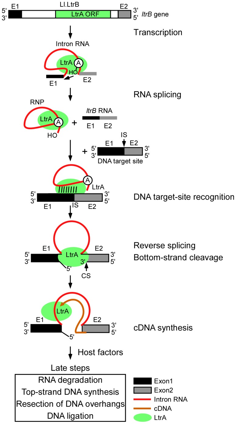
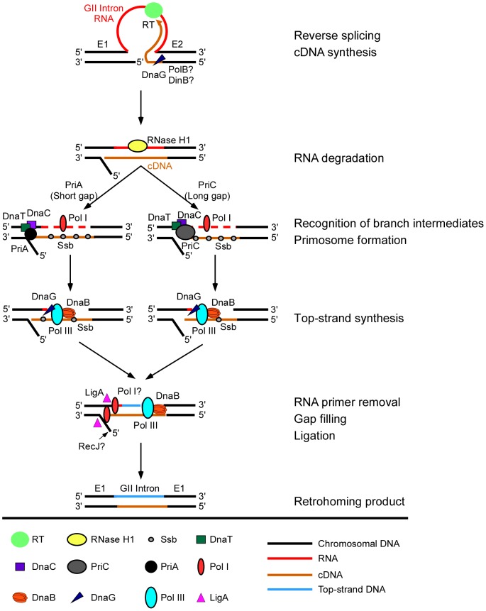
Mobile group II introns retrohome by an RNP-based mechanism in which the intron RNA reverse splices into a DNA site and is reverse transcribed by the associated intron-encoded protein. The resulting intron cDNA is then integrated into the genome by cellular mechanisms that have remained unclear. Here, we used an Escherichia coli genetic screen and Taqman qPCR assay that mitigate indirect effects to identify host factors that function in retrohoming. We then analyzed mutants identified in these and previous genetic screens by using a new biochemical assay that combines group II intron RNPs with cellular extracts to reconstitute the complete retrohoming reaction in vitro. The genetic and biochemical analyses indicate a retrohoming pathway involving degradation of the intron RNA template by a host RNase H and second-strand DNA synthesis by the host replicative DNA polymerase. Our results reveal ATP-dependent steps in both cDNA and second-strand synthesis and a surprising role for replication restart proteins in initiating second-strand synthesis in the absence of DNA replication. We also find an unsuspected requirement for host factors in initiating reverse transcription and a new RNA degradation pathway that suppresses retrohoming. Key features of the retrohoming mechanism may be used by human LINEs and other non-LTR-retrotransposons, which are related evolutionarily to mobile group II introns. Our findings highlight a new role for replication restart proteins, which function not only to repair DNA damage caused by mobile element insertion, but have also been co-opted to become an integral part of the group II intron retrohoming mechanism.


Conflict of interest statement

The authors have declared that no competing interests exist.

Figures

Figure 1
Figure 1. Retrohoming pathway of Ll.LtrB intron lariat RNA in bacteria.
The Ll.LtrB intron, found in a relaxase gene (ltrB) in an L. lactis conjugative element, encodes a multi-functional RT (LtrA protein) with RT, RNA splicing, DNA-binding, and DNA endonuclease activities. Transcription of the ltrB gene yields a precursor RNA containing the intron flanked by 5′ and 3′ exons (E1 and E2, respectively). LtrA is translated from within the intron using its own Shine-Dalgarno sequence and then binds to the intron in the precursor RNA to promote formation of the catalytically active RNA structure for RNA splicing. RNA splicing occurs via two sequential RNA-catalyzed transesterification reactions that are initiated by nucleophilic attack of the 2′ OH of a branch point A-residue near the 3′ end of the intron at the 5′-splice site and results in ligated ltrB exons and an excised intron lariat RNA with a 2′-5′ phosphodiester linkage. After splicing, LtrA remains tightly bound to the excised intron lariat RNA in an RNP. RNPs initiate retrohoming by recognizing a DNA target site (the ligated ltrB E1–E2 sequence), using both the IEP and base pairing of the intron RNA. The intron RNA then inserts via reversal of the two transesterification reactions used for RNA splicing (referred to as “full reverse splicing”) into the intron-insertion site (IS) at the ligated-exon junction in the top strand of the DNA target site. LtrA uses its DNA endonuclease activity to cleave the bottom strand at a site (CS) between positions +9 and +10 of E2 and uses the 3′ DNA end at the cleavage site as a primer for reverse transcription of the inserted intron RNA. The resulting intron cDNA is then integrated into the genome by host enzymes in late steps that minimally include degradation of the intron RNA template strand, second (top)-strand DNA synthesis, resection of DNA overhangs, and sealing of DNA strand nicks .
Figure 2
Figure 2. Genetic and Taqman qPCR assays used to identify E. coli mutants deficient in retrohoming.
(A) Genetic assay. The CamR intron-donor plasmid pALG3 uses a T7lac promoter and phage Φ10 Shine-Dalgarno (SD) sequence to express an ltrB/GFP fusion cassette. This cassette consists of a 0.9-kb Ll.LtrB-ΔORF intron and flanking 5′- and 3′-exons (E1 and E2, respectively) , with the intron carrying a trimethoprim-resistance retrotransposition-activated genetic marker (TpR-RAM), and E2 linked in-frame to an ORF encoding GFP. The LtrA ORF preceded by its own Shine-Dalgarno sequence is co-transcribed from a position downstream of the GFP ORF. The AmpR recipient plasmid contains a 45-bp Ll.LtrB target site (ligated E1–E2 sequence) upstream of a promoterless tetR gene. T1 and T2 are E. coli rrnB transcription terminators, and TΦ is a phage T7 transcription terminator. (B) Taqman qPCR assay. The assay quantifies 5′- and 3′-intron integration junctions resulting from retrohoming of a retargeted Ll.LtrB-ΔORF intron into a site in the rhlE gene in the E. coli chromosome. Retrohoming events are quantified by Taqman qPCR, which utilizes the 5′→3′ exonuclease activity of Taq DNA polymerase to cleave a fluorescently labeled DNA probe that base pairs to an internal region of a PCR amplicon. Digestion of the probe by Taq DNA polymerase releases the FAM label (red star) free of the MGB quencher (green star), resulting in a quantifiable fluorescence signal for each amplification event. The numbers of 5′- and 3′-intron integration junctions relative to the number of rhlE targets were determined by quantifying the fluorescence signals in three separate PCRs relative to standard curves generated from serial dilutions of a reference plasmid (Materials and Methods). Primers for these PCRs are depicted by arrows with numbers indicating the positions of the 5′ nucleotide of the upstream primer and 3′ nucleotide of the downstream primer relative to the intron-integration junction.
Figure 3
Figure 3. E. coli extract assay for bottom-strand (cDNA) and top-strand DNA synthesis.
(A) Time courses. Group II intron RNPs and labeled DNA substrates (73 bp) containing the Ll.LtrB-insertion site (ligated E1–E2 sequence) were incubated with E. coli HMS174(DE3) extract in the presence of 1 mM dNTPs, 1.5 mM ATP, and an ATP-regenerating system (phosphoenolpyruvate+pyruvate kinase) at 37°C. The DNA substrates were labeled at the 5′ end of either the top (T) or bottom (B) strands to separately assay top- and bottom-strand DNA synthesis. After terminating portions of the reaction at the indicated times, samples were split into halves, which were incubated without or with RNases A+H, and the products were analyzed in a denaturing 6% polyacrylamide gel, which was dried and scanned with a PhosphorImager. RNase-sensitive top-strand products contain the reverse-spliced intron RNA. Schematics below the gels depict bottom- and top-strand synthesis on the DNA substrates (intron and exons not drawn to scale; star indicates 5′ 32P-label). (B) Primer extension analysis. DNA products synthesized in a time course were digested with RNase A+H, purified in a 1% agarose gel (0.85–1.2 kb gel slice), and analyzed by primer extension using 5′ -labeled primers to detect bottom-strand cDNAs (primer FB); the top-strand 5′-intron-integration junction (primer 5T); and top-strand DNAs (primer FT). Major products are diagrammed below the gel. (C) Requirements for bottom- and top-strand DNA synthesis. Reactions with the indicated components were incubated at 37°C for 30 min and then processed and analyzed in a denaturing 6% polyacrylamide gel, as described above. (D) Bottom- and top-strand products obtained with RNPs containing wild-type LtrA protein or an RT-deficient mutant LtrA (RT; YADD motif changed to YAAA). For simplicity, the bottom part of the gel with the labeled DNA substrate (S) is shown only for panel D. Asterisks indicate gels bands of the size expected for full-length bottom- and top-strand products.
Figure 4
Figure 4. Assays of top- and bottom-strand DNA synthesis with extracts from E. coli mutants.
DNA substrate labeled with 32P at the 5′-end of the top (T) or bottom (B) strand were incubated with group II intron RNPs for 15 min at 37°C in reaction medium containing extracts from: (A) Keio deletion mutants and their parental wild-type strain BW25113. (B) Mutant strain C0719, which contains a mariner-transposon at the site of a predicted sRNA, a pnp mutant, a priB deletion (non-Keio), and their parental wild-type strains; and (C) temperature-sensitive mutants and their parental wild-type strains. After phenol-CIA extraction and proteinase K digestion, samples were split into halves, which were incubated without or with RNase A+H at 37°C for 30 min. The products were analyzed in a denaturing 6% polyacrylamide gel, which was dried and scanned with a PhosphorImager. Extracts were confirmed to contain equal amounts of protein by SDS-polyacrylamide gels stained with Coomassie blue (not shown). The amount of radiolabel in the indicated product band or bands was normalized for the amount of substrate (S) in each lane and expressed as a percent of that in the parental wild-type strain (Table 2). At least two assays were done for each mutant and were reproducible to within <30%.
Figure 5
Figure 5. Model for function of host factors in group II intron retrohoming in E. coli.
In initial steps, the group II intron lariat RNA reverse splices into the top strand of the DNA target site, while the intron-encoded RT cuts the bottom DNA strand and uses the 3′ end of the cleaved strand as a primer for target DNA-primed reverse transcription of the intron RNA. During or after cDNA synthesis, a host RNase H (RNase H1) degrades the intron RNA template strand. Extension of the intron cDNA into the 5′ exon displaces the bottom-DNA strand resulting in a branched intermediate that is recognized by the replication restart proteins PriA or PriC, with PriA preferentially recognizing intermediates with short gaps in the bottom strand and PriC preferentially recognizing intermediates with long gaps in the bottom strand. PriA and PriC then initiate a replisome loading cascade involving the sequential recruitment of the replicative helicase DnaB, the primase DnaG, and the replicative polymerase Pol III for second-strand DNA synthesis. Ssb stabilizes single-stranded DNA in gapped regions and interacts with PriA to stimulate the loading of DnaB. The 5′→3′ exonuclease activity of Pol I contributes to the removal of residual RNA primers and its DNA polymerase activity may contribute to filling in gaps, and a host DNA ligase (LigA) seals nicks in the top and bottom strands. Although bottom-strand synthesis is completely dependent on group II RT activity (Figure 3D), biochemical assays show that it is strongly inhibited in a DNA primase (DnaG) mutant and moderately inhibited in repair DNA polymerase DinB and PolB mutants, suggesting a previously unsuspected role for host factors in initiating bottom-strand (cDNA) synthesis. Deletion of RecJ moderately inhibits synthesis of full-length bottom strands in extracts, consistent with a role in resection of the 5′-overhang resulting from the staggered cleavage of the DNA substrate by group II intron RNPs .

Similar articles

Cited by

References

    1. Lambowitz AM, Zimmerly S (2011) Group II introns: mobile ribozymes that invade DNA. Cold Spring Harb Perspect Biol 3: a003616 doi:10.1101/cshperspect.a003616. - DOI - PMC - PubMed
    1. Zimmerly S, Guo H, Eskes R, Yang J, Perlman PS, et al. (1995) A group II intron RNA is a catalytic component of a DNA endonuclease involved in intron mobility. Cell 83: 529–538. - PubMed
    1. Zimmerly S, Guo H, Perlman PS, Lambowitz AM (1995) Group II intron mobility occurs by target DNA-primed reverse transcription. Cell 82: 545–554. - PubMed
    1. Yang J, Zimmerly S, Perlman PS, Lambowitz AM (1996) Efficient integration of an intron RNA into double-stranded DNA by reverse splicing. Nature 381: 332–335. - PubMed
    1. Cousineau B, Smith D, Lawrence-Cavanagh S, Mueller JE, Yang J, et al. (1998) Retrohoming of a bacterial group II intron: mobility via complete reverse splicing, independent of homologous DNA recombination. Cell 94: 451–462. - PubMed

Publication types

LinkOut - more resources