. 2013 Apr;9(4):e1003473.
doi: 10.1371/journal.pgen.1003473. Epub 2013 Apr 18.

Scavenger receptors mediate the role of SUMO and Ftz-f1 in Drosophila steroidogenesis

Affiliations

Scavenger receptors mediate the role of SUMO and Ftz-f1 in Drosophila steroidogenesis

Ana Talamillo et al. PLoS Genet. 2013 Apr.

Abstract

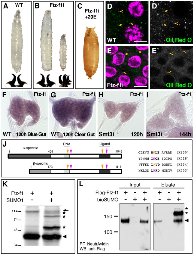
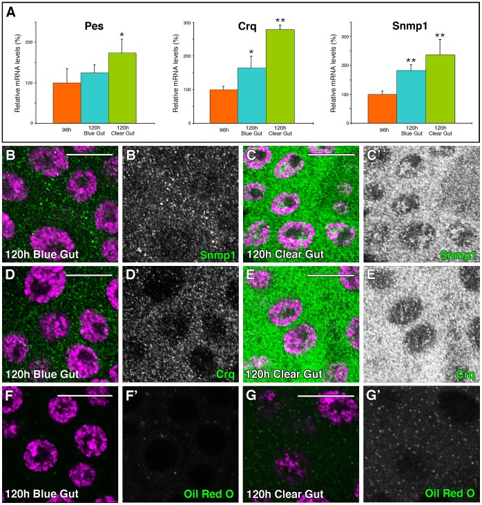
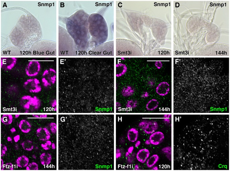
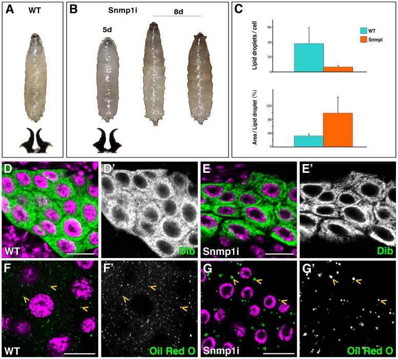
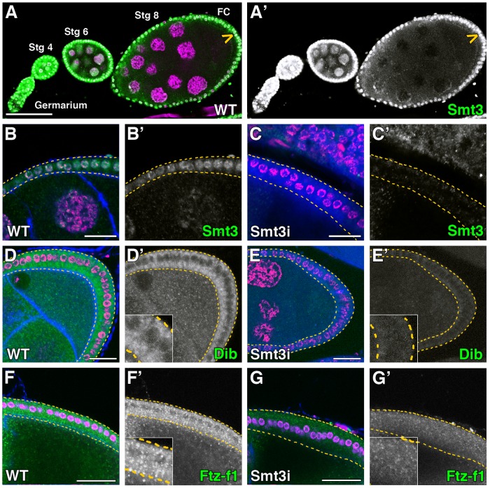
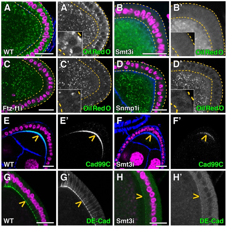
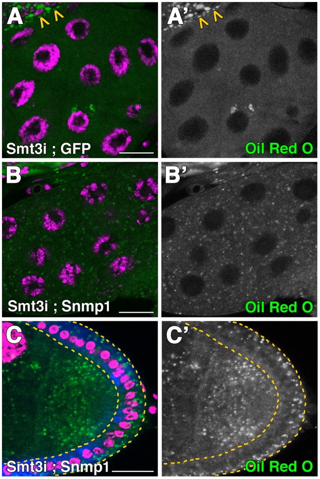
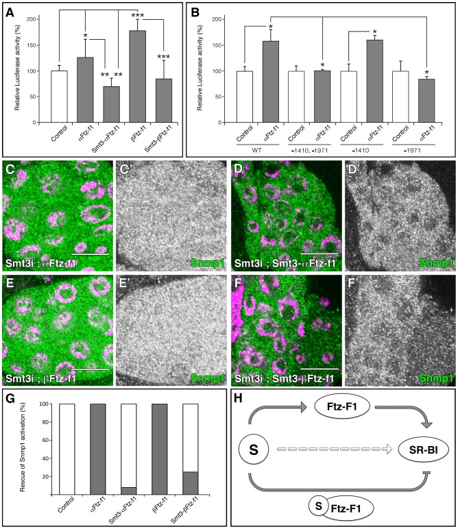
SUMOylation participates in ecdysteroid biosynthesis at the onset of metamorphosis in Drosophila melanogaster. Silencing the Drosophila SUMO homologue smt3 in the prothoracic gland leads to reduced lipid content, low ecdysone titers, and a block in the larval-pupal transition. Here we show that the SR-BI family of Scavenger Receptors mediates SUMO functions. Reduced levels of Snmp1 compromise lipid uptake in the prothoracic gland. In addition, overexpression of Snmp1 is able to recover lipid droplet levels in the smt3 knockdown prothoracic gland cells. Snmp1 expression depends on Ftz-f1 (an NR5A-type orphan nuclear receptor), the expression of which, in turn, depends on SUMO. Furthermore, we show by in vitro and in vivo experiments that Ftz-f1 is SUMOylated. RNAi-mediated knockdown of ftz-f1 phenocopies that of smt3 at the larval to pupal transition, thus Ftz-f1 is an interesting candidate to mediate some of the functions of SUMO at the onset of metamorphosis. Additionally, we demonstrate that the role of SUMOylation, Ftz-f1, and the Scavenger Receptors in lipid capture and mobilization is conserved in other steroidogenic tissues such as the follicle cells of the ovary. smt3 knockdown, as well as ftz-f1 or Scavenger knockdown, depleted the lipid content of the follicle cells, which could be rescued by Snmp1 overexpression. Therefore, our data provide new insights into the regulation of metamorphosis via lipid homeostasis, showing that Drosophila Smt3, Ftz-f1, and SR-BIs are part of a general mechanism for uptake of lipids such as cholesterol, required during development in steroidogenic tissues.


Conflict of interest statement

The authors have declared that no competing interests exist.

Figures

Figure 1
Figure 1. Ftz-f1 is required for pupariation and lipid uptake and is modified by SUMOylation in vivo and in vitro.
(A) Wild type (WT) L3 larva and mouth hooks. (B) Most of the phm-Gal4>UAS-ftz-f1 RNAi (ftz-f1i) larvae arrested development at L3, while some of them stop at L2–L3 transition, as shown by the mouth hook morphology. (C) L3 ftz-f1i larvae fed with 20E were able to pupariate. (D, E) Single plane confocal micrographs showing nuclei marked with DAPI (purple) and lipid droplets, indicated by yellow arrowheads, stained with Oil Red O (green) in L3 control (D) and ftz-f1i PGs (E). (D′, E′) Single green channels are shown in black and white. L3 ftz-f1i PG cells contained reduced number of lipid droplets. All pictures were taken under the same intensity settings. Scale bars indicate 10 µm. (F–I) Micrographs of ftz-f1 mRNA in situ hybridization in PGs from WT (F, G) or phm-Gal4>UAS-smt3 RNAi (smt3i) larvae (H, I) at the indicated hours AEL. ftz-f1 expression increases in clear-gut respect to blue-gut larvae (G versus F, respectively). In smt3i larvae ftz-f1 expression is downregulated (H, I). All the in situ reactions were stopped at the same time and pictures were taken at the same magnification. (J) Schematic representation of α- and βFtz-f1 isoforms indicating the predicted SUMOylation sites (arrows) and the sequences for the high-scoring sites found in Ftz-f1. Pink arrows indicate the SUMOylation sites conserved in insects and orange arrows the SUMOylation sites conserved from insects to vertebrate NR5A2. Pink and orange Ks indicate the lysines where SUMO attachment could take place. Numbering is based on the βFtz-f1 isoform. (K) In vitro SUMOylation assay. Incubation of the Ftz-f1 protein in the presence (+) of SUMO1 changes the motility of the protein (asterisks). Arrowhead indicates the unmodified protein and arrow indicates an unspecific band. Molecular weight markers are shown to the left. (L) In vivo SUMOylation assay. S2R+ cell extracts containing the indicated plasmids are shown. Arrowhead indicates the unmodified Ftz-f1 protein, while the asterisks indicate the high molecular weight SUMOylated forms. Input and eluate of the pull down reaction (PD) are shown. Note that the unmodified protein interacts with the NeutrAvidin beads (arrowhead).
Figure 2
Figure 2. SR-BIs expression and lipid uptake is upregulated at late L3.
(A) Graphical representation of the qPCR results showing the upregulation of the Drosophila SR-BI members pes, crq and Snmp1 from 96 hours AEL to 120 hours AEL blue-gut and 120 hours AEL clear-gut larvae. Error bars indicate standard deviation. One asterisk (p<0.001) and two asterisks (p<0.0001) indicate significant upregulation respect to 96 hours larvae. (B–G) Single plane confocal micrographs taken under the same intensity settings. Nuclei are marked with DAPI (purple). (B–C) Expression of Snmp1 (green) is upregulated in WT PGs from 120 hours AEL blue-gut larvae (B) to 120 hours AEL clear-gut larvae (C). (D–E) Crq expression (green) is upregulated in WT PGs from 120 hours AEL blue-gut larvae (D) to 120 hours AEL clear-gut larvae (E). (F, G) The number of lipid droplets shows simultaneous increase in the PGs during the same time, as shown by Oil Red O staining (green). (B′–G′) Single green channel are shown in black and white. Scale bars indicate 20 µm.
Figure 3
Figure 3. Expression of SR-BIs is reduced in smt3i and ftz-f1i PG cells.
(A–D) Micrographs of Snmp1 mRNA in situ hybridization in PGs from WT (A, B) or phm-Gal4>UAS-smt3 RNAi (smt3i) larvae (C, D) at the indicated hours AEL. Snmp1 mRNA is upregulated in WT clear-gut larvae (B, compare with A). However, no expression is visible in smt3i PGs (C, D). All the in situ reactions were stopped at the same time and pictures were taken at the same magnification. (E–H) Single plane confocal micrographs taken under the same intensity settings. Nuclei are marked with DAPI (purple) and Snmp1 (E–G) or Crq (H) are shown in green. (E, F) smt3i PG cells have basal but reduced levels of Snmp1 at 120 hours AEL larvae (compare to WT in Figure 2B) but they do not upregulate SR-BIs expression at 120–144 hours AEL larvae. (G, H) ftz-f1i PG cells show Snmp1 (G) or Crq (H) expression highly reduced compared to Figure 2C or 2E, respectively. (E′–H′) Single green channels are presented in black and white. Scale bars indicate 20 µm.
Figure 4
Figure 4. Snmp1 is required in the PG for pupariation and lipid uptake.
(A) Wild type (WT) L3 larva and mouth hooks. (B) phm-Gal4>UAS-Snmp1 RNAi (Snmp1i) larvae arrested development at L3, as shown by morphology of mouth hooks, and were able to survive for several days. (C) Graphical representation of the average number of lipid droplets per cell and the area per lipid droplet in WT versus Snmp1i PG cells. (D–G) Single plane confocal micrographs taken under the same intensity settings. Nuclei are marked with DAPI (purple). (D, E) Dib expression is similar in WT (D) and Snmp1i PG cells (E). (F–G) Oil Red O staining shows the lipid droplets (yellow arrowheads) content in WT (F) and Snmp1i PG cells (G). Note that the number of lipid droplets was highly reduced, while the size of the lipid droplet was larger in Snmp1i compared to control PG cells. (D′–G′) Single green channels are shown in black and white. Scale bars indicate 20 µm.
Figure 5
Figure 5. Smt3 is necessary for the expression of Dib and Ftz-f1 in follicle cells.
(A–G) Single plane confocal micrographs were taken under the same intensity settings in smt3 knockdown and control ovaries. Nuclei are labelled with DAPI (purple). F-actin cytoskeleton is shown in blue in B, D, F and G. GFP is shown in blue in C and E. Follicle cells are indicated by yellow dotted lines in B–G. (A′–G′) Single green channels are shown in black and white. Close-up of the follicle cells are shown in insets in D′–G′. Scale bars indicate 20 µm. (A) Expression of Smt3 (green) in the Drosophila germarium and egg chambers. Arrowhead points to follicle cells (FC). (B–C) Smt3 (green) expression levels in follicle cells in T155-Gal4>UAS-GFP;UAS-smt3i (Smt3i) (C) is severely reduced compared with the control (B). (D–G) Stage 8 and early stage 9 egg chambers showing in green the expression of Dib (D, E) or Ftz-f1 (F, G) in T155-Gal4>UAS-GFP;UAS-smt3i or T155-Gal4>UAS-smt3i (E, G) compared with the controls (D, F).
Figure 6
Figure 6. Smt3 and Snmp1 are necessary for lipid uptake in follicle cells.
(A–H) Single plane confocal micrographs taken under the same intensity settings. Nuclei are labelled with DAPI (purple). F-actin cytoskeleton is shown in blue. Follicle cells are indicated by yellow dotted lines. (A′–H′) Single green channels are shown in black and white. Close-up of the follicle cells are shown in insets in A′–D′. Scale bars indicate 20 µm. (A–D) Lipid droplets shown by Oil Red O staining (green) in follicle cells of WT (A), T155-Gal4>UAS-smt3i (B), T155-Gal4>UAS-ftz-f1i (C) or T155-Gal4>UAS-Snmp1i (D) adults. (E, F) Cad99C (green; yellow arrowheads) is very much reduced in T155-Gal4>UAS-smt3i (F) respect to WT (E). (G, H) DE-Cad (green; yellow arrowheads) is also reduced in T155-Gal4>UAS-smt3i (H) respect to WT (G).
Figure 7
Figure 7. Snmp1 restores the lipid droplets content in smt3i cells.
(A–B′) Single plane confocal micrographs taken under the same intensity settings showing Oil Red O staining (green) in PGs of phm-Gal4>UAS-GFP;UAS-smt3i (A) or phm-Gal4>UAS-Snmp1;UAS-smt3i (B). Arrowheads in A indicate the brain hemisphere with normal levels of lipid droplets, as smt3 was not silenced there. (C) Confocal micrograph showing Oil Red O staining (green) of follicle cells T155-Gal4>UAS-Snmp1;UAS-smt3i. GFP is shown in blue. Follicle cells are indicated by yellow dotted lines. (A′–C′) Single green channels are shown in black and white. Nuclei are labelled with DAPI (purple). Scale bars indicate 20 µm.
Figure 8
Figure 8. SUMOylation of Ftz-f1 modulates Snmp1 expression.
(A, B) Graphical representations of the luciferase activity from WT Snmp1-Luc (A) or the indicated mutant vectors (B) cotransfected in S2R+ Drosophila cells with plasmids expressing the indicated proteins. As control, empty pUASTattB vector was used. A single asterisk indicates p<0.05, two asterisks p<0.001 and three asterisks p<10−6. (C–F) Single plane confocal micrographs taken under the same intensity settings showing Snmp1 staining (green) in PGs of phm-Gal4>UAS-smt3i;UAS-αFtz-f1 (C), phm-Gal4>UAS-smt3i;UAS-Smt3-αFtz-f1 (D), phm-Gal4>UAS-smt3i;UAS-βFtz-f1 (E) or phm-Gal4>UAS-smt3i;UAS-Smt3-βFtz-f1 (F). Nuclei are labelled with DAPI (purple). (C′–F′) Single green channels are shown in black and white. Scale bars indicate 20 µm. (G) Graphical representation of the proportion of PGs that show Snmp1 expression in an smt3i background (grey bars). White bars indicate no Snmp1 expression rescue. The transgenes used for the rescue experiments are indicated. (H) Schematic summary of the results, where S means Smt3. Grey arrows indicate the requirement of SUMOylation for Ftz-f1 and SR-BI expression, and the regulation of SR-BI by Ftz-f1 or Smt3-Ftz-f1. Broken open arrow indicates hypothesized posttranslational modification of Scavengers by Smt3.

Similar articles

Cited by

References

    1. Warren JT, Yerushalmi Y, Shimell MJ, O'Connor MB, Restifo LL, et al. (2006) Discrete pulses of molting hormone, 20-hydroxyecdysone, during late larval development of Drosophila melanogaster: correlations with changes in gene activity. Dev Dyn 235: 315–326. - PMC - PubMed
    1. Gilbert LI (2004) Halloween genes encode P450 enzymes that mediate steroid hormone biosynthesis in Drosophila melanogaster . Mol Cell Endocrinol 215: 1–10. - PubMed
    1. Rewitz KF, Rybczynski R, Warren JT, LI G (2006) The Halloween genes code for cytochrome P450 enzymes mediating synthesis of the insect moulting hormone. Biochem Soc Trans 34: 1256–1260. - PubMed
    1. Thummel CS (2001) Molecular mechanisms of developmental timing in C. elegans and Drosophila . Dev Cell 1: 453–465. - PubMed
    1. Ruaud (2010) The Drosophila nuclear receptors DHR3 and betaFTZ-F1 control overlapping developmental responses in late embryos. Development 137: 123–131. - PMC - PubMed

Publication types

MeSH terms

Associated data