. 2013 Apr 26:7:49.
doi: 10.3389/fncom.2013.00049. eCollection 2013.

A model of electrophysiological heterogeneity in periglomerular cells

Affiliations

A model of electrophysiological heterogeneity in periglomerular cells

Praveen Sethupathy et al. Front Comput Neurosci. .

Abstract

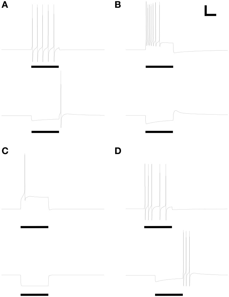
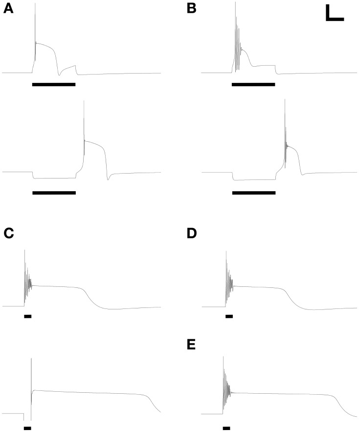
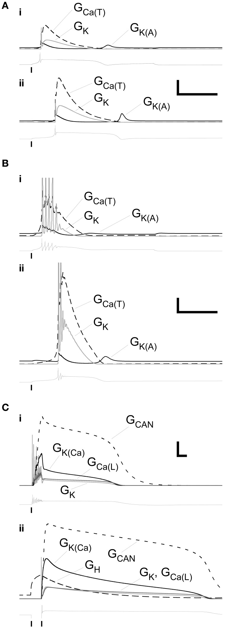
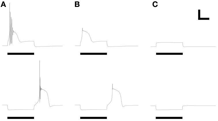
Olfactory bulb (OB) periglomerular (PG) cells are heterogeneous with respect to several features, including morphology, connectivity, patterns of protein expression, and electrophysiological properties. However, these features rarely correlate with one another, suggesting that the differentiating properties of PG cells may arise from multiple independent adaptive variables rather than representing discrete cell classes. We use computational modeling to assess this hypothesis with respect to electrophysiological properties. Specifically, we show that the heterogeneous electrophysiological properties demonstrated in PG cell recordings can be explained solely by differences in the relative expression levels of ion channel species in the cell, without recourse to modifying channel kinetic properties themselves. This PG cell model can therefore be used as the basis for diverse cellular and network-level analyses of OB computations. Moreover, this simple basis for heterogeneity contributes to an emerging hypothesis that glomerular-layer interneurons may be better described as a single population expressing distributions of partially independent, potentially plastic properties, rather than as a set of discrete cell classes.

Keywords: NEURON simulator; acetylcholine; computational model; glomerulus; juxtaglomerular neurons; olfactory bulb.


Figures

Figure 1
Figure 1
Periglomerular cell model. (A) Schematic representation of six-section PG cell model, comprising one gemmule (receiving synaptic input), a gemmule shaft, two dendrites (one to which the gemmule is attached), a soma, and an axon. (B) Expression profile of different channel types within model sections. Channel types marked as present in a given section (×) may have zero conductance under certain parameter sets (Table 3); channel types marked as absent in a given section (–) were always absent. The nicotinic cholinergic receptor channel (Gnic), an ohmic cation channel, was expressed only in the gemmule compartment (see text for details).
Figure 2
Figure 2
Model periglomerular cells display a range of simple spiking properties, each closely resembling their counterparts in Figure 1 of McQuiston and Katz (2001). Maximal conductance values for each membrane mechanism differ between panels (Table 3) but are identical within each depolarized/hyperpolarized pair of traces. (A) Non-accommodating spike train response to 3.5 pA depolarizing current (upper panel) and the corresponding response of the same neuron to release from a 1.2 pA hyperpolarizing current (lower panel). (B) Accommodating spike train response to 22 pA depolarizing current (upper panel) and the corresponding response of the same neuron to release from a 22 pA hyperpolarizing current (lower panel). (C) Single spike response to 25 pA depolarizing current (upper panel) and the corresponding response of the same neuron to release from a 25 pA hyperpolarizing current (lower panel). (D) Irregular spiking response to 7.5 pA depolarizing current (upper panel) and the corresponding anode break burst response of the same neuron to release from a 20 pA hyperpolarizing current (lower panel). Stimulus durations were all 600 ms (horizontal bars). Scale bars for all panels: 20 mV, 250 ms.
Figure 3
Figure 3
Model periglomerular cells display low threshold calcium spikes or persistent plateau potentials. Panels each closely resemble their counterparts in Figure 2 of McQuiston and Katz (2001). Maximal conductance values for each membrane mechanism differ between panels (Table 3) but are identical within each depolarized/hyperpolarized pair of traces. (A) Depolarizing current injection (10 pA, 600 ms) activated a low-threshold spike (LTS) with a single action potential (upper panel); release from 10 pA of hyperpolarizing current also evoked a similar LTS. (B) With modestly modified conductance ratios, injection of 10 pA depolarizing current (upper panel) or release from 10 pA of hyperpolarizing current (lower panel) evoked an LTS crowned with a train of decrementing spikes. (C) To replicate the rare persistent plateau potential response illustrated by McQuiston and Katz, additional current mechanisms were implemented. As in the original experimental results, depolarization (30 pA, 200 ms) led to a train of decrementing spikes followed by a plateau potential, whereas release from hyperpolarization (20 pA) generated only a single spike but also evoked a persistent plateau potential. (D) Elimination of the T-type calcium conductance had no appreciable effect on the depolarization-induced plateau potential. (E) Modest reduction of the delayed rectifier potassium current (Table 3) extended the duration of the plateau. Adjustment of ICAN and IK(Ca) conductances also could regulate plateau duration. Scale bars for panels (A,B): 20 mV, 250 ms; for panels (C–E): 20 mV, 500 ms.
Figure 4
Figure 4
Profiles of selected membrane conductances within primary dendrite during the LTS and plateau responses illustrated in Figures 2A–C. (A) Conductance timeseries associated with the responses illustrated in the upper panel (depolarizing stimulus, (i) and lower panel (release from hyperpolarization), (ii) of Figure 2A. Scale bars: 2.0 mS/cm2, 200 ms. (B) Conductance timeseries associated with the responses illustrated in the upper panel (i) and lower panel (ii) of Figure 2B. Scale bars: 0.2 mS/cm2, 200 ms. (C) Conductance timeseries associated with the responses illustrated in the upper panel (i) and lower panel (ii) of Figure 2C. Scale bars: 0.2 mS/cm2, 200 ms. Copies of the voltage timeseries are displayed below each group of conductance profiles. Vertical lines denote the onset or offset times of injected currents.
Figure 5
Figure 5
Replication of LTS pharmacology. Panels reflect their counterparts in Figures 3, 4 of McQuiston and Katz (2001). (A) LTS response under control conditions, using parameters of Figure 2B. (B) Evoked response after reducing the fast sodium conductance to zero, mimicking the application of intracellular QX314 or substitution of bath sodium with choline. (C) Evoked response after reducing the T-type calcium conductance to zero, mimicking the application of nickel ions or α-methyl-α-phenylsuccinimide (MPS). Stimulus durations were all 600 ms (horizontal bars). Scale bars: 20 mV, 250 ms.

Similar articles

Cited by

References

    1. Allen Z. J., 2nd., Waclaw R. R., Colbert M. C., Campbell K. (2007). Molecular identity of olfactory bulb interneurons: transcriptional codes of periglomerular neuron subtypes. J. Mol. Histol. 38, 517–525 10.1007/s10735-007-9115-4 - DOI - PubMed
    1. Aungst J. L., Heyward P. M., Puche A. C., Karnup S. V., Hayar A., Szabo G., et al. (2003). Centre-surround inhibition among olfactory bulb glomeruli. Nature 426, 623–629 10.1038/nature02185 - DOI - PubMed
    1. Brill M. S., Ninkovic J., Winpenny E., Hodge R. D., Ozen I., Yang R., et al. (2009). Adult generation of glutamatergic olfactory bulb interneurons. Nat. Neurosci. 12, 1524–1533 10.1038/nn.2416 - DOI - PMC - PubMed
    1. Cadetti L., Belluzzi O. (2001). Hyperpolarisation-activated current in glomerular cells of the rat olfactory bulb. Neuroreport 12, 3117–3120 - PubMed
    1. Carlin K. P., Jones K. E., Jiang Z., Jordan L. M., Brownstone R. M. (2000). Dendritic L-type calcium currents in mouse spinal motoneurons: implications for bistability. Eur. J. Neurosci. 12, 1635–1646 10.1046/j.1460-9568.2000.00055.x - DOI - PubMed

LinkOut - more resources