. 2013 Apr 26:5:4.
doi: 10.3389/fnene.2013.00004. eCollection 2013.

Astroglial networking contributes to neurometabolic coupling

Affiliations

Astroglial networking contributes to neurometabolic coupling

Carole Escartin et al. Front Neuroenergetics. .

Abstract

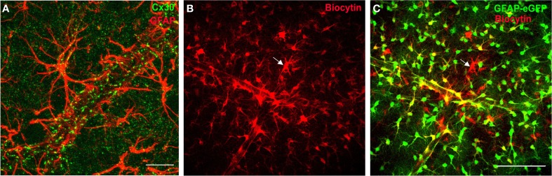
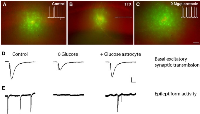
The strategic position of astrocytic processes between blood capillaries and neurons, provided the early insight that astrocytes play a key role in supplying energy substrates to neurons in an activity-dependent manner. The central role of astrocytes in neurometabolic coupling has been first established at the level of single cell. Since then, exciting recent work based on cellular imaging and electrophysiological recordings has provided new mechanistic insights into this phenomenon, revealing the crucial role of gap junction (GJ)-mediated networks of astrocytes. Indeed, astrocytes define the local availability of energy substrates by regulating blood flow. Subsequently, in order to efficiently reach distal neurons, these substrates can be taken up, and distributed through networks of astrocytes connected by GJs, a process modulated by neuronal activity. Astrocytic networks can be morphologically and/or functionally altered in the course of various pathological conditions, raising the intriguing possibility of a direct contribution from these networks to neuronal dysfunction. The present review upgrades the current view of neuroglial metabolic coupling, by including the recently unravelled properties of astroglial metabolic networks and their potential contribution to normal and pathological neuronal activity.

Keywords: astrocytes; astroglial networks; energy metabolism; epilepsy; gap junctions; neurodegenerative diseases; neuroglial interactions; neurometabolic coupling.


Figures

Figure 1
Figure 1
Connexins define a functional network of astrocytes at the gliovascular interface. (A) Staining of Cx30, one of the two main GJ protein in astrocytes, in mouse hippocampus co-localizes with astrocyte endfeet, labeled with the glial fibrillary acidic protein marker (GFAP), enwrapping blood vessels. Scale bar, 25 μm. (B,C) Functional coupling of perivascular astrocytes in GFAP-eGFP mice visualized by diffusion of biocytin (red, B; overlay with GFAP-eGFP, C), a tracer permeable to GJ channels, dialyzed for 20 min by whole-cell recording of a perivascular astrocyte (see arrow), revealing an extensive coupling of neighboring astrocytes. Note that some EGFP-positive cells near the dialyzed cell do not stain for biocytin, which indicates the presence of a preferential network of astrocytes. Scale bar, 100 μm. Adapted, with permission, from Giaume et al. (2010) (A) and Rouach et al. (2008) (B,C).
Figure 2
Figure 2
Activity-dependent glucose trafficking through astroglial networks sustain normal and pathological neuronal activity. (A) Sample pictures showing that the fluorescent glucose derivative 2-[N-(7-nitrobenz-2-oxa-1,3-diazol-4-yl)amino]-2-deoxyglucose (2-NBDG, green) trafficking in astrocytes is decreased when neuronal activity is inhibited with tetrodotoxin (TTX) (B) and increased during epileptiform activity (in 0 Mg2+-picrotoxin) (C) as compared with control conditions (A). Scale bar, 50 μm. Insets, corresponding spontaneous activity of hippocampal CA1 pyramidal cells recorded in current clamp in control, TTX (0.5 μm, 1–4 h), and 0 Mg2+-picrotoxin (100 μm, 1–4 h) conditions. Scale bar, 20 mV, 9 s. (D,E) Glucose supply through astrocytic networks sustains basal synaptic transmission and epileptiform activity during exogenous glucose deprivation (EGD). Sample traces of extracellular field potentials recorded in hippocampal slices showing that intracellular glucose (20 mM) delivery to astrocytic networks (+Glucose astrocytes) through the patch pipette inhibits the depression of fEPSP amplitude (D) and epileptiform activity (E) induced by exogenous glucose deprivation (0 glucose, 30 min) in wild-type mice. Scale bars (D) 0.2 mV, 5 ms and (E) 0.3 mV, 20 s. Adapted, with permission, from Rouach et al. (2008) (A–E).

References

    1. Allaman I., Belanger M., Magistretti P. J. (2011). Astrocyte-neuron metabolic relationships: for better and for worse. Trends Neurosci. 34, 76–87 10.1016/j.tins.2010.12.001 - DOI - PubMed
    1. Attwell D., Buchan A. M., Charpak S., Lauritzen M., Macvicar B. A., Newman E. A. (2010). Glial and neuronal control of brain blood flow. Nature 468, 232–243 10.1038/nature09613 - DOI - PMC - PubMed
    1. Ball K. K., Gandhi G. K., Thrash J., Cruz N. F., Dienel G. A. (2007). Astrocytic connexin distributions and rapid, extensive dye transfer via gap junctions in the inferior colliculus: implications for [(14)C]glucose metabolite trafficking. J. Neurosci. Res. 85, 3267–3283 10.1002/jnr.21376 - DOI - PMC - PubMed
    1. Belanger M., Allaman I., Magistretti P. J. (2011). Brain energy metabolism: focus on astrocyte-neuron metabolic cooperation. Cell Metab. 14, 724–738 10.1016/j.cmet.2011.08.016 - DOI - PubMed
    1. Bennett M. V., Garre J. M., Orellana J. A., Bukauskas F. F., Nedergaard M., Saez J. C. (2012). Connexin and pannexin hemichannels in inflammatory responses of glia and neurons. Brain Res. 1487, 3–15 10.1016/j.brainres.2012.08.042 - DOI - PMC - PubMed

LinkOut - more resources