. 2013 Apr 26;8(4):e60682.
doi: 10.1371/journal.pone.0060682. Print 2013.

The ocular conjunctiva as a mucosal immunization route: a profile of the immune response to the model antigen tetanus toxoid

Affiliations

The ocular conjunctiva as a mucosal immunization route: a profile of the immune response to the model antigen tetanus toxoid

Talin Barisani-Asenbauer et al. PLoS One. .

Abstract

Background: In a quest for a needle-free vaccine administration strategy, we evaluated the ocular conjunctiva as an alternative mucosal immunization route by profiling and comparing the local and systemic immune responses to the subcutaneous or conjunctival administration of tetanus toxoid (TTd), a model antigen.

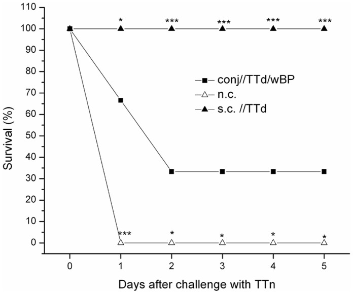
Materials and methods: BALB/c and C57BL/6 mice were immunized either subcutaneously with TTd alone or via the conjunctiva with TTd alone, TTd mixed with 2% glycerol or TTd with merthiolate-inactivated whole-cell B. pertussis (wBP) as adjuvants. Mice were immunized on days 0, 7 and 14 via both routes, and an evaluation of the local and systemic immune responses was performed two weeks after the last immunization. Four weeks after the last immunization, the mice were challenged with a lethal dose (2 × LD50) of tetanus toxin.

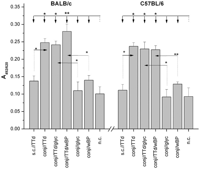
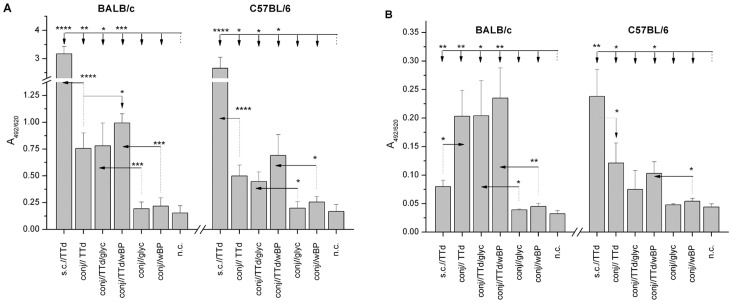
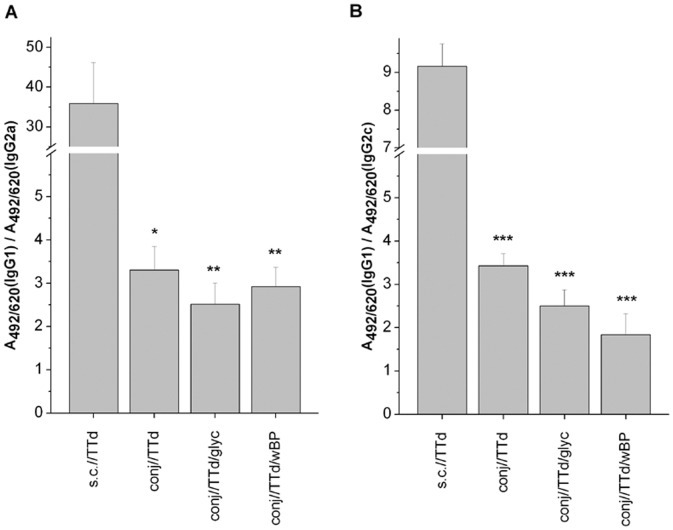
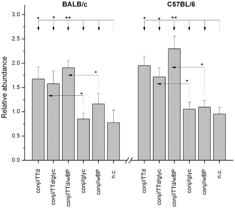
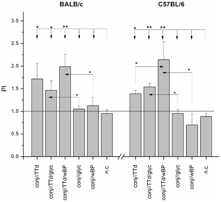
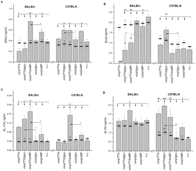
Results: The conjunctival application of TTd in BALB/c mice induced TTd-specific secretory IgA production and skewed the TTd-specific immune response toward a Th1/Th17 profile, as determined by the stimulation of IFNγ and IL-17A secretion and/or the concurrent pronounced reduction of IL-4 secretion, irrespective of the adjuvant. In conjunctivaly immunized C57BL/6 mice, only TTd administered with wBP promoted the establishment of a mixed Th1/Th17 TTd-specific immune response, whereas TTd alone or TTd in conjunction with glycerol initiated a dominant Th1 response against TTd. Immunization via the conjunctiva with TTd plus wBP adjuvant resulted in a 33% survival rate of challenged mice compared to a 0% survival rate in non-immunized animals (p<0.05).

Conclusion: Conjunctival immunization with TTd alone or with various adjuvants induced TTd-specific local and systemic immune responses, predominantly of the Th1 type. The strongest immune responses developed in mice that received TTd together with wBP, which implies that this alternative route might tailor the immune response to fight intracellular bacteria or viruses more effectively.


Conflict of interest statement

Competing Interests: The authors have declared that no competing interests exist.

Figures

Figure 1
Figure 1. Levels of TTd-specific SIgA in tear washes from BALB/c and C57BL/6 mice that were immunized according to the assigned protocols.
Samples were collected two weeks after the completion of the immunizations and were assayed by ELISA (dilution 1∶2). The results are presented as the mean A492/620± SE (n = 10). The significance of the observed differences was calculated by t-test (P<0.05*, P<0.005**). The reference group is indicated by a dotted line, and the comparison group is indicated by an arrow.
Figure 2
Figure 2. Levels of TTd-specific IgG (A) and IgA (B) in the sera of BALB/c and C57BL/6 mice immunized according to the assigned protocols.
Serum samples were collected two weeks after the completion of the immunizations and were assayed by ELISA (dilution 1∶100). The results are presented as the mean A492/620± SE (n = 10). The significance of the observed differences was calculated by t-test (P<0.05*, P<0.005**, P<0.0005***, P<0.00005). The reference group is indicated by a dotted line, and the comparison group is indicated by an arrow.
Figure 3
Figure 3. IgG1/IgG2a (A) and IgG1/IgG2c (B) ratios calculated for TTd-specific antibodies in the sera of TTd-immunized BALB/c and C57BL/6 mice, respectively.
Serum samples were collected two weeks after the completion of the immunizations and were assayed by ELISA (dilution 1∶100). Ratios were determined using the A405 values obtained upon the measurement of TTd-specific IgG1 and IgG2a or IgG2c in sera. The results are presented as the mean A405(IgG1)/A405(IgG2a,c) ± SE (n = 10). The significance of the differences between syngeneic mice immunized subcutaneously with TTd (reference group) and those immunized via the conjunctiva was calculated by t-test (P<0.05*, P<0.005**, P<0.0005***).
Figure 4
Figure 4. The relative abundance (RA) of TTd-specific mIgG+ B cells within the total population of mIgG+ B cells in SMLN upon completion of the assigned immunization protocols.
The RA of the TTd-specific mIgG+ B cell population was calculated for each mouse. The results are presented as the mean RA ± SE for each experimental group of mice (n = 5). The statistical significance of the observed differences in TTd-specific mIgG+ B cell pool abundances was determined by t-test (P<0.05*, P<0.005**, P<0.0005***). The reference group is indicated by a dotted line, and an arrow indicates the comparison group.
Figure 5
Figure 5. The proliferation indices (PI) of TTd-stimulated SMLN cells from BALB/c and C57BL/6 mice immunized via the conjunctiva according to the assigned immunization protocol.
The numbers of viable SMLN cells were assessed by MTT assay following a 48 h cultivation in 10% FCS/50 µM β-mercaptoethanol/RPMI 1640 medium supplemented or not with TTd (5 µg/ml). PIs were calculated for each mouse. The results are presented as the mean PI ± SE for each experimental group (n = 10). The statistical significance of the differences in PIs between groups treated according to the assigned protocols was determined by t-test (P<0.05*, P<0.005**). The reference group is indicated by a dotted line, and the comparison group is indicated by an arrow.
Figure 6
Figure 6. Levels of IFNγ (A), IL-4 (B), IL-17A (C) and IL-10 (D) in the supernatants from in vitro TTd-stimulated SMLN cells obtained from age-matched control mice (n.c.) and mice immunized via the conjunctiva according to the assigned protocol (bars).
SMLN cells were cultivated at 37°C in 5% CO2 for 48 h in 10% FCS/RPMI 1640/50 µM β-mercaptoethanol supplemented with 5 µg/ml TTd. The levels of cytokines in the supernatants of corresponding SMLN cells incubated in 10% FCS/RPMI 1640/50 µM β-mercaptoethanol under similar conditions (non-stimulated cells) are indicated by solid lines. The results are presented as the mean concentration ± SE (n = 10). Concentrations of cytokines in supernatants of TTd-stimulated cultures were compared by t-tests. The reference group is indicated by a dotted line, and the comparison group is indicated by an arrow. A t-test was also used for the comparison of cytokine concentration in supernatants of corresponding non-stimulated and TTd-stimulated cultures, and the statistical significance of the differences is marked next to the solid bar, indicating the level of the cytokine within the non-stimulated culture. The levels of statistical significance are assigned as follows: P<0.05*, P<0.005**, P<0.0005*** .
Figure 7
Figure 7. Survival rate of BALB/c and C57BL/6 mice immunized with TTd subcutaneously (s.c.//TTd), TTd mixed with wBP via the conjunctiva (conj//TTd/wBP) and non-treated age-matched mice (n.c.) upon challenge with tetanus toxin (TTn).
Mice were immunized on days 0, 7 and 14 (100 µg TTd per dose) and four weeks after the completion of the specified immunization protocol, mice were challenged by i.p. administration of 2×LD50 of TTn. In both strains, survival rates were the same; hence, a representative plot is provided. Survival rates were monitored daily. The statistical significance of the differences in survival rates between conj//TTd/wBP (reference group) and s.c.//TTd or n.c. groups was determined by t-test (P<0.05*, P<0.005**, P<0.0005***). The results are representative of two independent experiments, each comprising 6 mice per group.

References

    1. Daniel Nelson J, Douglas Cameron J (2010) The conjunctiva: anatomy and physiology. In: Krachmer JH, Mannis MJ, EJ H, editors. Cornea. 3 ed: Mosby, Elsevier. pp. 25–32.
    1. Gukasyan HJ, Kim K-J, Lee VHL (2008) The Conjunctival Barrier in Ocular Drug Delivery. In: Ehrhardt C, Kim K-J, editors. Drug Absorption Studies In situ, In vitro, In Silico Models: Springer US. pp. 310–312.
    1. Knop E, Knop N (2005) The role of eye-associated lymphoid tissue in corneal immune protection. J Anat 206: 271–285. - PMC - PubMed
    1. Steven P, Gebert A (2009) Conjunctiva-associated lymphoid tissue - current knowledge, animal models and experimental prospects. Ophthalmic research 42: 2–8. - PubMed
    1. Kageyama M, Nakatsuka K, Yamaguchi T, Owen RL, Shimada T (2006) Ocular defense mechanisms with special reference to the demonstration and functional morphology of the conjunctiva-associated lymphoid tissue in Japanese monkeys. Arch Histol Cytol 69: 311–322. - PubMed

Publication types

MeSH terms