. 2013 Apr 18;8(4):e60796.
doi: 10.1371/journal.pone.0060796. Print 2013.

Protection of cells against oxidative stress by nanomolar levels of hydroxyflavones indicates a new type of intracellular antioxidant mechanism

Affiliations

Protection of cells against oxidative stress by nanomolar levels of hydroxyflavones indicates a new type of intracellular antioxidant mechanism

Emanuele Lombardo et al. PLoS One. .

Abstract

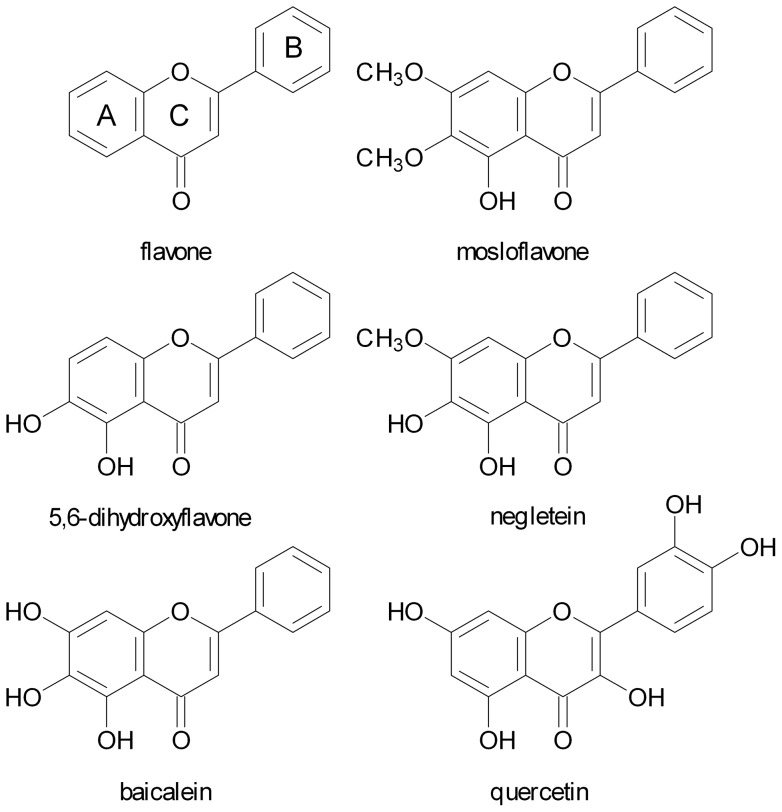
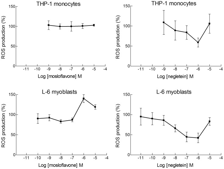
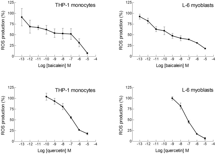
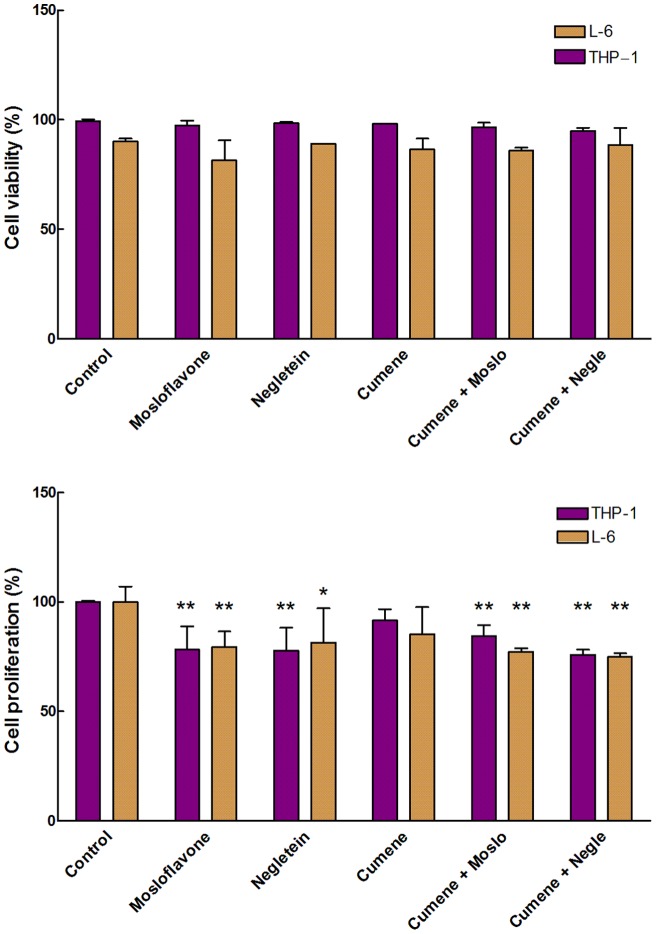
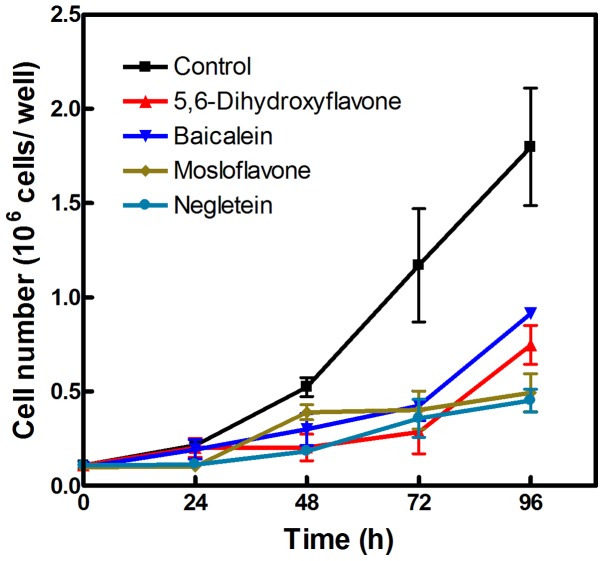
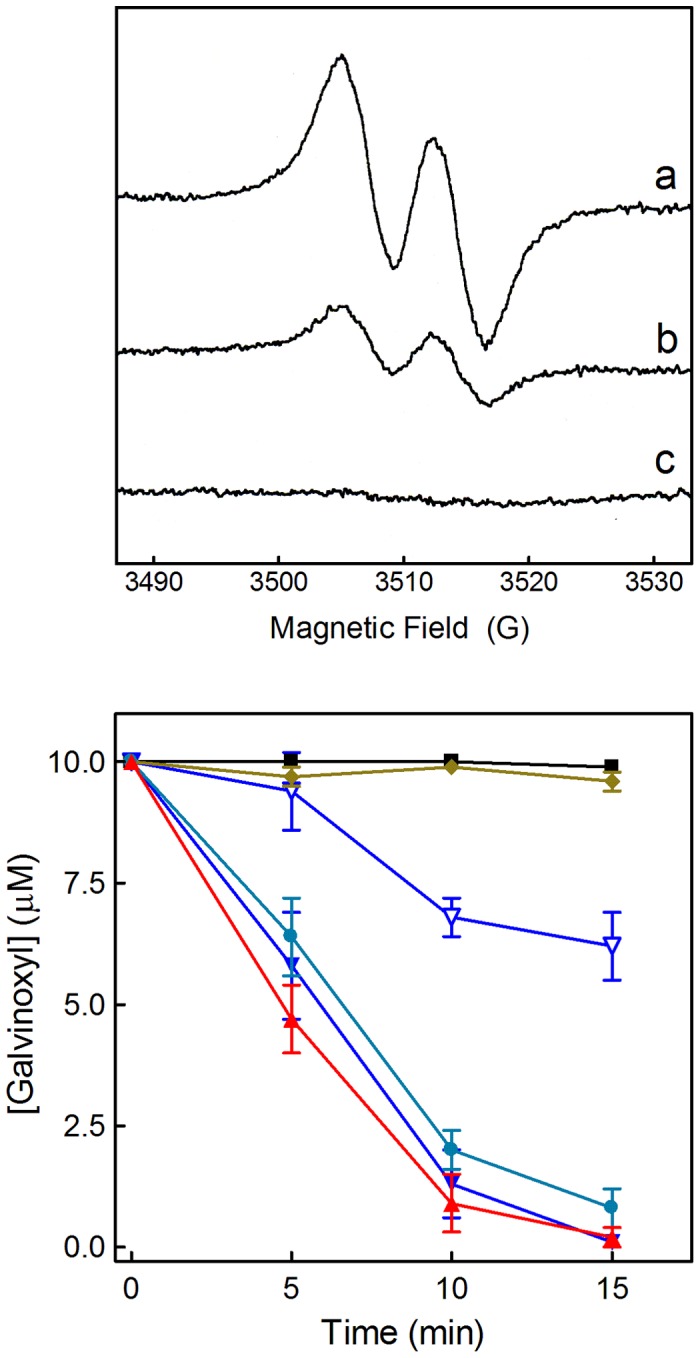
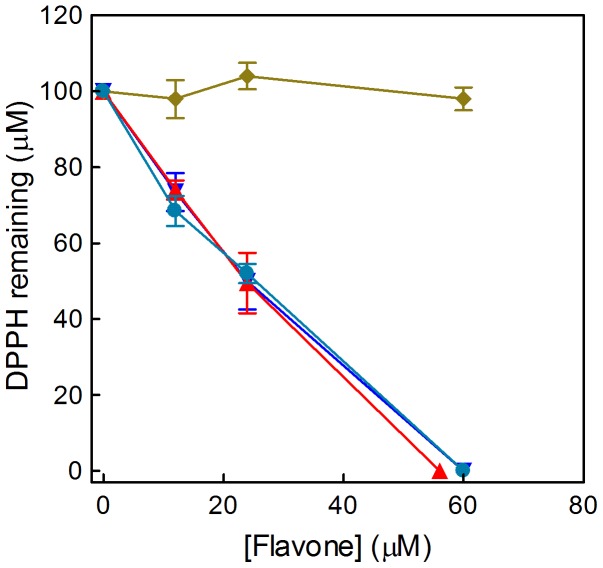
Natural polyphenol compounds are often good antioxidants, but they also cause damage to cells through more or less specific interactions with proteins. To distinguish antioxidant activity from cytotoxic effects we have tested four structurally related hydroxyflavones (baicalein, mosloflavone, negletein, and 5,6-dihydroxyflavone) at very low and physiologically relevant levels, using two different cell lines, L-6 myoblasts and THP-1 monocytes. Measurements using intracellular fluorescent probes and electron paramagnetic resonance spectroscopy in combination with cytotoxicity assays showed strong antioxidant activities for baicalein and 5,6-dihydroxyflavone at picomolar concentrations, while 10 nM partially protected monocytes against the strong oxidative stress induced by 200 µM cumene hydroperoxide. Wide range dose-dependence curves were introduced to characterize and distinguish the mechanism and targets of different flavone antioxidants, and identify cytotoxic effects which only became detectable at micromolar concentrations. Analysis of these dose-dependence curves made it possible to exclude a protein-mediated antioxidant response, as well as a mechanism based on the simple stoichiometric scavenging of radicals. The results demonstrate that these flavones do not act on the same radicals as the flavonol quercetin. Considering the normal concentrations of all the endogenous antioxidants in cells, the addition of picomolar or nanomolar levels of these flavones should not be expected to produce any detectable increase in the total cellular antioxidant capacity. The significant intracellular antioxidant activity observed with 1 pM baicalein means that it must be scavenging radicals that for some reason are not eliminated by the endogenous antioxidants. The strong antioxidant effects found suggest these flavones, as well as quercetin and similar polyphenolic antioxidants, at physiologically relevant concentrations act as redox mediators to enable endogenous antioxidants to reach and scavenge different pools of otherwise inaccessible radicals.


Conflict of interest statement

Competing Interests: The authors have declared that no competing interests exist.

Figures

Figure 1
Figure 1. Antioxidant molecules.
Structures of the flavone ring system, the four flavones tested, and the flavonol quercetin.
Figure 2
Figure 2. Dose-dependence curves for elimination of ROS by mosloflavone and negletein.
Wide concentration range dose-responses of mosloflavone (Left) and negletein (Right) effects on ROS production measured with the dichlorodihydrofluorescein (DCF) method in L-6 myoblasts and THP-1 monocytes. Oxidative stress was induced by cumene hydroperoxide (200 µM); the change in fluorescence was measured for 10 min and reported as percentage with respect to the variation of fluorescence caused by cumene hydroperoxide in the absence of flavones. Data are reported as mean ± SD of 5–10 different experiments. For negletein p<0.05 at least, as from a Student's t test, from 10−8 M to 10−6 M for L-6 cells, and p<0.01 at 10−6 M for THP-1 cells.
Figure 3
Figure 3. Dose-dependence curves for elimination of ROS by 5,6-dihydroxyflavone.
Wide concentration range dose-responses of the antioxidant activity of 5,6-dihydroxyflavone in THP-1 monocytes (Upper panel) and in L-6 myoblasts (Lower panel). The oxidative stress was induced and measured as in Fig. 2. In addition to monocytes (▪) the upper panel also includes results obtained for differentiated macrophages after PMA treatment for 24 h (▴) or 72 h (▾). Data are reported as mean ± SD of 5–10 different experiments. p<0.05 at least, as from a Student's t test, starting from 10−10 M for both L-6 cells and THP-1 cells, at any stage of differentiation.
Figure 4
Figure 4. Dose-dependence curves for elimination of ROS by baicalein and quercetin.
Wide concentration range dose-response curves for the antioxidant activity of baicalein (Upper panels) and quercetin (Lower panels) in THP-1 monocytes and L-6 myoblasts. The oxidative stress was induced and measured as in Fig. 2. Data are reported as mean ± SD of 5–10 different experiments. p<0.01 at least, as from a Student's t test, starting from 10−12 M for baicalein with both L-6 and THP-1 cells, and starting from 10−8 M for quercetin with both L-6 and THP-1 cells.
Figure 5
Figure 5. Cytotoxicity of flavones in monocytes.
Effects of the four hydroxyflavones on the survival of THP-1 monocytes, as determined by the MTT assay. Cells were plated in 6-wells plates using 1×10⁶ cells/well and treated with the flavones (10 µM or 10 nM) for 30 min at 37° C, then cumene hydroperoxide was added at the final concentration of 200 µM for additional 30 min at 37°C. Then MTT solution (0.5 mg/ml final concentration) was added and incubation was carried out at 37°C for 3–4 h. Results represent cell viability and are given as percentage relative to untreated controls. Data are mean ± SD of 3 different experiments. The statistical significance was evaluated by a Student's t test; where not indicated the differences were not significant, except for the difference of Control vs. Cumene hydroperoxide (Cu) that was always significant. #not statistically different from Control; *p<0.05 with respect to cumene hydroperoxide alone; **p<0.01 with respect to cumene hydroperoxide alone, ***p<0.001 with respect to cumene hydroperoxide alone.
Figure 6
Figure 6. Cytotoxicity of flavones in myoblasts.
Effects of the four hydroxyflavones on the survival of L-6 cells, as determined by the MTT assay. The concentration of cumene hydroperoxide was 27.5 µM, since preliminary experiments showed that higher concentrations were too toxic for these cells, and incubation with MTT was carried out for 24 h. Data are given as optical density (O.D.) rather than viability, because the values measured also include a component due to proliferation of the myoblasts. The bar graph shows data from a representative experiment with measurements carried out in triplicate. Data are mean ± SD of 3 different experiments. #not statistically different from Control; **p<0.01 with respect to cumene hydroperoxide alone.
Figure 7
Figure 7. Direct cytotoxic effects of flavones.
Direct effects of the four hydroxyflavones on THP-1 monocytes (Upper panels) and L-6 myoblasts (Lower panels) in the absence of any oxidative stress. Both cell viability (Left panels) and cell proliferation (Right panels) are shown as determined by the Trypan Blue assay; in these experiments cumene hydroperoxide was not added to the samples. The flavone concentration was 10 µM or 40 µM as indicated. Two types of control samples were included, one without any additions (control) and one with addition of a volume of the vehicle (DMSO) corresponding to the quantity added with the flavones. The data are shown as the mean ± SD of 3 different experiments with measurements carried out in triplicate, ***p<0.001 with respect to the corresponding control cells. For cell proliferation with 10 µM flavones a single representative experiment measured in triplicate is shown (statistical significance not calculated).
Figure 8
Figure 8. Cytotoxicity of flavones combined with cumene peroxide.
Viability (Upper panel) and proliferation (Lower panel) of THP-1 monocytes and L-6 myoblasts treated with 10 µM of either mosloflavone or negletein with or without cumene hydroperoxide (27.5 µM), measured as described in Fig. 7. **p<0.01 with respect to the corresponding control; *p<0.05 with respect to the corresponding control.
Figure 9
Figure 9. Long-term effects of flavones on proliferation.
L-6 cells were grown for four days in the presence of 10 µM of either baicalein (▾), 5,6-dihydroxyflavone (▴), mosloflavone (♦) or negletein (•); an equivalent volume of DMSO was added to the control cells (▪).
Figure 10
Figure 10. Antioxidant activity measured by EPR spectroscopy.
Upper panel: EPR spectra showing the reaction between galvinoxyl and antioxidants after 5 min incubation. (a) 10 µM galvinoxyl in ethanol; (b) with the addition of 2 µM 5,6-dihydroxyflavone; (c) with the addition of 1.0 µM quercetin. For each spectrum 4 scans were accumulated . The reduced product, galvinol, is not a radical, therefore it does not show an EPR spectrum. In theory it should be possible to see also the radical of the oxidized antioxidant, but almost all radicals of flavonoids and most other polyphenolic compounds are too reactive to be detected in this assay; their EPR spectra are not visible when the antioxidants are used in the micromolar range. The capacity of a compound to eliminate the galvinoxyl radical gives a relative measure of its general radical scavenging activity, although the intracellular antioxidant effect may depend on other factors and mechanisms. Lower panel: Kinetics of antioxidant activities of the four flavones measured by the EPR technique. The data show the concentration of galvinoxyl remaining in the samples at different times after addition of flavones, and are given as mean ± SD of 3 experiments. Control 10 µM galvinoxyl in ethanol (▪), and after addition of mosloflavone 10 µM (♦), negletein 1 µM (•), 5,6-dihydroxyflavone 1 µM (▴), baicalein 1 µM (▾), or baicalein 0.1 µM (▵). In samples containing 10 µM of either negletein, 5,6-dihydroxyflavone or baicalein the galvinoxyl signal disappeared completely within 30 s.
Figure 11
Figure 11. Antioxidant activity measured by the DPPH assay.
The dose dependent radical scavenging activity of the four flavones was measured by the decrease in absorption at 517 nm. The data show the concentration of DPPH remaining in the samples 20 min after addition of mosloflavone (♦), negletein (•), 5,6-dihydroxyflavone (▴) or baicalein (▾). In control samples containing only 100 µM DPPH in 95% ethanol no change in the spectrum was detected within the time of the measurements.

References

    1. Rice-Evans CA, Miller NJ (1996) Antioxidant activities of flavonoids as bioactive components of food. Biochem Soc Trans 24: 790–795. - PubMed
    1. Rice-Evans CA, Miller NJ, Paganga G (1996) Structure-antioxidant activity relationships of flavonoids and phenolic acids. Free Radic Biol Med 20: 933–956. - PubMed
    1. Halliwell B (2009) The wanderings of a free radical. Free Radic Biol Med 46: 531–542. - PubMed
    1. Gutteridge JMC, Halliwell B (2010) Antioxidants: Molecules, medicines, and myths. Biochem Biophys Res Commun 393: 561–564. - PubMed
    1. Clifford MN (2004) Diet-derived phenols in plasma and tissues and their implications for health. Planta Med 70: 1103–1114. - PubMed

Publication types