. 2013 Apr 18;8(4):e61309.
doi: 10.1371/journal.pone.0061309. Print 2013.

Vertical transmission of respiratory syncytial virus modulates pre- and postnatal innervation and reactivity of rat airways

Affiliations

Vertical transmission of respiratory syncytial virus modulates pre- and postnatal innervation and reactivity of rat airways

Giovanni Piedimonte et al. PLoS One. .

Abstract

Background: Environmental exposure to respiratory syncytial virus (RSV) is a leading cause of respiratory infections in infants, but it remains unknown whether this infection is transmitted transplacentally from the lungs of infected mothers to the offspring. We sought to test the hypothesis that RSV travels from the respiratory tract during pregnancy, crosses the placenta to the fetus, persists in the lung tissues of the offspring, and modulates pre- and postnatal expression of growth factors, thereby predisposing to airway hyperreactivity.

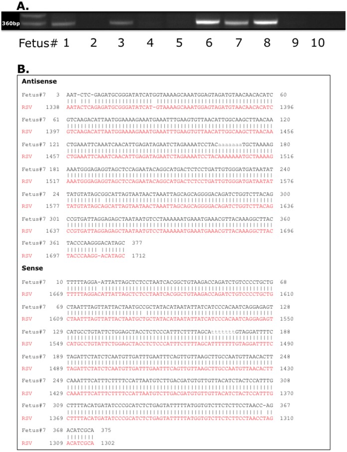
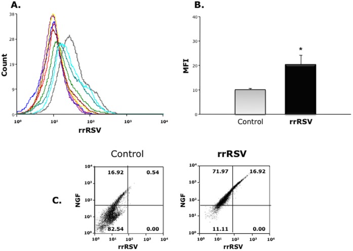
Methodology: Pregnant rats were inoculated intratracheally at midterm using recombinant RSV expressing red fluorescent protein (RFP). Viral RNA was amplified by RT-PCR and confirmed by sequencing. RFP expression was analyzed by flow cytometry and viral culture. Developmental and pathophysiologic implications of prenatal infection were determined by analyzing the expression of genes encoding critical growth factors, particularly neurotrophic factors and receptors. We also measured the expression of key neurotransmitters and postnatal bronchial reactivity in vertically infected lungs, and assessed their dependence on neurotrophic signaling using selective biological or chemical inhibition.

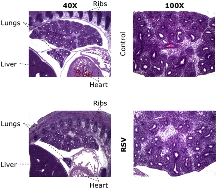
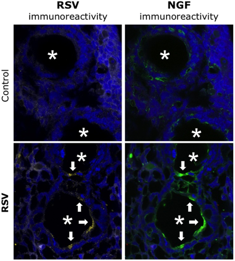
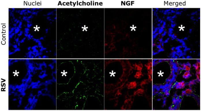
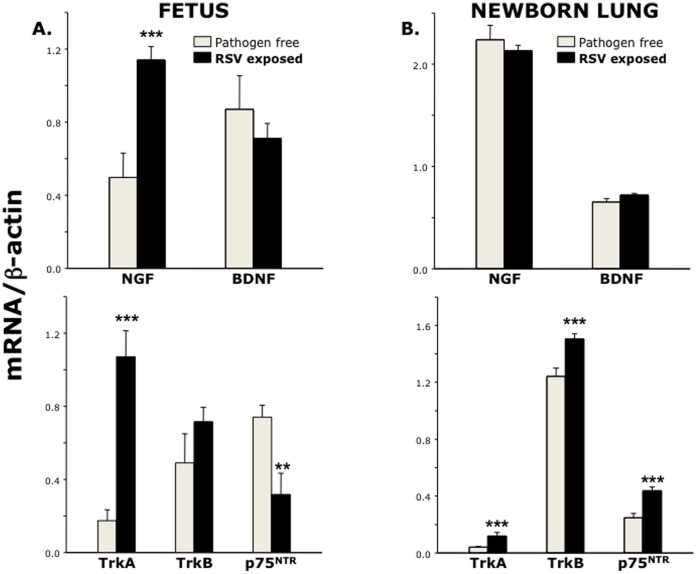
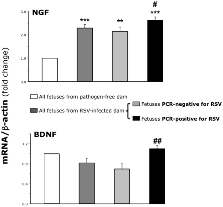
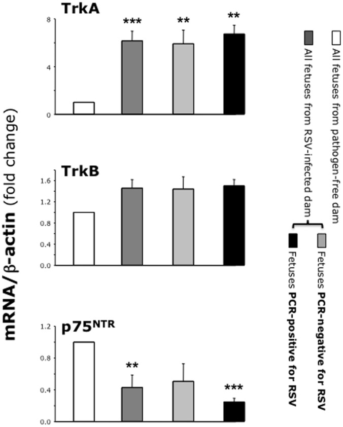
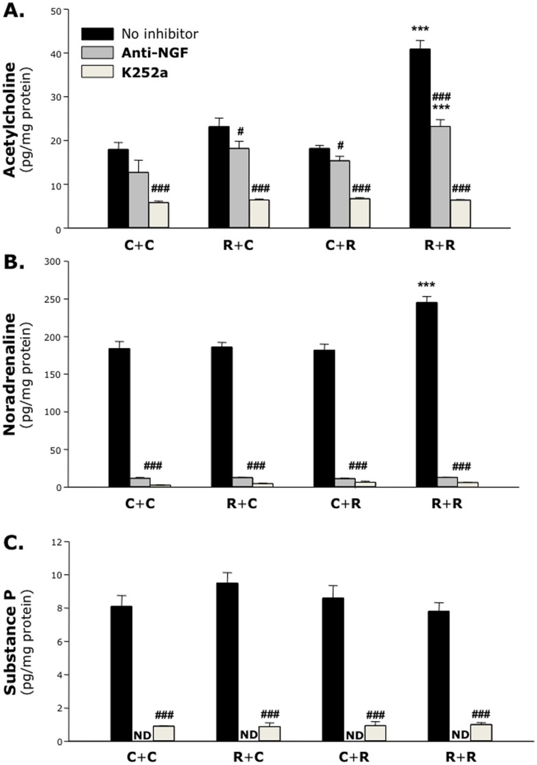
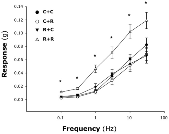
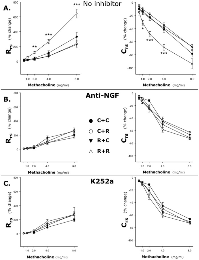
Principal findings: RSV genome was found in 30% of fetuses, as well as in the lungs of 40% of newborns and 25% of adults. RFP expression was also shown by flow cytometry and replicating virus was cultured from exposed fetuses. Nerve growth factor and its TrkA receptor were upregulated in RSV- infected fetal lungs and co-localized with increased cholinergic innervation. Acetylcholine expression and smooth muscle response to cholinergic stimulation increased in lungs exposed to RSV in utero and reinfected after birth, and blocking TrkA signaling inhibited both effects.

Conclusions/significance: Our data show transplacental transmission of RSV from mother to offspring and persistence of vertically transmitted virus in lungs after birth. Exposure to RSV in utero is followed by dysregulation of neurotrophic pathways predisposing to postnatal airway hyperreactivity upon reinfection with the virus.


Conflict of interest statement

Competing Interests: The authors have declared that no competing interests exist.

Figures

Figure 1
Figure 1. Vertical transmission of RSV genome.
( A ) RT-PCR analysis of fetuses harvested from RSV-infected dams using primers specific for the viral nucleocapsid protein. The bands in the first lane of the gel are DNA molecular weight standards; each of the other lanes shows RNA amplified from an individual fetus identified by the number indicated below the gel. RSV genomic sequences were found in one-third of the fetuses (#6, 7, and 8), whereas all matched control fetuses from pathogen-free dams were negative (not shown). ( B ) Sequence analysis of the amplicon from Fetus #7. All positive specimens yielded a 367-bp product whose sequence shared 98% homology with the RSV-A2 genome.
Figure 2
Figure 2. Histopathology of RSV-infected fetal lungs.
Sagittal sections of an RSV- infected E17 rat fetus stained with hematoxylin and eosin, showing the lungs at two different magnifications (40X and 100X). Histological analysis revealed late pseudoglandular development of the bronchial tree, which was appropriate for gestational age, and absence of acute or chronic inflammatory cell infiltrates. Similarly, the aerated newborn lung bronchi and alveolar air spaces were essentially normal without acute or chronic inflammatory cell infiltrates, and no definitive histologic differences were identified between the pups delivered from RSV-infected dams and those from pathogen-free dams (not shown).
Figure 3
Figure 3. Replication of vertically transmitted RSV.
( A ) Fluorescence-activated sorting of fetal cells from dams infected with rrRSV or pathogen-free control dams. Each curve has a different computer-generated color corresponding to a different fetus. RFP was detected in two-thirds of the fetuses harvested from dams infected with rrRSV, whereas the other third overlapped the control fetuses harvested from pathogen-free dams. ( B ) Geometric mean fluorescent intensity (MFI) averaged from the same flow cytometry measurements confirmed a significant increase in red fluorescence in fetuses from rrRSV- infected dams. * = p<0.05 compared to control fetuses from pathogen-free dams. ( C ) FACS analysis showed that exposure to rrRSV in utero was associated to fetal overexpression of NGF protein.
Figure 4
Figure 4. Propagation of vertically transmitted RSV.
Extracts of whole fetuses delivered from dams inoculated with rrRSV or from pathogen-free controls and co-cultured with human airway epithelial cells. After 48 h of incubation, the cells were then stained with a polyclonal anti-NGF antibody. The micrographs show red fluorescence in the cytoplasm of cells exposed to fetal extracts from rrRSV-infected dams, confirming the presence of actively replicating infectious virus that is associated with markedly increased green NGF immunoreactivity. As RFP expression reflects virus infection and production of the transgene rather than virus spread and plaque formation, this methodology is more accurate than the standard plaque assay to assess RSV infectivity. Magnification = 60X.
Figure 5
Figure 5. Localization of vertically transmitted RSV.
Sections of fetuses delivered from dams inoculated with rrRSV or from pathogen-free controls co-stained with polyclonal antibodies against RFP and NGF. Immunohistochemical analysis of fetal airways (asterisks) by confocal microscopy localized increased green NGF immunoreactivity to the epithelial layer, overlapping areas of yellow RSV immunoreactivity (arrows). No significant fluorescence was detected in any other fetal structures. Magnification = 60X.
Figure 6
Figure 6. Cholinergic innervation of RSV-infected fetal airways.
Sections of fetuses delivered from dams inoculated with rrRSV or from pathogen-free controls co-stained with polyclonal antibodies against acetylcholine and NGF. Immunohistochemical analysis by confocal microscopy found increased acetylcholine immunoreactivity in the subepithelial neural plexus lining the lumen of fetal airways exposed in utero to RSV and co-expressing NGF immunoreactivity (asterisks). In contrast, noradrenaline and substance P immunoreactivity was very weak in all fetuses.
Figure 7
Figure 7. Neurotrophins expression after vertical RSV infection.
( A ) RT-PCR of fetuses (n = 9–10 per group) from RSV-infected dams showed upregulation of NGF and its high-affinity receptor TrkA, with downregulation of the low affinity pan-neurotrophin receptor p75NTR. ( B ) All neurotrophin receptors were upregulated in the lungs of newborns (n = 5 per group) delivered from RSV-infected dams. Data are expressed as mean ± SEM of the densitometry score normalized by the β-actin control. ** = p<0.01; *** = p<0.001 compared to age-matched controls delivered from pathogen-free dams.
Figure 8
Figure 8. Neurotrophic factors in PCR-positive vs. PCR-negative fetuses.
RT-PCR analysis of neurotrophic factors in fetuses harvested from the same RSV-infected dam. Expression of both NGF and BDNF genes was higher in fetuses RSV-positive by PCR compared to PCR-negative fetuses. However, fetuses from the RSV-infected dam had higher NGF expression compared to the control fetuses from pathogen-free dam, even if no RSV RNA could be detected. Data are expressed as the mean ± SEM (n = 9–10 per group). ** = p<0.01; *** = p<0.001 compared to fetuses delivered from pathogen-free control dam. # = p<0.05; ## = p<0.01 compared to RSV-negative fetuses delivered from same RSV-infected dam.
Figure 9
Figure 9. Neurotrophic receptors in PCR-positive vs. PCR-negative fetuses.
RT-PCR analysis of neurotrophic receptors in fetuses harvested from the same RSV-infected dam. Fetuses from the RSV-infected dam had higher TrkA and lower p75NTR expression compared to the control fetuses from pathogen-free dam, even if no RSV RNA could be detected. Data are expressed as the mean ± SEM (n = 9–10 per group). ** = p<0.01; *** = p<0.001 compared to fetuses delivered from pathogen-free control dam.
Figure 10
Figure 10. Neurotransmitters in the lung tissues.
Compared to rats kept pathogen-free during and after gestation ( group C+C ), cholinergic, adrenergic, and NANC innervation did not change significantly after maternal ( group R+C ) or neonatal ( group C+R ) infection. However, when pups were exposed to RSV both in utero and after birth ( group R+R ) acetylcholine expression in their lungs doubled. This effect was reduced or abolished by inhibiting neurotrophic signaling. Data are expressed as the mean ± SEM (n = 6–8 per group). *** = p<0.001 compared to C+C control. # = p<0.05; ### = p<0.001 compared to no-inhibitor control.
Figure 11
Figure 11. RSV-induced airway hyperreactivity in vitro.
Smooth muscle contraction evoked by EFS in tracheas from pathogen-free pups delivered from pathogen-free dams ( group C+C ) was not affected by either prenatal ( group R+C ) or postnatal RSV infection ( group C+R ). However, when pups delivered from RSV-infected dams were reinfected postnatally ( group R+R ) their airways became significantly hyperresponsive to any frequency of EFS. Data are expressed as the mean ± SEM (n = 8 per group). * = p<0.05 compared to C+C control.
Figure 12
Figure 12. RSV-induced airway hyperreactivity in vivo.
Ten-day old rats delivered from pathogen-free or RSV-infected dams were given either sterile medium ( C+C and R+C groups ) or RSV suspension ( C+R and R+R groups ), and then challenged with inhaled methacholine 5 days later. Airway reactivity in vivo increased only when the exposure to RSV in utero was followed by postnatal reinfection with the same virus (A). RSV-induced airway hyperreactivity was abolished by selective immunologic inhibition of NGF activity by a blocking antibody (B) or chemical inhibition of receptor tyrosine kinase signaling by the specific antagonist K252a (C). Data are expressed as the mean ± SEM (n = 6–8 per group). * = p<0.05; ** = p<0.01; *** = p<0.001 compared to C+C control.

Similar articles

Cited by

References

    1. Wright M, Piedimonte G (2011) Respiratory syncytial virus prevention and therapy: Past, present, and future. Pediatr Pulmonol 46: 324–347. - PubMed
    1. Othumpangat S, Gibson LF, Samsell L, Piedimonte G (2009) NGF is an essential survival factor for bronchial epithelial cells during respiratory syncytial virus infection. PLoS One 4: e6444. - PMC - PubMed
    1. Iankevich OD, Dreizin RS, Makhlinovskaia NL, Gorodnitskaia NA (1975) Viremia in respiratory syncytial virus infection. Vopr Virusol 4: 455–458. - PubMed
    1. Liu XM, Wang Z, Guo Y (2007) Respiratory syncytial virus nephropathy in rats. Kidney Int 71: 388–396. - PubMed
    1. Eisenhut M (2006) Extrapulmonary manifestations of severe respiratory syncytial virus infection - a systematic review. Crit Care 10: R107. - PMC - PubMed

Publication types

MeSH terms