. 2013 Apr 18;8(4):e61372.
doi: 10.1371/journal.pone.0061372. Print 2013.

Microgravity induces pelvic bone loss through osteoclastic activity, osteocytic osteolysis, and osteoblastic cell cycle inhibition by CDKN1a/p21

Affiliations

Microgravity induces pelvic bone loss through osteoclastic activity, osteocytic osteolysis, and osteoblastic cell cycle inhibition by CDKN1a/p21

Elizabeth A Blaber et al. PLoS One. .

Abstract

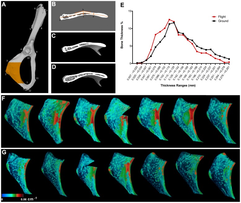
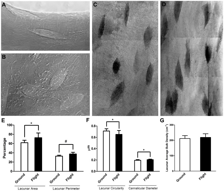
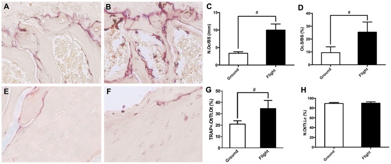
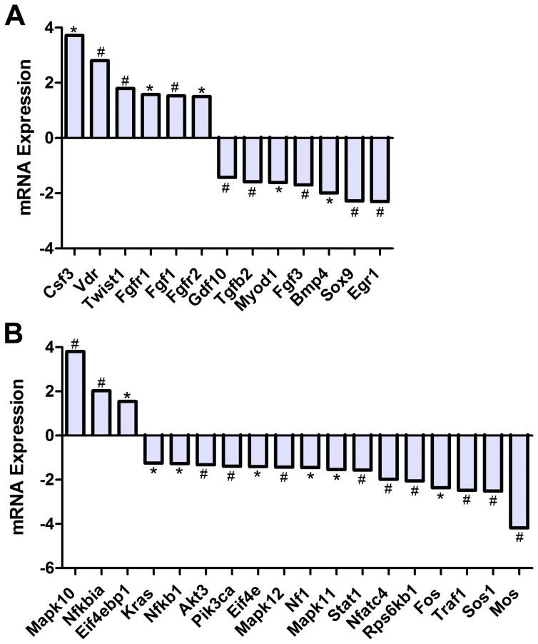
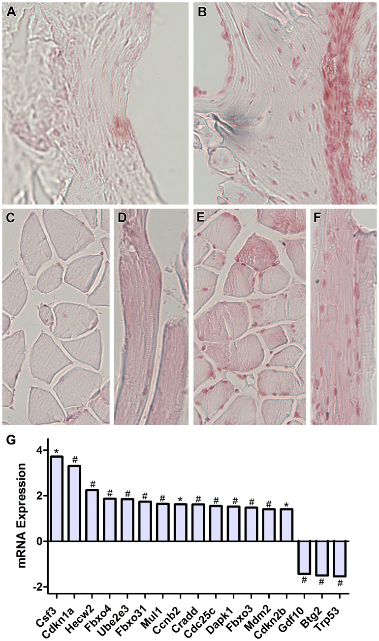
Bone is a dynamically remodeled tissue that requires gravity-mediated mechanical stimulation for maintenance of mineral content and structure. Homeostasis in bone occurs through a balance in the activities and signaling of osteoclasts, osteoblasts, and osteocytes, as well as proliferation and differentiation of their stem cell progenitors. Microgravity and unloading are known to cause osteoclast-mediated bone resorption; however, we hypothesize that osteocytic osteolysis, and cell cycle arrest during osteogenesis may also contribute to bone loss in space. To test this possibility, we exposed 16-week-old female C57BL/6J mice (n = 8) to microgravity for 15-days on the STS-131 space shuttle mission. Analysis of the pelvis by µCT shows decreases in bone volume fraction (BV/TV) of 6.29%, and bone thickness of 11.91%. TRAP-positive osteoclast-covered trabecular bone surfaces also increased in microgravity by 170% (p = 0.004), indicating osteoclastic bone degeneration. High-resolution X-ray nanoCT studies revealed signs of lacunar osteolysis, including increases in cross-sectional area (+17%, p = 0.022), perimeter (+14%, p = 0.008), and canalicular diameter (+6%, p = 0.037). Expression of matrix metalloproteinases (MMP) 1, 3, and 10 in bone, as measured by RT-qPCR, was also up-regulated in microgravity (+12.94, +2.98 and +16.85 fold respectively, p<0.01), with MMP10 localized to osteocytes, and consistent with induction of osteocytic osteolysis. Furthermore, expression of CDKN1a/p21 in bone increased 3.31 fold (p<0.01), and was localized to osteoblasts, possibly inhibiting the cell cycle during tissue regeneration as well as conferring apoptosis resistance to these cells. Finally the apoptosis inducer Trp53 was down-regulated by -1.54 fold (p<0.01), possibly associated with the quiescent survival-promoting function of CDKN1a/p21. In conclusion, our findings identify the pelvic and femoral region of the mouse skeleton as an active site of rapid bone loss in microgravity, and indicate that this loss is not limited to osteoclastic degradation. Therefore, this study offers new evidence for microgravity-induced osteocytic osteolysis, and CDKN1a/p21-mediated osteogenic cell cycle arrest.


Conflict of interest statement

Competing Interests: The authors have declared that no competing interests exist.

Figures

Figure 1
Figure 1. Micro-Computed Tomography (μCT) Analysis of Spaceflown Ischium.
The ischium of the pelvis, shown in orange (A), was analyzed using μCT (720 slices = 4.89 mm). The anatomical markers used for μCT analysis were (1) caudal apex of obturator foramen, (2) dorsal-most point of the ventral ramus of ischium, and (3) the ischial tuberosity (A) (For full details on anatomical markers see [74]). The ischial cross-sectional geometry (B) was analyzed by length (a), the width at the midpoint (b) and at 1/3 distance from the obturator foramen (c); and the bend angle (d). Flight samples (D) exhibited a more open cross-sectional geometry compared to the ground control (C), indicating a possible reduction in the pull force applied to the bone. Ground control samples (F) also exhibited greater thickness (orange/red) then the flight samples (G), indicating a reduction in overall bone thickness in spaceflight samples.
Figure 2
Figure 2. Nano-Computed Tomography Analysis of Lacunar Enlargement Following Spaceflight.
Figures 2A and 2B show X-ray phase contrast images of osteocytes viewed laterally and from the top, respectively and illustrate the flattened shape of osteocytic lacunae. For quantification of lacunar size we used X-Ray absorption images of lateral views of lacunae in ischial cortical bone from ground control (C) and spaceflight (D) animals (n = 7). We observed a 17% increase in lacunae area and a 14% increase in lacunae perimeter of flight animals compared to ground controls (E). We also found a 13% increase in lacunae canalicular diameter and a 9% decrease in lacunae circularity of flight animals compared to ground controls (F). However, bulk density analysis showed no statistical difference between flight and ground control animals that is in agreement with µCT analysis (G). *indicates p<0.05, #indicates p<0.01.
Figure 3
Figure 3. TRAP Staining of Osteoclast and Osteocyte-Mediated Bone Resorption Following Spaceflight.
Panel A displays a representative cancellous region near the femoral head growth plate (40×) from ground control mice, which is mostly free of TRAP-positive osteoclasts, whilst B displays a similar region from flight animals with numerous TRAP-positive osteoclasts. Analysis of osteoclastic activity in the trabecular region below the femoral head of the femur showed an increase in osteoclast numbers in the bone surface of the growth plate of flight samples compared to ground controls (9.99 Oc/mm and 3.36 Oc/mm respectively) (C). The bone surface covered by osteoclasts was also increased in flight animals compared to ground controls (25.40% and 9.99% respectively, D). The number of TRAP-positive osteocytes in cortical bone from the femoral shaft proximal to the femoral head was increased in response to spaceflight (E) compared to ground controls (D) (34.43% and 20.94% respectively, F). However, we found no differences in the number of empty lacunae in cortical bone between flight and ground controls (10.6% and 10.0% empty lacunae respectively, H). * indicates p<0.05, # indicates p<0.01.
Figure 4
Figure 4. Spaceflight Causes Up-Regulation of Matrix Degradation Molecules.
RT-PCR analysis of ilium revealed significant up-regulation of matrix degradation molecules MMP1a, MMP3, and MMP10 as well as small changes in a number of extracellular matrix molecules in flight samples compared to ground controls (A). Immunohistochemical analysis localized over-expression of MMP10 to osteocytes in the shaft of the proximal femur in flight samples (C) but not in ground controls (B), indicating a role for osteocytes in lacunae degradation. * indicates p<0.05, # indicates p<0.01.
Figure 5
Figure 5. Spaceflight Alters mRNA Expression of Genes Associated with Osteogenic Growth and Mitogenic Signal Transduction Pathways.
RT-PCR analysis of revealed altered expression levels of key genes involved in osteogenic growth and proliferation including growth factors, Bmp4 and Tgfβ2, and transcription factors Vdr and Sox9 (A). Analysis of key mitogenic signal transduction pathways revealed alterations in gene expression of the MAPK pathway, whilst Pi3K and Akt signaling molecules were not changed statistically. We also observed significant up-regulation of the NFκB inhibitor, NFκBIa/IκBα (B).
Figure 6
Figure 6. Spaceflight Causes Overexpression of the Cell Cycle Arrest Molecule, p21, Independently of p53 Activation.
RT-PCR analysis revealed significant alterations in many cell cycle molecules including a 3.31 fold up-regulation of p21 and down-regulation of p53 (G). Immunohistochemical analysis localized this overexpression of p21 to osteoblasts along the periosteal surface of the proximal femur (A, ground control, B, flight). Interestingly, we also observed p21-positive nuclei in cross-sections and longitudinal sections of muscle fibers adjacent to the femur (C–D, ground control, E–F, flight). *indicates p<0.05, # indicates p<0.01.

Similar articles

Cited by

References

    1. Nabavi N, Khandani A, Camirand A, Harrison RE (2011) Effects of microgravity on osteoclast bone resorption and osteoblast cytoskeletal organization and adhesion. Bone 49: 965–974. - PubMed
    1. Hughes-Fulford M (2003) Function of the cytoskeleton in gravisensing during spaceflight. Adv Space Res 32: 1585–1593. - PubMed
    1. Ohshima H (2006) [Bone loss and bone metabolism in astronauts during long-duration space flight]. Clin Calcium 16: 81–85. - PubMed
    1. Smith SM, Heer M (2002) Calcium and bone metabolism during space flight. Nutrition 18: 849–852. - PubMed
    1. Zayzafoon M, Meyers VE, McDonald JM (2005) Microgravity: the immune response and bone. Immunol Rev 208: 267–280. - PubMed

Publication types

Substances