. 2013 Apr 26;8(4):e61389.
doi: 10.1371/journal.pone.0061389. Print 2013.

Gaze holding in healthy subjects

Affiliations

Gaze holding in healthy subjects

Giovanni Bertolini et al. PLoS One. .

Abstract

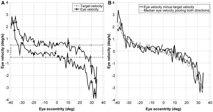
Eccentric gaze in darkness evokes minor centripetal eye drifts in healthy subjects, as cerebellar control sufficiently compensates for the inherent deficiencies of the brainstem gaze-holding network. This behavior is commonly described using a leaky integrator model, which assumes that eye velocity grows linearly with gaze eccentricity. Results from previous studies in patients and healthy subjects suggest caution when this assumption is applied to eye eccentricities larger than 20 degrees. To obtain a detailed characterization of the centripetal gaze-evoked drift, we recorded horizontal eye position in 20 healthy subjects. With their head fixed, they were asked to fixate a flashing dot (50 ms every 2 s)that was quasi-stationary displacing(0.5 deg/s) between ± 40 deg horizontally in otherwise complete darkness. Drift velocity was weak at all angles tested. Linearity was assessed by dividing the range of gaze eccentricity in four bins of 20 deg each, and comparing the slopes of a linear function fitted to the horizontal velocity in each bin. The slopes of single subjects for gaze eccentricities of ± 0-20 deg were, in median,0.41 times the slopes obtained for gaze eccentricities of ± 20-40 deg. By smoothing the individual subjects' eye velocity as a function of gaze eccentricity, we derived a population of position-velocity curves. We show that a tangent function provides a better fit to the mean of these curves when large eccentricities are considered. This implies that the quasi-linear behavior within the typical ocular motor range is the result of a tuning procedure, which is optimized in the most commonly used range of gaze. We hypothesize that the observed non-linearity at eccentric gaze results from a saturation of the input that each neuron in the integrating network receives from the others. As a consequence, gaze-holding performance declines more rapidly at large eccentricities.


Conflict of interest statement

Competing Interests: The authors have declared that no competing interests exist.

Figures

Figure 1
Figure 1. Raw data recorded in a single trial from a typical subject.
Panel A - Left eye position plotted as function of time. Positive angles correspond to right eccentricities as seen by the subject. In this trial the dot was moving (0.5 deg/s) rightward at first. Inset 1: At extreme eccentricities the centrifugal beating nystagmus is clearly visible and the slow phase shows the tendency of the eye to return toward the primary position. Inset 2 and 3: Difference in the slope of the position trace at the same eccentricities when the dot is moving outward or inward. Panel B and C - Position (panel B) and velocity (panel C) of the eye at eccentricities larger than 10 deg right. The eye velocity begins to decrease from its baseline before the onset of the nystagmus, showing the growing centrifugal drift. Note that the baseline velocity is not zero but is positive between 10 and 25 deg of gaze eccentricity. When returning to 10 deg, however, the velocity is negative, showing the asymmetry in the baseline velocity showing the subject's attempt to match the target displacement velocity.
Figure 2
Figure 2. Position-Velocity plot considering target direction.
Panel A - Black lines: Medians of the eye velocity within a 1 deg-wide bin plotted as a function of gaze eccentricity keeping the direction of target displacement separated. Gray lines: velocity of the target as a function of its position during the whole recording period. Note that the eye velocity matches the target velocity from the beginning suggesting that the offset observed around the straight ahead gaze is not due to a memory effect. The arrows show the directions of target and eye displacement. Panel B - Black lines: Velocity traces from the left panel after subtracting the correspondent target velocity. Gray line: Medians of the eye velocity within a 1 deg-wide bin plotted as a function of gaze eccentricity after pooling data from different directions of target displacement.
Figure 3
Figure 3. Position-Velocity plot and linear fit.
Gray dots: Instantaneous velocity plotted as a function of the eye eccentricity. Light gray dots: Velocity in the 0–20 deg bins; dark gray dots: Velocity in the 20–40 deg bins; black line: linear fit of the velocity in the 0–20 deg bins and in the 20–40 deg bins.
Figure 4
Figure 4. Smoothed Position -Velocity plot of the whole population.
Dashed gray lines: Individual position-velocity curves obtained after smoothing and interpolating instantaneous velocity as a function of eye eccentricity; solid thick gray line: mean of the smoothed individual position-velocity curves; solid medium gray line: mean ±1 standard deviation of the smoothed individual position-velocity curves; dashed black line: tangent fit of the mean of the smoothed individual position-velocity curves.
Figure 5
Figure 5. Simulation of the network without tuning.
Panel A shows the output of the synaptic activation function of each neuron (thin lines) as a function of the internal representation of eye eccentricity (Δ), the zero of each line indicates the neuron threshold, i.e. the eccentricity at which the inhibitory cutoff takes place. The thick lines are the cumulative output of both sides of the network, obtained by combining all the synaptic activation functions with their factor η. Panel B shows the presynaptic factor η of each neuron, indexed according to the threshold shown in the upper panel, here set to the same value for all neurons to illustrate the general features of a non-tuned network. Panel C shows the PV plot for the internal representation of eye position.
Figure 6
Figure 6. Simulation of the network tuned with non-uniform error function.
The Gaussian function of the eye eccentricity represented by the solid line in the central panel has been multiplied to the resulting drift, i.e. the error to minimize, during the optimization procedure. The contents of the panels are as in figure 1A.
Figure 7
Figure 7. Simulation of the network tuned favoring leakiness against instability.
The inverse of the Gaussian function of the eye eccentricity shown by the solid line in the central panel has been used as a gaze-dependent threshold for the non-penalized drift during the optimization procedure. The contents of the panels are as in figure 1A.
Figure 8
Figure 8. Simulation of the network using nonlinear synaptic activation functions.
The contents of the panels are as in figure 5.
Figure 9
Figure 9. PV plots of random perturbations of tuned network.
Effect of modifying the tuned values of η by a random fraction of 5% of the value used in figure 7. Dashed gray lines shows 20 different perturbations. The black solid line represents their mean.

References

    1. Leigh RJ, Zee DS (2006) The neurology of eye movements. Oxford Press.
    1. Abel LA, Parker L, Daroff RB, Dell'Osso LF (1978a) End-point nystagmus, Invest. Ophthalmol. Vis Sci 17: 539–544. - PubMed
    1. Eizenman M, Cheng P, Sharpe JA, Frecker RC (1990) End-point nystagmus and ocular drift: an experimental and theoretical study. Vision Res 30(6): 863–877. - PubMed
    1. Shallo-Hoffmann J, Schwarze H, Simonsz HJ, Mühlendych H (1990) A reexamination of end-point and rebound nystagmus in normal. Invest Ophthalmol Vis Sci 31(2): 388–392. - PubMed
    1. Whyte CA, Petrock AM, Rosenberg M (2010) Occurrence of physiologic gaze-evoked nystagmus at small angles of gaze. . Invest Ophthalmol Vis Sci 51: 2476–2478. - PubMed

Publication types