. 2013 Apr 18;8(4):e61749.
doi: 10.1371/journal.pone.0061749. Print 2013.

Primary breast cancer tumours contain high amounts of IgA1 immunoglobulin: an immunohistochemical analysis of a possible carrier of the tumour-associated Tn antigen

Affiliations

Primary breast cancer tumours contain high amounts of IgA1 immunoglobulin: an immunohistochemical analysis of a possible carrier of the tumour-associated Tn antigen

Charlotte Welinder et al. PLoS One. .

Abstract

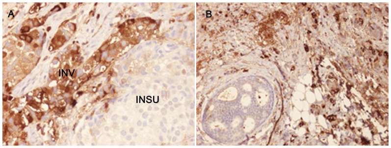
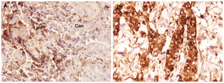
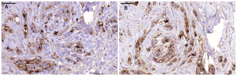
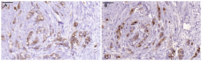
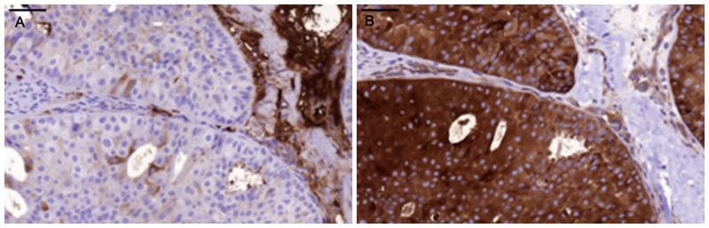
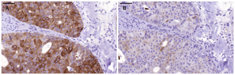
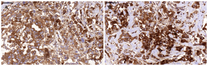
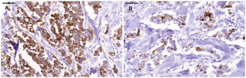
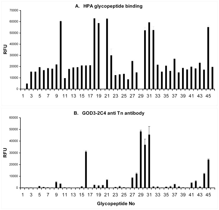
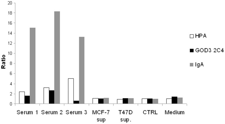
The Tn antigen (GalNAc alpha-O-Ser/Thr) as defined by the binding of the lectin, helix pomatia agglutinin (HPA) or anti-Tn monoclonal antibodies, is known to be exposed in a majority of cancers, and it has also been shown to correlate positively with the metastatic capacity in breast carcinoma. The short O-glycan that forms the antigen is carried by a number of different proteins. One potential carrier of the Tn antigen is immunoglobulin A1 (IgA1), which we surprisingly found in tumour cells of the invasive parts of primary breast carcinoma. Conventional immunohistochemical analysis of paraffin-embedded sections from primary breast cancers showed IgA1 to be present in the cytoplasm and plasma membrane of 35 out of 36 individual primary tumours. The immunohistochemical staining of HPA and anti-Tn antibody (GOD3-2C4) did to some extent overlap with the presence of IgA1 in the tumours, but differences were seen in the percentage of stained cells and in the staining pattern in the different breast cancers analysed. Anti-Tn antibody and HPA were also shown to specifically bind to a number of possible constellations of the Tn antigen in the hinge region of IgA1. Both reagents could also detect the presence of Tn positive IgA in serum. On average 51% of the tumour cells in the individual breast cancer tumour sections showed staining for IgA1. The overall amount of staining in the invasive part of the tumour with the anti Tn antibody was 67%, and 93% with HPA. The intra-expression or uptake of IgA1 in breast cancer makes it a new potential carrier of the tumour associated and immunogenic Tn antigen.


Conflict of interest statement

Competing Interests: The authors have declared that no competing interests exist.

Figures

Figure 1
Figure 1. Immunohistochemical staining of breast tumour specimens showing the presence of IgA1 in the invasive part of the tumour.
A) In Sample 2, 40% of the tumour cells in the invasive part were stained with anti-IgA1 with a relative intensity of 3. Strong cytoplasmic and plasma membrane staining of IgA1 is observed in the invasive part of the section (INV) but only very weak staining in the in situ part (INSU). B) In Sample 7, 80% of the cells were IgA1-positive with a relative intensity of 3.
Figure 2
Figure 2. Immunohistochemical staining of specimens from individual breast cancer tumours showing the presence of IgA1 in both lymphocytes and tumour cells.
A) A section showing weak positive staining of cancer cells (Can) and intensively stained lymphocytes (lym). B) Intense anti- IgA1 staining of cancer cells in Sample 28, an ER/PGR-negative tumour in which 100% of the invasive part of the section was regarded as being maximally stained for IgA1 with an intensity of 3.
Figure 3
Figure 3. Intermediate staining percentage of IgA1-positive tumour cells in Sample 1 compared to staining for HPA.
Scale bar = 50 µm A) Anti-IgA1, B) HPA.
Figure 4
Figure 4. Staining with anti-Tn and anti-pIgR in Sample 1.
Scale bar = 50 µm. A) Anti-Tn, B) Anti-pIgR.
Figure 5
Figure 5. Very low percentage of IgA1-positive tumour cells in tumour Sample 33.
Scale bar = 50 µm. A) Anti.IgA1, B) HPA.
Figure 6
Figure 6. Staining with anti-Tn and anti-pIgR in Sample 33.
Scale bar = 50 µm. A) Anti-Tn, B) Anti-pIgR.
Figure 7
Figure 7. High percentage of IgA1-positive tumour cells compared with staining with HPA in tumour Sample 30.
Scale bar = 50 µm. A) Anti-IgA1, B) HPA.
Figure 8
Figure 8. Staining with anti-Tn and anti-pIgR in Sample 30.
Scale bar = 50 µm A) Anti-Tn, B) Anti-pIgR.
Figure 9
Figure 9. Helix Pomatia Lectin and GOD3-2C4 (anti-Tn antibody) glycopeptide array.
Relative fluorescence signal (RFU). The mean value of five individual measurements is given for each glycopeptide. The numbers of each specific Tn peptide are listed in Table 1. A) HPA, B) GOD3-2C4.
Figure 10
Figure 10. Results of sandwich IgA ELISA of human serum and cultivation supernatant from dense cultures of MCF-7 and T47D breast carcinoma cell lines.
Serum or conditioned culture supernatant from two different breast carcinoma cell lines (MCF-7 sup. and T47D sup.) were incubated in test plates coated with HPA, GOD3-2C4 (Tn) or polyclonal anti-IgA antibody (IGA). A polyclonal anti-IgA HRP conjugated antibody was used for detection, and the results are presented as the quotient between a coated non-binding control and the relevant catcher reagent. Mouse serum (CTRL) and fresh RPMI 1640 cultivation medium (Medium) were used as negative controls.

Similar articles

Cited by

References

    1. Ando H, Matsushita T, Masako Wakitani, Takashi Sato, Kodama-Nishida S, et al. (2008) Mouse-Human Chimeric Anti-Tn IgG1 Induced Anti-tumor Activity against Jurkat Cells in Vitro and in Vivo. . Biol Pharm Bull 31: 1739–44. - PubMed
    1. Freire T, Medeiros A, Reis CA, Real FX, Osinaga E (2003) Biochemical characterization of soluble Tn glycoproteins from malignant effusions of patients with carcinomas. Oncol Rep 10: 1577–85. - PubMed
    1. Cazet A, Julien S, Bobowski M, Burchell J, Delannoy P (2010) Tumour-associated carbohydrate antigens in breast cancer. Breast Cancer Res 12: 204. - PMC - PubMed
    1. Hoja-Lukowicz D, Przybylo M, Pochec E, Drabik A, Silberring J, et al. (2009) The new face of nucleolin in human melanoma. Cancer Immunol Immunother 58: 1471–1480. - PMC - PubMed
    1. Dwek MV, Ross HA, Streets AJ, Brooks SA, Adam E, et al. (2001) Helix pomatia agglutinin lectin-binding oligosaccharides of aggressive breast cancer. Int J Cancer Mar 20;95 2: 79–85. - PubMed

Publication types