. 2013 Apr 24;8(4):e61773.
doi: 10.1371/journal.pone.0061773. Print 2013.

Serine proteolytic pathway activation reveals an expanded ensemble of wound response genes in Drosophila

Affiliations

Serine proteolytic pathway activation reveals an expanded ensemble of wound response genes in Drosophila

Rachel A Patterson et al. PLoS One. .

Abstract

After injury to the animal epidermis, a variety of genes are transcriptionally activated in nearby cells to regenerate the missing cells and facilitate barrier repair. The range and types of diffusible wound signals that are produced by damaged epidermis and function to activate repair genes during epidermal regeneration remains a subject of very active study in many animals. In Drosophila embryos, we have discovered that serine protease function is locally activated around wound sites, and is also required for localized activation of epidermal repair genes. The serine protease trypsin is sufficient to induce a striking global epidermal wound response without inflicting cell death or compromising the integrity of the epithelial barrier. We developed a trypsin wounding treatment as an amplification tool to more fully understand the changes in the Drosophila transcriptome that occur after epidermal injury. By comparing our array results with similar results on mammalian skin wounding we can see which evolutionarily conserved pathways are activated after epidermal wounding in very diverse animals. Our innovative serine protease-mediated wounding protocol allowed us to identify 8 additional genes that are activated in epidermal cells in the immediate vicinity of puncture wounds, and the functions of many of these genes suggest novel genetic pathways that may control epidermal wound repair. Additionally, our data augments the evidence that clean puncture wounding can mount a powerful innate immune transcriptional response, with different innate immune genes being activated in an interesting variety of ways. These include puncture-induced activation only in epidermal cells in the immediate vicinity of wounds, or in all epidermal cells, or specifically in the fat body, or in multiple tissues.


Conflict of interest statement

Competing Interests: The authors have declared that no competing interests exist.

Figures

Figure 1
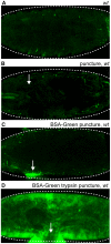
Figure 1. Localized endogenous proteolytic activity occurs at clean puncture wound sites.
Confocal images of Bovine Serum Albumin conjugated-Green (BSA-Green) wounded wild-type embryos. (A) Unwounded wild-type embryos display no fluorescence. (B) Puncture wounded wild-type embryos display no fluorescence at the wound site. (C) Wild-type embryos puncture wounded with BSA-Green exhibit green fluorescence surrounding wound site at 30 minutes after wounding, indicating proteolysis of BSA. (D) Simultaneous puncture wounding of trypsin along with BSA-Green results in whole body cavity green fluorescence 30 minutes after wounding. Arrows mark the wound site. Dashed lines in the data panels mark the outlines of embryos.
Figure 2
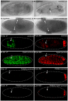
Figure 2. Serine proteases are required and sufficient for Ddc.47 and ple-WE1 activation.
Bright field images of wild-type stage 15–17 embryos. Ddc.47 and ple-WE1 are fluorescent reporters that include wound-induced DNA enhancers from the Ddc and ple loci, respectively. (A, C, D) A melanized wound site is not observed in unwounded, Pefabloc wounded, or trypsin wounded embryos. (B) Melanization at the wound site occurs after puncture-only wounding of wild-type embryos. Confocal images of Ddc.47 and ple-WE1 embryos 6 hours post wounding. (E, F) Control puncture, water puncture, and HCl (trypsin buffer) puncture wounded Ddc.47 and ple-WE1 embryos all exhibit localized reporter activation at epidermal wound sites. (G, H) Trypsin puncture wounded Ddc.47 and ple-WE1 embryos exhibit global reporter activation. (I, J) Pefabloc puncture wounded Ddc.47 and ple-WE1 embryos do not activate reporter at the wound site. (K, L) Pefabloc trypsin puncture wounded Ddc.47 and ple-WE1 embryos do not activate any epidermal wound reporter expression. The anal pad expression provided by the enhancer in the ple-WE1 wound reporter controls for developmental stage. Arrows mark the wound site. Dashed lines in the data panels mark the outlines of embryos.
Figure 3
Figure 3. Trypsin treatment does not compromise epidermal barrier integrity.
Ddc.47 is a fluorescent reporter that includes a wound-induced DNA enhancer from the Ddc locus. Confocal images of Ddc. 47 (green) embryos injected with fluorescent Rhodamine Dextran (red) to assess epidermal integrity and reporter activation after trypsin puncture wounding. (A, B) Perivitelline injection of trypsin along with Rhodamine Dextran globally activates the Ddc.47 wound reporter without compromising the epidermal barrier since Rhodamine Dextran is limited to the perivitelline space. (C, D) Embryos punctured with trypsin and Rhodamine Dextran globally activate Ddc.47 wound reporter, but epidermal integrity is lost as Rhodamine Dextran is observed within the embryonic body cavity. (E, F) Control embryos that have been injected in the perivitelline space with Rhodamine Dextran in carrier solution do not activate the Ddc.47 wound reporter. Arrows mark the wound site. Dashed lines in the data panels mark the outlines of embryos.
Figure 4
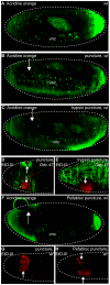
Figure 4. Trypsin or Pefabloc treatments do not cause widespread epidermal cell death.
Confocal images of embryos stained with acridine orange (apoptosis marker) and Ethidium homodimer-III (EtD-III, necrosis marker) two to five hours after wounding. Ddc.47 is a fluorescent reporter that includes a wound-induced DNA enhancer from the Ddc locus. (A) Wild-type unwounded embryos exhibit normal acridine orange (green) staining in the ventral nerve cord and brain region. (B, C, F) Similar acridine orange staining is observed in puncture (both water and HCl trypsin buffer), trypsin puncture wounded, and Pefabloc puncture wounded embryos. Anterior and posterior pole staining is an artifact. (D) Puncture-only wounded Ddc.47 embryos activate reporter (green) around the wound site in the epidermis while EtD-III (red) stain is localized to the melanized scab. (E) Puncture-trypsin wounded embryos activate reporter globally throughout the epidermis, but EtD-III staining remains relatively localized to the puncture wound site. (G) Wild-type embryos puncture wounded with water exhibit EtD-III (red) stain localized to the wound site. (H) Pefabloc puncture wounded embryos exhibit a slight expansion of EtD-III staining around the wound site compared to puncture wounded without Pefabloc (G). vnc = ventral nerve cord. Arrows mark the wound site. Dashed white lines outline embryos.
Figure 5
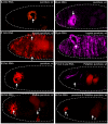
Figure 5. Serine proteases are sufficient and required for epidermal wound response gene transcription.
Confocal images of wild-type embryos after in situ hybridization with fluorescently labeled RNA probes made to detect transcripts from ple (magenta) and Ddc (red). (A, B) 30 minutes after HCl (trypsin buffer) puncture wounding, Ddc and ple transcripts accumulate in the epidermis around the wound site. (C, D) 30 minutes after puncture-trypsin wounding, ple and Ddc transcript accumulation can be observed throughout a large region of the epidermis. (E) One hour after water puncture wounding, wild-type embryos activate Ddc transcripts in the epidermis surrounding the wound site. (F) One hour after Pefabloc puncture wounding, no Ddc transcripts are activated in the epidermis surrounding the wound site in wild-type embryos, but normal developmental expression of ple in the cells that secrete the head skeleton is observed (magenta). Gut autofluorescence is seen in red. (G) In wild-type, double water puncture wounded embryos, 60 minutes after the first water puncture wound, a moderately wide zone of Ddc transcripts in the epidermis around wound sites is observed, while 30 minutes after the second wound, a narrow zone of Ddc transcript accumulation is observed around the wound site. (H) In wild-type double puncture wounded embryos with Pefabloc injected at the second site, 60 minutes after the first puncture wound, a narrow zone of accumulation of Ddc transcripts in the localized epidermis is observed around the first wound site, while 30 minutes after the second puncture wound with Pefabloc, no Ddc transcript accumulation is observed at the wound site. Arrows mark the wound sites. “1″ and “2″ indicate first and second wounds, respectively. Dashed lines in the data panels mark the outlines of embryos.
Figure 6
Figure 6. Serine protease activity is downstream of hydrogen peroxide with respect to wound reporter activation.
Confocal images of Ddc.47 and ple-WE1 embryos that have been double puncture wounded with hydrogen peroxide and/or Pefabloc. (A, B) Embryos that have been water puncture wounded first and then wounded with hydrogen peroxide second exhibit global reporter activation. (C, D) Embryos that have been wounded first with Pefabloc and second with hydrogen peroxide do not activate reporter at either wound site. Ple-WE1 developmental anal pad expression is observed in each treatment. The numbers “1″ and “2″ indicate the first and second wound sites, respectively. Arrows mark the wound site(s). Dashed lines outline the embryos. Ddc.47 and ple-WE1 are fluorescent reporters that include wound-induced DNA enhancers from the Ddc and ple loci, respectively.
Figure 7
Figure 7. Serine protease-mediated wound reporter activation is upstream of grainyhead function.
Confocal images of control Ddc.47 and grhIM mutants; Ddc.47 or grhIM; ple -WE1 activation about six hours after puncture and trypsin puncture wounding. Ddc.47 and ple-WE1 are fluorescent reporters that include wound-induced DNA enhancers from the Ddc and ple loci, respectively. (A, B) Ddc.47 embryos puncture wounded with carrier solution activate localized reporter at the wound site, but dramatically reduced localized reporter activation is observed in grh mutants after the same treatment. (C, D) Ple-WE1 embryos puncture wounded with carrier solution activate reporter around the wound site, while grh mutants exhibit only slightly reduced reporter activation at the wound site. The developmental anal pad expression from the ple-WE1 reporter construct is observed in each treatment. (E, F) Puncture-trypsin wounded Ddc.47 embryos activate reporter globally, while grh mutants exhibit dramatically reduced and scattered wound reporter activation after trypsin treatment. (G, H) Trypsin-treated ple-WE1 embryos activate reporter globally, while grh mutants activate lower, patchier, but still easily detectable global reporter activation after trypsin treatment. Developmental ple -WE1 anal pad expression is observed in every treatment. The pathway is shown on the right side of the figure. Arrows mark the wound site. Dashed lines in the data panels mark the outlines of embryos.
Figure 8
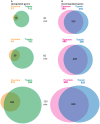
Figure 8. Abundant overlap of differentially regulated genes after puncture and trypsin puncture wounding.
At each of the three time points (30 minutes, 60 minutes, and 120 minutes) after puncture and trypsin puncture wounding, a comparison of the statistically significant (FDR <0.01) regulated genes was performed using Microsoft Excel software. (A) 30 minutes after puncture and trypsin puncture wounding, 15 and 112 significant genes respectively, were upregulated 1.8 fold or greater; 14 genes were commonly upregulated after both wounding treatments. 60 minutes post puncture and trypsin puncture wounding, 64 and 332 significant genes respectively, were upregulated 1.8 fold or greater; 58 genes were commonly upregulated after both wounding treatments. 120 minutes post puncture and trypsin puncture wounding, 210 and 624 significant genes respectively, were upregulated 1.8 fold or greater; 186 genes were commonly upregulated after both wounding treatments. (B) 30 minutes post puncture and trypsin puncture wounding, 389 and 378 significant genes respectively, were downregulated −1.8 fold or lower; 333 genes were commonly down regulated after both wounding treatments. 60 minutes post puncture and trypsin puncture wounding, 496 and 508 significant genes respectively, were downregulated −1.8 fold or lower; 429 genes were commonly downregulated after both wounding treatments. 120 minutes post puncture and trypsin puncture wounding, 698 and 826 significant genes respectively, were downregulated −1.8 fold or lower; 624 genes were commonly downregulated after both wounding treatments.
Figure 9
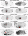
Figure 9. Novel localized epidermal, global epidermal, and fat body wound response genes in late stage Drosophila embryos.
Alkaline phosphatase in situ hybridization with probes targeting RNA of candidate wound response genes. Puncture wounded and wild-type stage 15–17 embryos were compared for tissue-specific transcript induction one hour after wounding. (A) Ady43A transcripts accumulate in broad zone in the epidermis around puncture wound sites. (B) Ets21C transcripts are observably upregulated in the narrow zone wound site, and are already present in unwounded embryos at an easily detectable level throughout the entire epidermis. (C) An increase in jra/jun transcripts is observed in a narrow zone around the epidermal wound site. (D) An increase in kay/fos transcripts are also detected in a moderately broad zone around the epidermal wound site. (E) Spz transcripts are detected in a broad zone around the epidermal wound site. (F) After puncture wounding, dorsal transcripts are detected in a moderately broad zone around the epidermal wound site. (G) After puncture wounding, rhomboid transcripts are detected around the epidermal wound site. (H) After puncture wounding Rel RNA is upregulated around the epidermal wound site and in the fat body. (I) IM1 RNA is upregulated throughout the fat body after puncture wounding, but not in the epidermis. (J) NijA RNA is upregulated throughout the entire epidermis after puncture wounding. Embryo bodies are outlined with dashed lines. The puncture wound site is indicated with a red asterisk. Black arrows point to fat body expression.

Similar articles

Cited by

References

    1. Wright TR (1996) The Wilhelmine E. Key 1992 Invitational lecture. Phenotypic analysis of the Dopa decarboxylase gene cluster mutants in Drosophila melanogaster. J Hered 87: 175–190. - PubMed
    1. Nemes Z, Steinert PM (1999) Bricks and mortar of the epidermal barrier. Exp Mol Med 31: 5–19. - PubMed
    1. Harden N (2005) Cell biology. Of grainy heads and broken skins. Science 308: 364–365. - PubMed
    1. Razzell W, Wood W, Martin P (2011) Swatting flies: modelling wound healing and inflammation in Drosophila. Dis Model Mech 4: 569–574. - PMC - PubMed
    1. Belacortu Y, Paricio N (2011) Drosophila as a model of wound healing and tissue regeneration in vertebrates. Dev Dyn 240: 2379–2404. - PubMed

Publication types