

Cholesteryl esters accumulate in the heart in a porcine model of ischemia and reperfusion
- PMID: 23637933
- PMCID: PMC3637450
- DOI: 10.1371/journal.pone.0061942
Cholesteryl esters accumulate in the heart in a porcine model of ischemia and reperfusion
Abstract
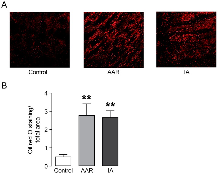
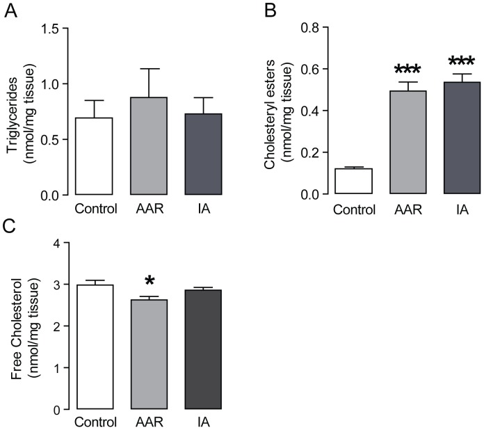
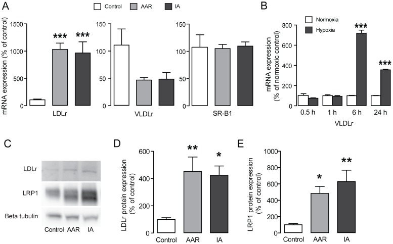
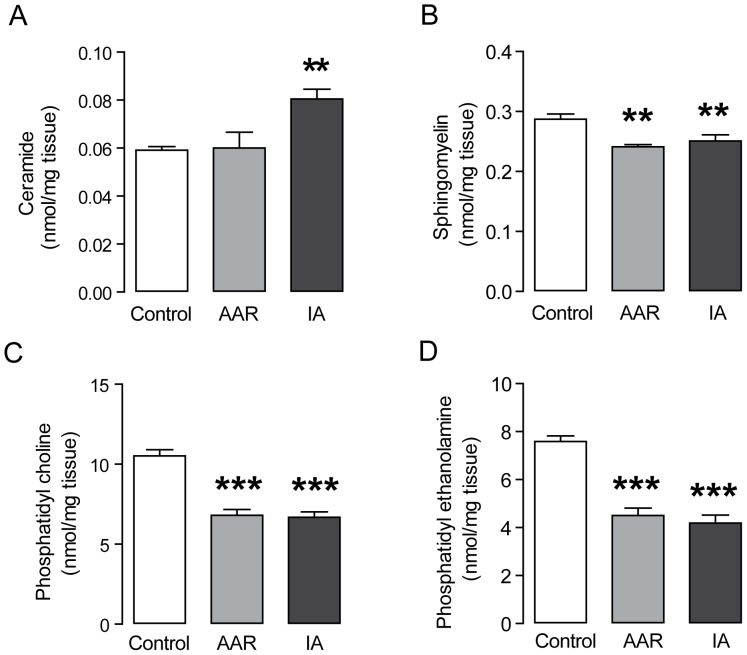
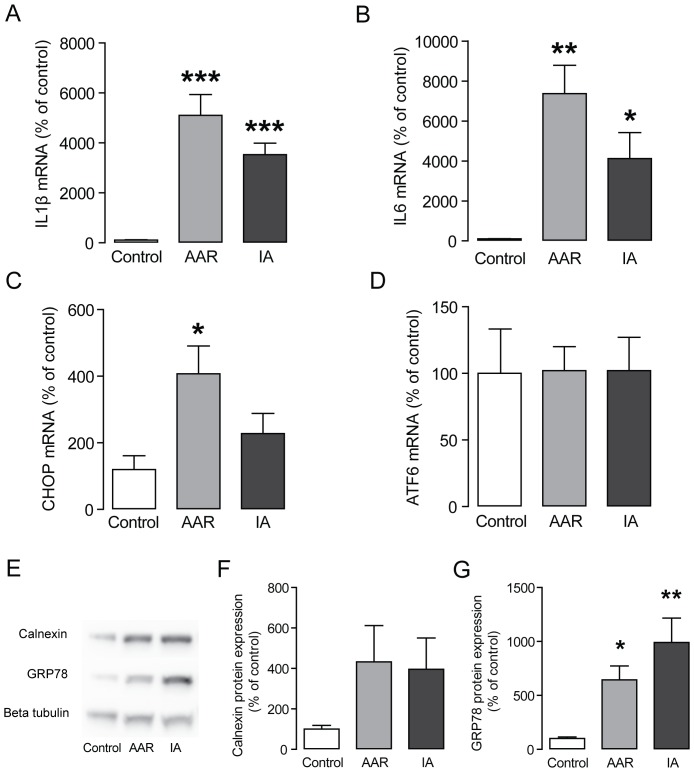
Myocardial ischemia is associated with intracellular accumulation of lipids and increased depots of myocardial lipids are linked to decreased heart function. Despite investigations in cell culture and animal models, there is little data available on where in the heart the lipids accumulate after myocardial ischemia and which lipid species that accumulate. The aim of this study was to investigate derangements of lipid metabolism that are associated with myocardial ischemia in a porcine model of ischemia and reperfusion. The large pig heart enables the separation of the infarct area with irreversible injury from the area at risk with reversible injury and the unaffected control area. The surviving myocardium bordering the infarct is exposed to mild ischemia and is stressed, but remains viable. We found that cholesteryl esters accumulated in the infarct area as well as in the bordering myocardium. In addition, we found that expression of the low density lipoprotein receptor (LDLr) and the low density lipoprotein receptor-related protein 1 (LRP1) was up-regulated, suggesting that choleteryl ester uptake is mediated via these receptors. Furthermore, we found increased ceramide accumulation, inflammation and endoplasmatic reticulum (ER) stress in the infarcted area of the pig heart. In addition, we found increased levels of inflammation and ER stress in the myocardium bordering the infarct area. Our results indicate that lipid accumulation in the heart is one of the metabolic derangements remaining after ischemia, even in the myocardium bordering the infarct area. Normalizing lipid levels in the myocardium after ischemia would likely improve myocardial function and should therefore be considered as a target for treatment.
Conflict of interest statement
Figures
Similar articles
-
Low-density lipoprotein receptor-related protein 1 mediates hypoxia-induced very low density lipoprotein-cholesteryl ester uptake and accumulation in cardiomyocytes.Cardiovasc Res. 2012 Jun 1;94(3):469-79. doi: 10.1093/cvr/cvs136. Epub 2012 Mar 27. Cardiovasc Res. 2012. PMID: 22454363
-
Low density lipoprotein receptor-related protein 1 expression correlates with cholesteryl ester accumulation in the myocardium of ischemic cardiomyopathy patients.J Transl Med. 2012 Aug 8;10:160. doi: 10.1186/1479-5876-10-160. J Transl Med. 2012. PMID: 22873206 Free PMC article.
-
Chronic Kidney Disease Exacerbates Myocardial Ischemia Reperfusion Injury: Role of Endoplasmic Reticulum Stress-Mediated Apoptosis.Shock. 2018 Jun;49(6):712-720. doi: 10.1097/SHK.0000000000000970. Shock. 2018. PMID: 28846567
-
Ischemia reperfusion injury, ischemic conditioning and diabetes mellitus.J Mol Cell Cardiol. 2016 Feb;91:11-22. doi: 10.1016/j.yjmcc.2015.12.020. Epub 2015 Dec 21. J Mol Cell Cardiol. 2016. PMID: 26718721 Review.
-
Cardiac magnetic resonance spectroscopy.Biochem Cell Biol. 1998;76(2-3):510-21. doi: 10.1139/bcb-76-2-3-510. Biochem Cell Biol. 1998. PMID: 9923721 Review.
Cited by
-
Plin2-deficiency reduces lipophagy and results in increased lipid accumulation in the heart.Sci Rep. 2019 May 6;9(1):6909. doi: 10.1038/s41598-019-43335-y. Sci Rep. 2019. PMID: 31061399 Free PMC article.
-
HMG-CoA Reductase Inhibitors Relieve Endoplasmic Reticulum Stress by Autophagy Inhibition in Rats With Permanent Brain Ischemia.Front Neurosci. 2018 Jun 19;12:405. doi: 10.3389/fnins.2018.00405. eCollection 2018. Front Neurosci. 2018. PMID: 29970982 Free PMC article.
-
The role of ER stress in lipid metabolism and lipotoxicity.J Lipid Res. 2016 Aug;57(8):1329-38. doi: 10.1194/jlr.R067595. Epub 2016 May 4. J Lipid Res. 2016. PMID: 27146479 Free PMC article. Review.
-
Glucosylceramide synthase deficiency in the heart compromises β1-adrenergic receptor trafficking.Eur Heart J. 2021 Nov 14;42(43):4481-4492. doi: 10.1093/eurheartj/ehab412. Eur Heart J. 2021. PMID: 34297830 Free PMC article.
-
Assessment of postoperative prognosis in patients with acute ST-segment elevation myocardial infarction after PCI using LRP1.Front Cardiovasc Med. 2025 Feb 27;12:1520696. doi: 10.3389/fcvm.2025.1520696. eCollection 2025. Front Cardiovasc Med. 2025. PMID: 40083817 Free PMC article.
References
-
- Neely JR, Morgan HE (1974) Relationship between carbohydrate and lipid metabolism and the energy balance of heart muscle. Annu Rev Physiol 36: 413–459. - PubMed
-
- Park TS, Yamashita H, Blaner WS, Goldberg IJ (2007) Lipids in the heart: a source of fuel and a source of toxins. Curr Opin Lipidol 18: 277–282. - PubMed
-
- Schaffer JE (2003) Lipotoxicity: when tissues overeat. Curr Opin Lipidol 14: 281–287. - PubMed
-
- Torffvit O, Agardh C (2000) The prognosis for type 2 diabetic patients with heart disease. A 10-year observation study of 385 patients. J Diabetes Complications 14: 301–306. - PubMed
Publication types
MeSH terms
Substances
LinkOut - more resources
Full Text Sources
Other Literature Sources
Research Materials
Miscellaneous
