. 2013 Apr 24;8(4):e62008.
doi: 10.1371/journal.pone.0062008. Print 2013.

Identification and validation of novel contraction-regulated myokines released from primary human skeletal muscle cells

Affiliations

Identification and validation of novel contraction-regulated myokines released from primary human skeletal muscle cells

Silja Raschke et al. PLoS One. .

Abstract

Proteins secreted by skeletal muscle, so called myokines, have been shown to affect muscle physiology and additionally exert systemic effects on other tissues and organs. Although recent profiling studies have identified numerous myokines, the amount of overlap from these studies indicates that the secretome of skeletal muscle is still incompletely characterized. One limitation of the models used is the lack of contraction, a central characteristic of muscle cells. Here we aimed to characterize the secretome of primary human myotubes by cytokine antibody arrays and to identify myokines regulated by contraction, which was induced by electrical pulse stimulation (EPS). In this study, we validated the regulation and release of two selected myokines, namely pigment epithelium derived factor (PEDF) and dipeptidyl peptidase 4 (DPP4), which were recently described as adipokines. This study reveals that both factors, DPP4 and PEDF, are secreted by primary human myotubes. PEDF is a contraction-regulated myokine, although PEDF serum levels from healthy young men decrease after 60 min cycling at VO2max of 70%. Most interestingly, we identified 52 novel myokines which have not been described before to be secreted by skeletal muscle cells. For 48 myokines we show that their release is regulated by contractile activity. This profiling study of the human skeletal muscle secretome expands the number of myokines, identifies novel contraction-regulated myokines and underlines the overlap between proteins which are adipokines as well as myokines.


Conflict of interest statement

Competing Interests: The authors have declared that no competing interests exist.

Figures

Figure 1
Figure 1. DPP4 and PEDF protein level during differentiation and release by human primary myotubes.
A. Primary human myoblasts were differentiated for 0 to 6 days. Protein level of DPP4 and PEDF were analyzed in total cell lysates by SDS-PAGE and Western blot. MHC served as a positive control for differentiation. B+C. To analyze the secretion mechanism, myotubes were incubated with 1 µg/µl BFA for 24 h on day 5 of differentiation. The release of DDP4 was analyzed by ELISA (B, n = 10) and the release of PEDF was analyzed by Western blot (C, n = 6, *p<0.001).
Figure 2
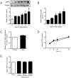
Figure 2. DPP4 protein level and release from myotubes and serum level after acute exercise.
A. Primary human myotubes were differentiated and DPP4 protein level during differentiation was analyzed by SDS-PAGE and Western blot. Data were normalized to the protein level of tubulin and are expressed relative to day 2 of differentiation. n = 4, *p<0.05 vs. day 2 of differentiation. B. Release of DPP4 during differentiation of myotubes was analyzed by ELISA. Data were normalized to day 2 of differentiation and are expressed relative to day 2 of differentiation. n = 9−10, *p<0.001 vs. day 2 of differentiation. C. Relative gene expression of DPP4 after 24 h EPS (1 Hz, 2 ms, 11.5 V) compared to non-stimulated cells was measured by real-time PCR as described, n = 4. D. DPP4 released by human myotubes was measured after 4, 8, 12 and 24 hours in non-stimulated cells compared to cells stimulated by EPS using ELISA, n≥11. White symbols, control; black symbols, EPS. E. Serum samples were taken before and after 60 min cycling (70% VO2max) at indicated time points. Serum DPP4 was analyzed by ELISA, n = 8. All data are mean values ± SEM.
Figure 3
Figure 3. PEDF expression, protein level, and release in myotubes and serum concentration after acute exercise.
A. Relative gene expression of PEDF during differentiation of myotubes was measured by real-time PCR as described and is expressed relative to day 2 of differentiation, n = 3−4, *p<0.05 vs. day 0 of differentiation. B. PEDF protein level of primary human myotubes was analyzed during differentiation by SDS-PAGE and Western blot. Data were normalized to the protein level of tubulin and are expressed relative to day 2 of differentiation. n = 5−6, *p<0.05 vs. day 0 of differentiation. C. Secretion of PEDF during differentiation of myotubes was analyzed by ELISA. Data are normalized to day 2 of differentiation, n = 5. D. Relative gene expression of PEDF after 24 h EPS (1 Hz, 2 ms, 11.5V) compared to non-stimulated cells was measured by real-time PCR as described, n = 4. E. PEDF secretion of human myotubes was measured after 4, 8, 12 and 24 hours in non-stimulated cells compared to cells stimulated by ELISA, n = 3−4, *p<0.05 vs. control. White symbols, control; black symbols, EPS. F. Serum samples were taken before and after 60 min cycling (70% VO2max) at indicated time points and PEDF level was analyzed by ELISA, n = 8, *p<0.05. All data are mean values ± SEM.
Figure 4
Figure 4. Assessment of contraction-regulated myokines by cytokine antibody arrays.
CM of control and EPS-treated cells were collected after 24 h and analyzed as described in Material and Methods. A. Cytokine antibody array membranes #5, #9 and #10 after incubation with CM of control and EPS-treated cells are shown. The encircled areas reflect spots corresponding to the myokines shown in Fig.4B. 1 = positive control; 2 = negative control; 3 = IL-6; 4 = IL-7; 5 = IL-8; 6 = VEGF. B. Known contraction-regulated myokines were analyzed by cytokine antibody arrays and quantified as positive controls. IL, interleukin; VEGF, vascular endothelial growth factor, n = 4, *p<0.01 vs. control, #p<0.05 vs. control. C. IL-6 and VEGF protein concentration were additionally analyzed by ELISA in CM of control and EPS-treated cells. IL-6, n = 6, *p<0.01. VEGF, n = 4, #p<0.05. White bars, control; black bars, EPS.

References

    1. Pedersen BK (2011) Muscles and their myokines. J Exp Biol 214: 337–346. - PubMed
    1. Holloszy JO, Coyle EF (1984) Adaptations of skeletal muscle to endurance exercise and their metabolic consequences. J Appl Physiol 56: 831–838. - PubMed
    1. Ross A, Leveritt M (2001) Long-term metabolic and skeletal muscle adaptations to short-sprint training: implications for sprint training and tapering. Sports Med 31: 1063–1082. - PubMed
    1. Petersen AM, Pedersen BK (2005) The anti-inflammatory effect of exercise. J Appl Physiol 98: 1154–1162. - PubMed
    1. Kersten S, Lichtenstein L, Steenbergen E, Mudde K, Hendriks HF, et al. (2009) Caloric restriction and exercise increase plasma ANGPTL4 levels in humans via elevated free fatty acids. Arterioscler Thromb Vasc Biol 29: 969–974. - PubMed

Publication types

MeSH terms

LinkOut - more resources