. 2013 Apr 29;8(4):e62083.
doi: 10.1371/journal.pone.0062083. Print 2013.

The genome sequence of Lone Star virus, a highly divergent bunyavirus found in the Amblyomma americanum tick

Affiliations

The genome sequence of Lone Star virus, a highly divergent bunyavirus found in the Amblyomma americanum tick

Andrea Swei et al. PLoS One. .

Abstract

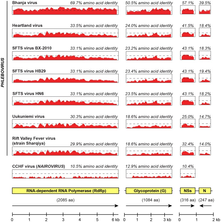
Viruses in the family Bunyaviridae infect a wide range of plant, insect, and animal hosts. Tick-borne bunyaviruses in the Phlebovirus genus, including Severe Fever with Thrombocytopenia Syndrome virus (SFTSV) in China, Heartland virus (HRTV) in the United States, and Bhanja virus in Eurasia and Africa have been associated with acute febrile illness in humans. Here we sought to characterize the growth characteristics and genome of Lone Star virus (LSV), an unclassified bunyavirus originally isolated from the lone star tick Amblyomma americanum. LSV was able to infect both human (HeLa) and monkey (Vero) cells. Cytopathic effects were seen within 72 h in both cell lines; vacuolization was observed in infected Vero, but not HeLa, cells. Viral culture supernatants were examined by unbiased deep sequencing and analysis using an in-house developed rapid computational pipeline for viral discovery, which definitively identified LSV as a phlebovirus. De novo assembly of the full genome revealed that LSV is highly divergent, sharing <61% overall amino acid identity with any other bunyavirus. Despite this sequence diversity, LSV was found by phylogenetic analysis to be part of a well-supported clade that includes members of the Bhanja group viruses, which are most closely related to SFSTV/HRTV. The genome sequencing of LSV is a critical first step in developing diagnostic tools to determine the risk of arbovirus transmission by A. americanum, a tick of growing importance given its expanding geographic range and competence as a disease vector. This study also underscores the power of deep sequencing analysis in rapidly identifying and sequencing the genomes of viruses of potential clinical and public health significance.


Conflict of interest statement

Competing Interests: This study was partly funded by an Abbott Viral Discovery Award. Charles Y. Chiu is the director of the UCSF-Abbott Viral Diagnostics and Discovery Center (VDDC). This does not alter the authors’ adherence to all PLOS ONE policies on sharing data and materials.

Figures

Figure 1
Figure 1. Time course of the development of cytopathic effects by Lone Star virus in human (HeLa) and monkey (Vero) cell cultures.
CPE is shown at 24, 48, 72, 96, and 120 hours post-inoculation (hpi). Uninfected controls at 120 hpi are also shown.
Figure 2
Figure 2. Identification and assembly of the LSV genome by unbiased deep sequencing.
(A) Using a rapid computational pipeline, reads identified as bunyaviruses by SNAP nucleotide alignment (orange) or RAPSearch amino acid alignment (dark red) were mapped to the assembled LSV genome. The coverage (y-axis) achieved at each position along the genome (x-axis) is plotted on a logarithmic scale. (B) De novo assembly of the LSV genome using the PRICE assembler (3 rounds of 15 cycles each) and LSV seed sequences (“S”) identified from (A). (C) The genome structure of LSV. Boxes represent open reading frames (ORFs) corresponding to the RdRp, G, N, and NSs proteins, flanked by noncoding regions, which are indicated by lines. Coding directions are indicated by arrows. (D) Mapping of the actual deep sequencing reads derived from LSV to the final assembled genome. The coverage (y-axis) achieved at each position along the genome (x-axis) is plotted on a logarithmic scale. GenBank accession numbers are reported in the text. Abbreviations: kb, kilobases; bp, base pairs.
Figure 3
Figure 3. Amino acid phylogenetic analysis of the four LSV protein sequences relative to those from representative phleboviruses and Gouleako virus.
For the RdRp, glycoprotein, and N protein, Gouleako virus is included as an outgroup to the phleboviruses (tan). Gouleako virus, the closest known bunyavirus relative to phleboviruses, is a member of a proposed new genus in the family Bunyaviridae . Also shown color-coded are the Uukuniemi (blue), Bhanja (red), and SFTS (green) clades of known tick-borne phleboviruses. GenBank accession numbers are reported in the text.
Figure 4
Figure 4. Amino acid pairwise identity of LSV relative to other representative bunyaviruses.
The amino acid identities are shown for the four LSV proteins (RdRp, G, N, and NSs). A sliding window of 50 bp was used. GenBank accession numbers are reported in the text.

References

    1. Walter CT, Barr JN (2011) Recent advances in the molecular and cellular biology of bunyaviruses. J Gen Virol 92: 2467–2484. - PubMed
    1. Ergonul O (2012) Crimean-Congo hemorrhagic fever virus: new outbreaks, new discoveries. Curr Opin Virol 2: 215–220. - PubMed
    1. Jonsson CB, Figueiredo LT, Vapalahti O (2010) A global perspective on hantavirus ecology, epidemiology, and disease. Clin Microbiol Rev 23: 412–441. - PMC - PubMed
    1. Guu TS, Zheng W, Tao YJ (2012) Bunyavirus: structure and replication. Adv Exp Med Biol 726: 245–266. - PubMed
    1. Xu B, Liu L, Huang X, Ma H, Zhang Y, et al. (2011) Metagenomic analysis of fever, thrombocytopenia and leukopenia syndrome (FTLS) in Henan Province, China: discovery of a new bunyavirus. PLoS Pathog 7: e1002369. - PMC - PubMed

Publication types

Associated data