

Microsporidia and its relation to Crohn's disease. A retrospective study
- PMID: 23637975
- PMCID: PMC3630148
- DOI: 10.1371/journal.pone.0062107
Microsporidia and its relation to Crohn's disease. A retrospective study
Abstract
Background: The cause of Crohn's Disease (CD) remains unknown. Recently a decrease in the global lymphocyte population in the peripheral blood of CD patients has been reported. This decrease was more evident in γδ T lymphocytes, especially γδ CD8+T subsets. Furthermore, a decrease of IL-7 was also observed in these patients. We propose the hypothesis that microsporidia, an obligate intracellular opportunistic parasite recently related to fungi, in CD patients can take advantage of the lymphocytes and IL-7 deficits to proliferate and to contribute to the pathophysiology of this disease.
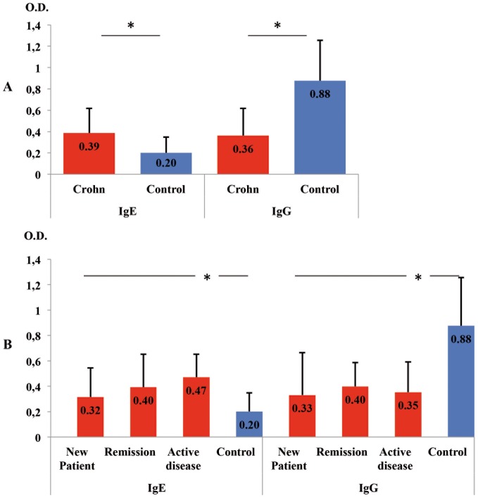
Methods and findings: In this case-control study, serum samples were collected from 36 CD patients and from 36 healthy individuals (controls), IgE and IgG anti-Encephalitozoon antibodies were determined by ELISA; and forty-four intestinal tissue samples were analyzed through real time Polymerase Chain Reaction (PCR), twenty CD patients, nine with others diseases and 15 healthy subjects. We observed that IgE anti-Encephalitozoon levels were significantly higher in patients with CD: 0.386(±0.256) vs control group, 0.201(±0.147), P<0.001. However, IgG anti-Encephalitozoon values were significantly lower in CD patients: 0.361(±0.256) vs control group, 0.876(±0.380), P<0.001. In the group of CD patients, 6/20 (30%) were positive by real time PCR for microsporidia and, all the patients of the control group were negative by real time PCR.
Conclusions: These results suggest that CD patients are a group at risk for microsporidiasis and, moreover that microsporidia may be involved as a possible etiologic factor of CD.
Conflict of interest statement
Figures
References
-
- Andreu-Ballester JC, Amigó-García V, Catalán-Serra I, Gil-Borrás R, Ballester F, et al. (2011) Deficit of gammadelta T lymphocytes in the peripheral blood of patients with Crohn's Disease. Dig Dis Sci 56: 2613–2622. - PubMed
-
- Andreu-Ballester JC, Pérez-Griera J, Garcia-Ballesteros C, Amigo V, Catalán-Serra I, et al.. (2012) Deficit of interleukin-7 in serum of patients with Crohn's disease. Inflamm Bowel Dis Feb 7. doi: 10.1002/ibd.22914. - PubMed
-
- Keeling PJ, Fast NM (2002) Microsporidia: Biology and evolution of highly reduced intracellular parasites. Annu Rev Microbiol 56: 93–116. - PubMed
MeSH terms
Substances
LinkOut - more resources
Full Text Sources
Other Literature Sources
Medical
Research Materials
