. 2013 Apr 24;8(4):e62170.
doi: 10.1371/journal.pone.0062170. Print 2013.

miR-221 promotes tumorigenesis in human triple negative breast cancer cells

Affiliations

miR-221 promotes tumorigenesis in human triple negative breast cancer cells

Rounak Nassirpour et al. PLoS One. .

Retraction in

Abstract

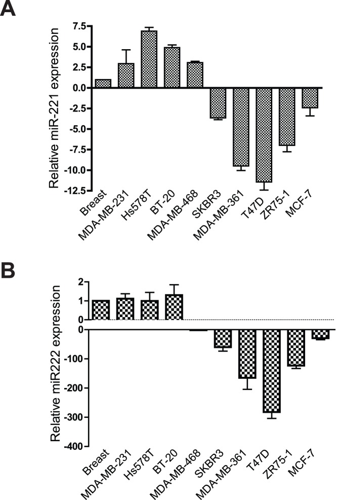
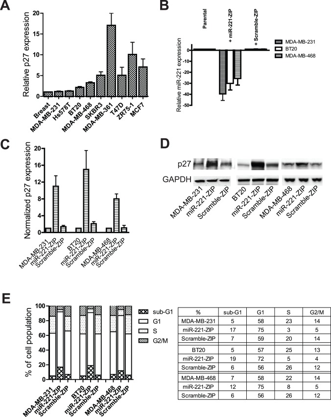
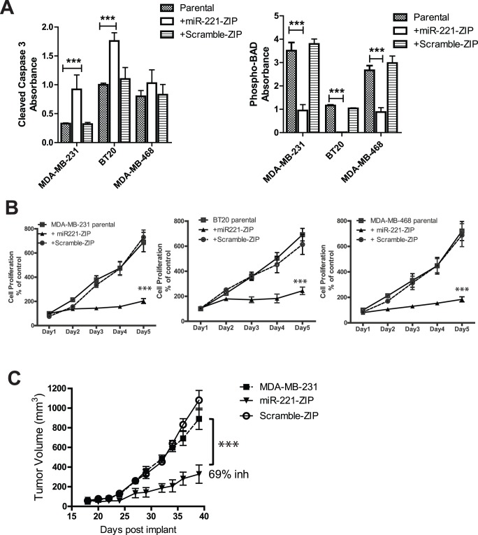
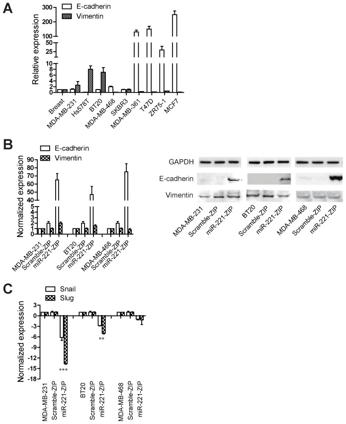
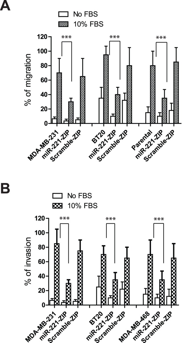
Patients with triple-negative breast cancers (TNBCs) typically have a poor prognosis. TNBCs are characterized by their resistance to apoptosis, aggressive cellular proliferation, migration and invasion, and currently lack molecular markers and effective targeted therapy. Recently, miR-221/miR-222 have been shown to regulate ERα expression and ERα-mediated signaling in luminal breast cancer cells, and also to promote EMT in TNBCs. In this study, we characterized the role of miR-221 in a panel of TNBCs as compared to other breast cancer types. miR-221 knockdown not only blocked cell cycle progression, induced cell apoptosis, and inhibited cell proliferation in-vitro but it also inhibited in-vivo tumor growth by targeting p27(kip1). Furthermore, miR-221 knockdown inhibited cell migration and invasion by altering E-cadherin expression, and its regulatory transcription factors Snail and Slug in human TNBC cell lines. Therefore, miR-221 functions as an oncogene and is essential in regulating tumorigenesis in TNBCs both in vitro as well as in vivo.


Conflict of interest statement

Competing Interests: All authors are current full-time employees of Pfizer Inc. This does not alter the authors' adherence to all the PLOS ONE policies on sharing data and materials.

Figures

Figure 1
Figure 1. miR-221 is over expressed in TNBCs.
qRT-PCR was performed to quantitatively measure RNA expression levels of miR-221 (A) and miR-222 (B) in a panel of breast cancer cell lines. All expression levels are displayed as fold changes normalized to the expression level in normal breast tissue.
Figure 2
Figure 2. miR-221 modulates cell cycle progression by targeting p27kip1.
(A) mRNA expression level of p27kip1 was measured in a panel of breast cancer cell lines. Data are displayed as fold changes normalized to the expression level in normal breast tissue. (B) TNBC lines, MDA-MB-231, BT-20, and MDA-MB-468 cells were established to stably express anti-miR-221 (miR-221-ZIP) or a control scramble miRNA (Scramble-ZIP). miR-221 expression level was measured and is displayed as fold changes normalized to the expression level of parental cell lines. (C) Transcript expression level of p27kip1 was measured and is displayed as fold changes normalized to the expression level of parental cell lines. (D) Western blot analysis of MDA-MB-231, BT-20, and MDA-MB-468 cells stably expressing anti-miR-221 or scramble miR-ZIP depicting changes in p27 protein level. GAPDH was used as loading control. (E) MDA-MB-231, BT-20, and MDA-MB-468 cells stably expressing anti-miR-221 or scramble miR-ZIP were cultured for 72 hours to reach 80–90% confluency before harvesting and cell cycle analysis was performed and displayed as percentages of each cell cycle stage. These experiments were repeated at least three times, and representative data is shown.
Figure 3
Figure 3. miR-221 knockdown induces apoptosis, inhibits cell proliferation and supresses tumor growth in mice.
MDA-MB-231, BT-20, and MDA-MB-468 cells stably expressing miR-221-ZIP or scramble-ZIP were used to perform apoptosis, cell proliferation, and in-vivo tumor growth assays. (A) Cleaved caspase 3 and phosphorylation of BAD were measured as apoptosis markers. All cell lines were seeded at similar density as the parental cell lines and cultured for 72 hours until the parental cell lines reached 80–95% confluencey, before cell lysates were prepared and subject to cleaved caspase 3 assays. (B) Cell proliferation measurements were normalized to the readings in parental cells at day 1. (C) Nude mice were implanted subcutaneously with MDA-MB-231 parental cells, and MDA-MB-231 cells stably expressing miR-221-ZIP or scramble-ZIP. Tumor measurements were recorded and tumor growth inhibition was calculated as described in Materials and Methods. T-test was performed in (A) and (B) and one way ANOVA was performed in (C) to compare the differences between parental cells versus miR-221-ZIP cells. *denotes p-value ≤0.05. ***denotes p-value ≤0.005.
Figure 4
Figure 4. Down regulation of miR-221 increases E-cadherin levels and decreases the expression levels of Snail and Slug.
(A) The RNA expression level of E-cadherin and vimentin was measured in a panel of breast cancer cell lines. Fold changes are recorded as normalized to normal breast tissue levels. (B) E-cadherin and vimentin expression levels were measured in MDA-MB-231, BT-20, MDA-MB-468 parental cells, as well cells harboring miR-221-ZIP, or scramble-ZIP. Data are normalized to the expression level in parental cells. Western blot analysis was also performed to examine the protein levels of E-cadherin and vimentin. (C) Snail and Slug expression levels were also examined in MDA-MB-231, BT-20, and MDA-MB-468 cells. Data were normalized to the expression level in parental cells and fold changes were plotted. ***denotes p<0.005. **denotes p<0.01.
Figure 5
Figure 5. Down regulation of miR-221 inhibits cell migration and invasion in TNBC lines.
(A) Migration and (B) Invasion assays were performed in the absence or presence of 10% FBS after 72 hours in culture. Data are displayed as the percentage of cells migrated or invaded. T-test was performed to compare the differences between parental cells versus miR-221-ZIP cells, ***denotes p<0.005.

Similar articles

Cited by

References

    1. Bartel DP (2004) MicroRNAs: genomics, biogenesis, mechanism, and function. Cell 116: 281–297. - PubMed
    1. Lim LP, Lau NC, Garrett-Engele P, Grimson A, Schelter JM, et al. (2005) Microarray analysis shows that some microRNAs downregulate large numbers of target mRNAs. Nature 433: 769–773. - PubMed
    1. Lewis BP, Shih IH, Jones-Rhoades MW, Bartel DP, Burge CB (2003) Prediction of mammalian microRNA targets. Cell 115: 787–798. - PubMed
    1. Esquela-Kerscher A, Slack FJ (2006) Oncomirs - microRNAs with a role in cancer. Nat Rev Cancer 6: 259–269. - PubMed
    1. Ambros V (2004) The functions of animal microRNAs. Nature 431: 350–355. - PubMed

Publication types

MeSH terms