Reduction of in-stent restenosis risk on nickel-free stainless steel by regulating cell apoptosis and cell cycle
- PMID: 23638002
- PMCID: PMC3637440
- DOI: 10.1371/journal.pone.0062193
Reduction of in-stent restenosis risk on nickel-free stainless steel by regulating cell apoptosis and cell cycle
Abstract
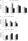
High nitrogen nickel-free austenitic stainless steel (HNNF SS) is one of the biomaterials developed recently for circumventing the in-stent restenosis (ISR) in coronary stent applications. To understand the ISR-resistance mechanism, we have conducted a comparative study of cellular and molecular responses of human umbilical vein endothelial cells (HUVECs) to HNNF SS and 316L SS (nickel-containing austenitic 316L stainless steel) which is the stent material used currently. CCK-8 analysis and flow cytometric analysis were used to assess the cellular responses (proliferation, apoptosis, and cell cycle), and quantitative real-time PCR (qRT-PCR) was used to analyze the gene expression profile of HUVECs exposed to HNNF SS and 316L SS, respectively. Flow cytometry analysis revealed that 316L SS could activate the cellular apoptosis more efficiently and initiate an earlier entry into the S-phase of cell cycle than HNNF SS. At the molecular level, qRT-PCR results showed that the genes regulating cell apoptosis and autophagy were overexpressed on 316L SS. Further examination indicated that nickel released from 316L SS triggered the cell apoptosis via Fas-Caspase8-Caspase3 exogenous pathway. These molecular mechanisms of HUVECs present a good model for elucidating the observed cellular responses. The findings in this study furnish valuable information for understanding the mechanism of ISR-resistance on the cellular and molecular basis as well as for developing new biomedical materials for stent applications.
Conflict of interest statement
Figures


Similar articles
-
EGCG Regulates Cell Apoptosis of Human Umbilical Vein Endothelial Cells Grown on 316L Stainless Steel for Stent Implantation.Drug Des Devel Ther. 2021 Feb 11;15:493-499. doi: 10.2147/DDDT.S296548. eCollection 2021. Drug Des Devel Ther. 2021. PMID: 33603339 Free PMC article.
-
Biological behaviour of human umbilical artery smooth muscle cell grown on nickel-free and nickel-containing stainless steel for stent implantation.Sci Rep. 2016 Jan 4;6:18762. doi: 10.1038/srep18762. Sci Rep. 2016. PMID: 26727026 Free PMC article.
-
In vivo and in vitro analyses of the effects of a novel high-nitrogen low-nickel coronary stent on reducing in-stent restenosis.J Biomater Appl. 2018 Jul;33(1):64-71. doi: 10.1177/0885328218773306. Epub 2018 May 2. J Biomater Appl. 2018. PMID: 29720017
-
A review on nickel-free nitrogen containing austenitic stainless steels for biomedical applications.Mater Sci Eng C Mater Biol Appl. 2013 Oct;33(7):3563-75. doi: 10.1016/j.msec.2013.06.002. Epub 2013 Jun 12. Mater Sci Eng C Mater Biol Appl. 2013. PMID: 23910251 Review.
-
Nickel-free austenitic stainless steels for medical applications.Sci Technol Adv Mater. 2010 Feb 26;11(1):014105. doi: 10.1088/1468-6996/11/1/014105. eCollection 2010 Feb. Sci Technol Adv Mater. 2010. PMID: 27877320 Free PMC article. Review.
Cited by
-
The development of carotid stent material.Interv Neurol. 2015 Mar;3(2):67-77. doi: 10.1159/000369480. Interv Neurol. 2015. PMID: 26019710 Free PMC article. Review.
-
EGCG Regulates Cell Apoptosis of Human Umbilical Vein Endothelial Cells Grown on 316L Stainless Steel for Stent Implantation.Drug Des Devel Ther. 2021 Feb 11;15:493-499. doi: 10.2147/DDDT.S296548. eCollection 2021. Drug Des Devel Ther. 2021. PMID: 33603339 Free PMC article.
-
Designing Better Cardiovascular Stent Materials - A Learning Curve.Adv Funct Mater. 2021 Jan 4;31(1):2005361. doi: 10.1002/adfm.202005361. Epub 2020 Nov 4. Adv Funct Mater. 2021. PMID: 33708033 Free PMC article.
-
Effect of Copper-Containing Stainless Steel on Apoptosis of Coronary Artery Smooth Muscle Cells.Iran J Public Health. 2021 Sep;50(9):1825-1831. doi: 10.18502/ijph.v50i9.7055. Iran J Public Health. 2021. PMID: 34722378 Free PMC article.
-
One-Year Outcomes of CGuard Double Mesh Stent in Carotid Artery Disease: A Systematic Review and Meta-Analysis.Medicina (Kaunas). 2024 Feb 8;60(2):286. doi: 10.3390/medicina60020286. Medicina (Kaunas). 2024. PMID: 38399573 Free PMC article.
References
-
- Waterhouse A, Yin Y, Wise SG, Bax DV, McKenzie DR, et al. (2010) The immobilization of recombinant human tropoelastin on metals using a plasma-activated coating to improve the biocompatibility of coronary stents. Biomaterials 31: 8332–8340. - PubMed
-
- Ren L, Xu L, Feng J, Zhang Y, Yang K (2012) In vitro study of role of trace amount of Cu release from Cu-bearing stainless steel targeting for reduction of in-stent restenosis. Journal of Materials Science: Materials in Medicine 23: 1235–1245. - PubMed
-
- Bauters C, Banos JL, Van Belle E, Mc Fadden EP, Lablanche JM, et al. (1998) Six-Month Angiographic Outcome After Successful Repeat Percutaneous Intervention for In-Stent Restenosis. Circulation 97: 318–321. - PubMed
-
- Inoue T, Node K (2009) Molecular basis of restenosis and novel issues of drug-eluting stents. Circ J 73: 615–621. - PubMed
-
- Lü X, Lu H, Zhao L, Yang Y, Lu Z (2010) Genome-wide pathways analysis of nickel ion-induced differential genes expression in fibroblasts. Biomaterials 31: 1965–1973. - PubMed
Publication types
MeSH terms
Substances
LinkOut - more resources
Full Text Sources
Other Literature Sources
Research Materials
Miscellaneous

