. 2013 Apr 24;8(4):e62263.
doi: 10.1371/journal.pone.0062263. Print 2013.

The evolutionary paradox of tooth wear: simply destruction or inevitable adaptation?

Affiliations

The evolutionary paradox of tooth wear: simply destruction or inevitable adaptation?

Stefano Benazzi et al. PLoS One. .

Abstract

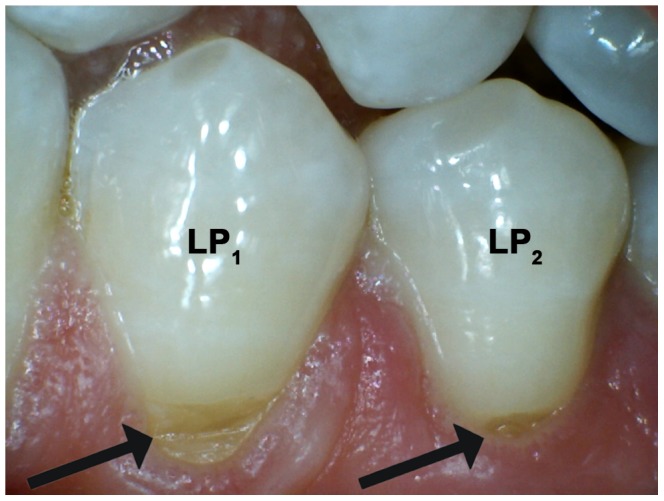
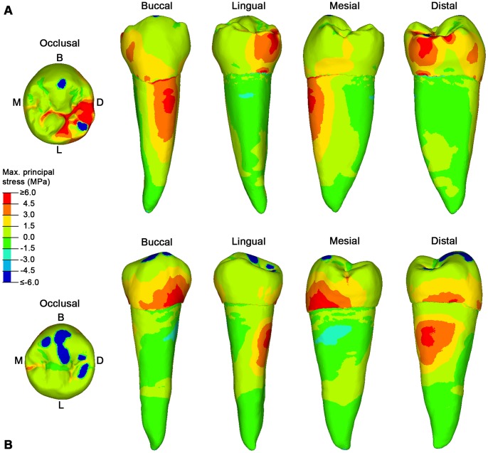
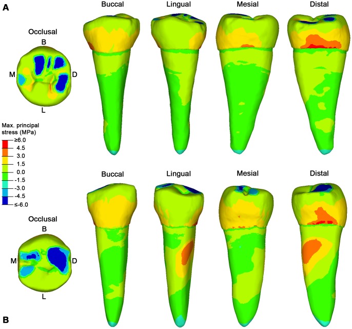
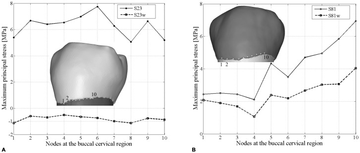
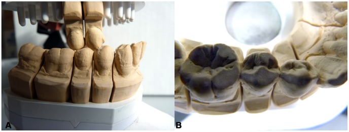
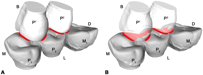
Over the last century, humans from industrialized societies have witnessed a radical increase in some dental diseases. A severe problem concerns the loss of dental materials (enamel and dentine) at the buccal cervical region of the tooth. This "modern-day" pathology, called non-carious cervical lesions (NCCLs), is ubiquitous and worldwide spread, but is very sporadic in modern humans from pre-industrialized societies. Scholars believe that several factors are involved, but the real dynamics behind this pathology are far from being understood. Here we use an engineering approach, finite element analysis (FEA), to suggest that the lack of dental wear, characteristic of industrialized societies, might be a major factor leading to NCCLs. Occlusal loads were applied to high resolution finite element models of lower second premolars (P2) to demonstrate that slightly worn P2s envisage high tensile stresses in the buccal cervical region, but when worn down artificially in the laboratory the pattern of stress distribution changes and the tensile stresses decrease, matching the results obtained in naturally worn P2s. In the modern industrialized world, individuals at advanced ages show very moderate dental wear when compared to past societies, and teeth are exposed to high tensile stresses at the buccal cervical region for decades longer. This is the most likely mechanism explaining enamel loss in the cervical region, and may favor the activity of other disruptive processes such as biocorrosion. Because of the lack of dental abrasion, our masticatory apparatus faces new challenges that can only be understood in an evolutionary perspective.


Conflict of interest statement

Competing Interests: The Dental Workshop Bensheim is a company owned by one of the co-authors (Mr. Dieter Schulz). There is no conflict of interests and this does not alter the authors’ adherence to all the PLOS ONE policies on sharing data and materials.

Figures

Figure 1
Figure 1. Modern lower premolars presenting non-carious cervical lesions (NCCLs).
Arrows point towards the NCCLs in the buccal cervical region of the lower left first and second premolar (LP1 and LP2, respectively).
Figure 2
Figure 2. The maximum principal stress distribution for specimen S23 and S81 lower right second premolars (RP2).
A, specimen S23 in occlusal, buccal, lingual, mesial and distal view. B. specimen S81 in occlusal, buccal, lingual, mesial and distal view. Blue spots in the occlusal surface (compressive stress) represent the contact areas with the antagonistic teeth, during maximum intercuspation (see Video S1 for specimen S23, and Video S2 for specimen S81), where the load was applied. Red spots represent tensile stresses. B, buccal; D, distal; L, lingual; M, mesial.
Figure 3
Figure 3. The maximum principal stress distribution for specimen S5 and S126 lower right second premolars (RP2).
A, specimen S5 in occlusal, buccal, lingual, mesial and distal view. B. specimen S126 in occlusal, buccal, lingual, mesial and distal view. Blue spots in the occlusal surface (compressive stress) represent the contact areas with the antagonistic teeth, during maximum intercuspation (see Video S3 for specimen S5, and Video S4 for specimen S126), where the load was applied. Red spots represent tensile stresses. B, buccal; D, distal; L, lingual; M, mesial.
Figure 4
Figure 4. The maximum principal stress distribution for specimen S23w and S81w lower right second premolars (RP2).
A, specimen S23w in occlusal, buccal, lingual, mesial and distal view. B. specimen S81w in occlusal, buccal, lingual, mesial and distal view. Blue spots in the occlusal surface (compressive stress) represent the contact areas with the antagonistic teeth, during maximum intercuspation (see Video S5 for specimen S23w, and Video S6 for specimen S81w), where the load was applied. Red spots represent tensile stresses. B, buccal; D, distal; L, lingual; M, mesial.
Figure 5
Figure 5. Differences in tensile stress values between the original (S23 and S81) and the artificially worn down (S23w and S81w) lower right second premolars.
A, the maximum principal stress values for specimen S23 and S23w based on 10 homologous nodes in the buccal cervical region. B, the maximum principal stress values for specimen S81 and S81w based on 10 homologous nodes in the buccal cervical region.
Figure 6
Figure 6. Cast of specimen S81 in the dental articulator (PROTAR, KaVo Dental GmbH).
A, buccal view of the specimen during the artificial attrition experiment of the RP2 based on the individual pattern of occlusal movements. B, occlusal view of specimen S81w RP1-RM1 crowns with artificially enlarged wear facets.
Figure 7
Figure 7. Collision detection for specimen S126 in the Occlusal Fingerprint Analyser (OFA) software.
A, mesiolingual view during maximum intercuspation between the lower right premolars and first molar (RP1-RM1) and the upper right premolars (RP1-RP2). B, the RP1-RP2 are transparent to better show the collision (red areas) on the occlusal surface of the RP2. See also Video S4. B = buccal; D = distal; L = lingual; M = mesial.
Figure 8
Figure 8. Loading position and direction for specimen S23, S23w, S81, S81w, S5 and S126.
For each lower right second premolar (RP2) only the volumetric mesh of the enamel is displayed. The load (black arrows) was distributed proportionally according to the occlusal contact areas detected in the Occlusal Fingerprint Analyser (OFA) software. D = distal; L = lingual; M = mesial.

References

    1. Wood I, Jawad Z, Paisley C, Brunton P (2008) Non-carious cervical tooth surface loss: a literature review. J Dent 36: 759–66. - PubMed
    1. Perez Cdos R, Gonzalez MR, Prado NA, de Miranda MS, Macêdo Mde A, et al. (2012) Restoration of noncarious cervical lesions: when, why, and how. Int J Dent 2012: 1–8. - PMC - PubMed
    1. Grippo JO, Simring M, Coleman TA (2012) Abfraction, abrasion, biocorrosion, and the enigma of noncarious cervical lesions: a 20-year perspective. J Esthet Restor Dent 24: 10–23. - PubMed
    1. Aubry M, Maffart B, Donat B, Brau JJ (2003) Brief communication: Study of noncarious cervical tooth lesions in samples of prehistoric, historic and modern populations from the South of France. Am J Phys Anthropol 121: 10–14. - PubMed
    1. Ritter AV, Grippo JO, Coleman TA, Morgan ME (2009) Prevalence of carious and non-carious cervical lesions in archaeological populations from North America and Europe. J Esthet Restor Dent 21: 324–335. - PubMed

Publication types

LinkOut - more resources