. 2013 Apr 25;8(4):e62302.
doi: 10.1371/journal.pone.0062302. Print 2013.

A novel beta-defensin antimicrobial peptide in Atlantic cod with stimulatory effect on phagocytic activity

Affiliations

A novel beta-defensin antimicrobial peptide in Atlantic cod with stimulatory effect on phagocytic activity

Jareeporn Ruangsri et al. PLoS One. .

Abstract

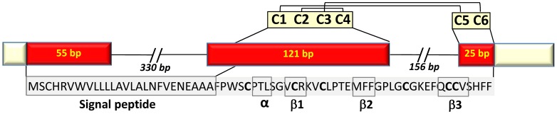
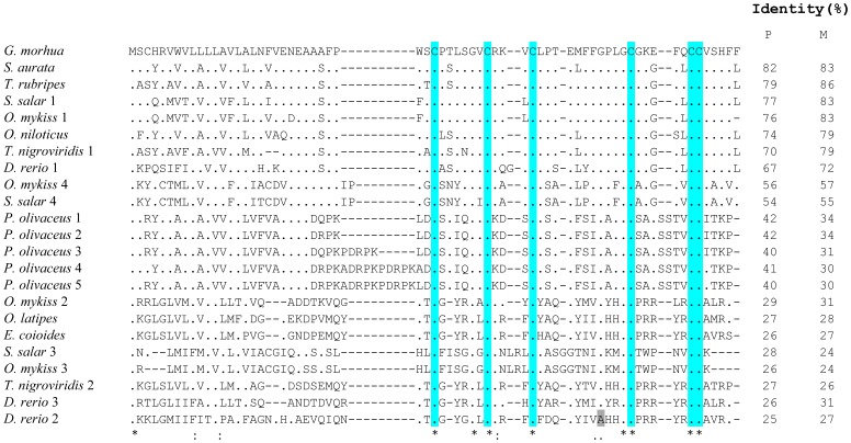
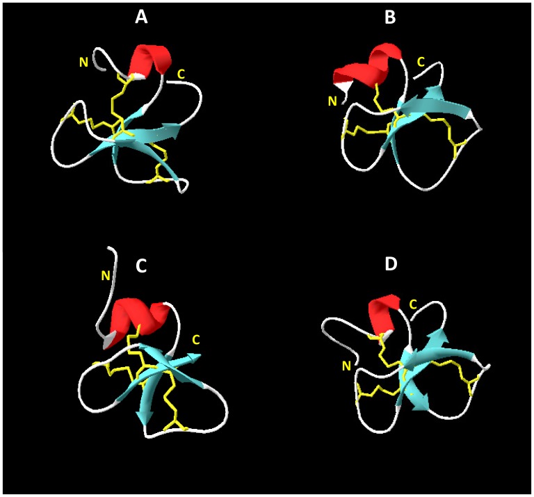
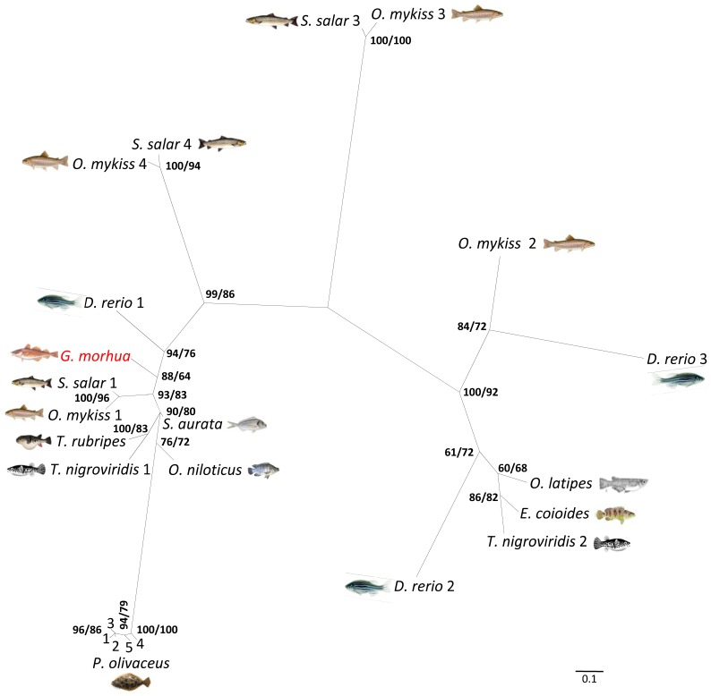
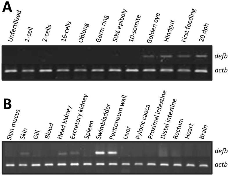
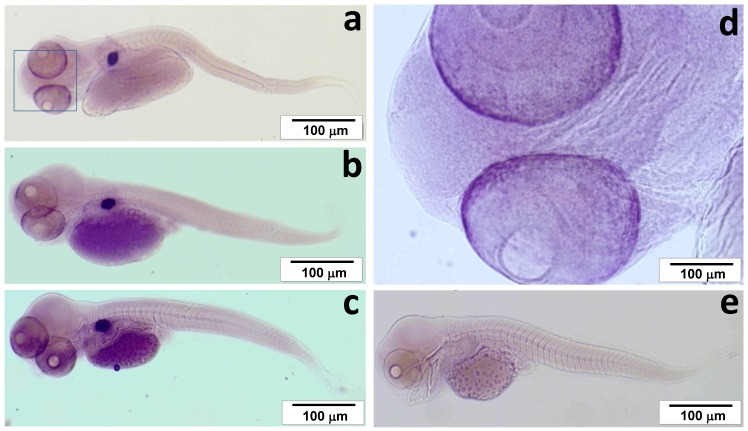
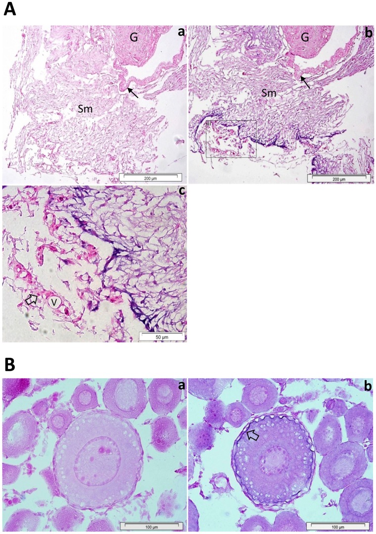
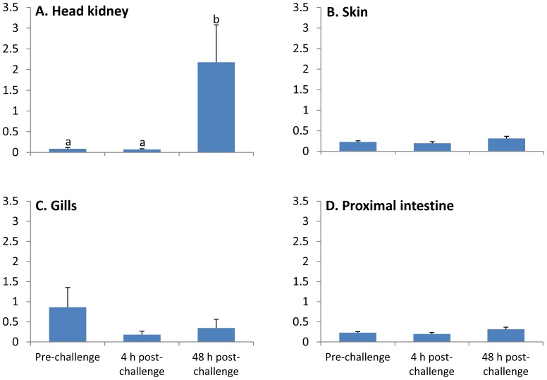
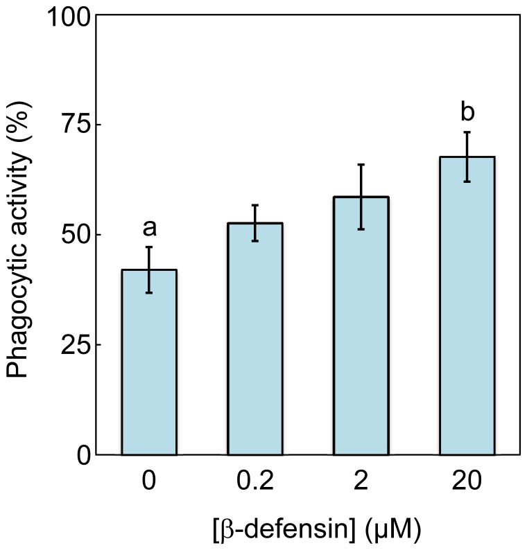
A novel defensin antimicrobial peptide gene was identified in Atlantic cod, Gadus morhua. This three exon/two intron defensin gene codes for a peptide precursor consisting of two domains: a signal peptide of 26 amino acids and a mature peptide of 40 residues. The mature cod defensin has six conserved cysteine residues that form 1-5, 2-4 and 3-6 disulphide bridges. This pattern is typical of beta-defensins and this gene was therefore named cod beta-defensin (defb). The tertiary structure of Defb exhibits an α/β fold with one α helix and β1β2β3 sheets. RT-PCR analysis indicated that defb transcripts were present mainly in the swim bladder and peritoneum wall but could also be detected at moderate to low levels in skin, head- and excretory kidneys. In situ hybridisation revealed that defb was specifically expressed by cells located in the swim bladder submucosa and the oocytes. During embryonic development, defb gene transcripts were detectable from the golden eye stage onwards and their expression was restricted to the swim bladder and retina. Defb was differentially expressed in several tissues following antigenic challenge with Vibrio anguillarum, being up-regulated up to 25-fold in head kidney. Recombinant Defb displayed antibacterial activity, with a minimal inhibitory concentration of 0.4-0.8 µM and 25-50 µM against the Gram-(+) bacteria Planococcus citreus and Micrococcus luteus, respectively. In addition, Defb stimulated phagocytic activity of cod head kidney leucocytes in vitro. These findings imply that beta-defensins may play an important role in the innate immune response of Atlantic cod.


Conflict of interest statement

Competing Interests: The authors have declared that no competing interests exist.

Figures

Figure 1
Figure 1. Schematic representation of the beta-defensin gene in Atlantic cod.
Pale yellow boxes represent partial 5′ and 3′untranslated regions. Red boxes indicate the coding region corresponding to the 66 amino acid residue peptide precursor. The predicted signal peptide and important secondary structure elements in the mature peptide are boxed. Conserved cysteine residues are highlighted in bold letters and predicted disulphide linkage patterns are also shown.
Figure 2
Figure 2. Multiple alignment of cod beta-defensin amino acid sequences with other beta-defensin proteins from teleosts.
Identical amino acid to cod beta-defensin are represented by dots, while identity, similarity and weak similarity residues across fish taxa are represented by (∗), (:) and (⋅), respectively. The six conservative cysteine residues are shaded in blue. Identity between cod beta-defensin of various beta-defensin peptide precursors (P) or mature peptides (M) are also shown on the right. Accession number of the sequences are listed on Table 2.
Figure 3
Figure 3. Three dimensional structures of cod Defb (A), crotamine (B), mBD8 (C) and zebrafish Defb1 (D).
The presence of an α-helix (red), three antiparallel β-strands (blue), and the disulphide linkages of 1–5, 2–4 and 3–6 pattern (yellow) were found in all species. Homology modelling of cod Defb was performed using the PBD structure of crotamine, a neurotoxin from rattlesnake, Crotalus durissus trrificus (PDB ID: 1H5O), and mouse beta-defensin 8 (mBD8, PDB ID: 1E4R) as templates.
Figure 4
Figure 4. Unrooted radiation tree illustrating the phylogenetic relationship between fish beta-defensins.
Cod beta-defensin (highlighted by red) falls into the same clade with a large group of other beta-defensins from different teleost taxa. Bayesian posterior probabilities and maximum likelihood values are indicated as percentages on the tree nodes, respectively.
Figure 5
Figure 5. Representative expression pattern of defb (A) during embryonic development and (B) in various juvenile cod tissues, as determined by semi-quantitative RT-PCR.
Beta-actin (actb) was used as a reference gene. Amplicon sizes are 139 bp and 175 bp for defb and actb, respectively.
Figure 6
Figure 6. Expression of defb in bladder (a), hindgut (b) and first feeding (c) cod larval stages.
Defb is highly expressed in the swim bladder and in the eye. The eye is magnified showing a strong positive signal on the edges of the retina (d). No positive signal is seen in negative controls with the sense probe (e).
Figure 7
Figure 7. Localization of defb in the swim bladder (A) and oocytes (B) of Atlantic cod.
In the swim bladder of juvenile fish, defb is expressed in the loose connective tissue of the submucosa (Sm) (Ab) but not in the secretory epithelial cells of the gas gland (G) and the epithelium of swim bladder wall (arrow). The sense mRNA probe (a) shows no defb positive signal (Aa). A portion of the swim bladder’s submucosa is magnified, (Ac) showing a strong defb positive signal in the loose connective tissue but not in red blood cells (open arrow) or blood vessels (V). In oocytes (B), defb transcripts are present along the egg membrane of early vitellogenic stage (Bb, open arrow). No signal was observed with the negative control probe (Ba).
Figure 8
Figure 8. Relative expression of defb in immune-related tissues of Atlantic cod upon challenge with pathogenic bacteria (Vibrio anguillarum, strain H610).
Each bar represents the mean (n = 6) with error bars indicating the SEM. Different letters above the bar indicate statistically significant expression differences between the challenged group of a particular treatment. Data are normalised against expression of ubi and eef1a for proximal intestine or rps9 and eef1a for head kidney, skin and gill.
Figure 9
Figure 9. Effect of cod beta-defensin on phagocytic activity of cod head kidney leucocytes.
Data are represented as mean ± SEM (n = 5) and different letters indicates significant between two groups (p<0.05).

References

    1. Stotz HU, Thomson JG, Wang Y (2009) Plant defensins: defense, development and application. Plant Signal Behav 4: 1010–1012. - PMC - PubMed
    1. Taylor K, Barran PE, Dorin JR (2008) Review: Structure-activity relationships in beta-defensin peptides. Biopolymers 90: 1–7. - PubMed
    1. Ganz T (2003) Defensins: antimicrobial peptides of innate immunity. Nat Rev Immunol 3: 710–720. - PubMed
    1. Fernandes JM, Saint N, Kemp GD, Smith VJ (2003) Oncorhyncin III: a potent antimicrobial peptide derived from the non-histone chromosomal protein H6 of rainbow trout, Oncorhynchus mykiss. Biochem J 373: 621–628. - PMC - PubMed
    1. Fernandes JM, Smith VJ (2002) A novel antimicrobial function for a ribosomal peptide from rainbow trout skin. Biochem Biophys Res Commun 296: 167–171. - PubMed

Publication types

LinkOut - more resources