. 2013 Apr 30;8(4):e62363.
doi: 10.1371/journal.pone.0062363. Print 2013.

In vivo tracking and comparison of the therapeutic effects of MSCs and HSCs for liver injury

Affiliations

In vivo tracking and comparison of the therapeutic effects of MSCs and HSCs for liver injury

Qiang Li et al. PLoS One. .

Abstract

Background: Mesenchymal stem cells (MSCs) and hematopoietic stem cells (HSCs) have been studied for damaged liver repair; however, the conclusions drawn regarding their homing capacity to the injured liver are conflicting. Besides, the relative utility and synergistic effects of these two cell types on the injured liver remain unclear.

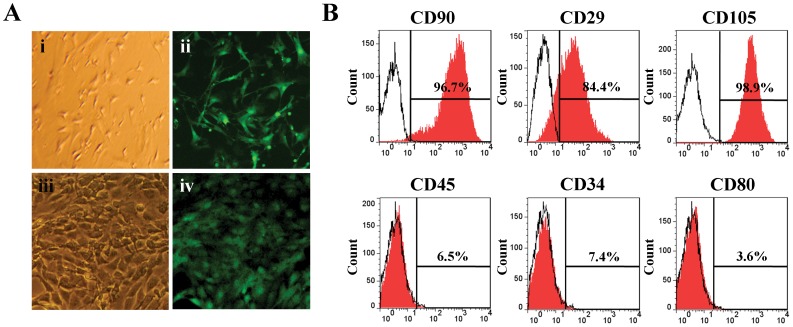
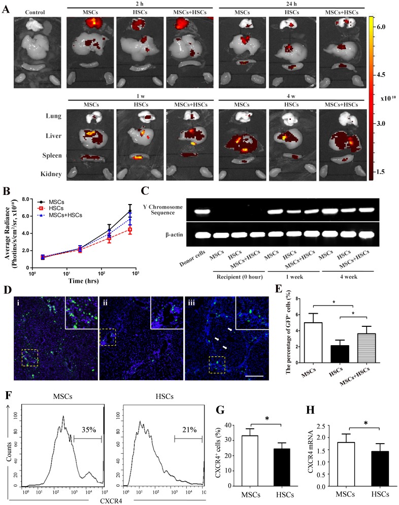
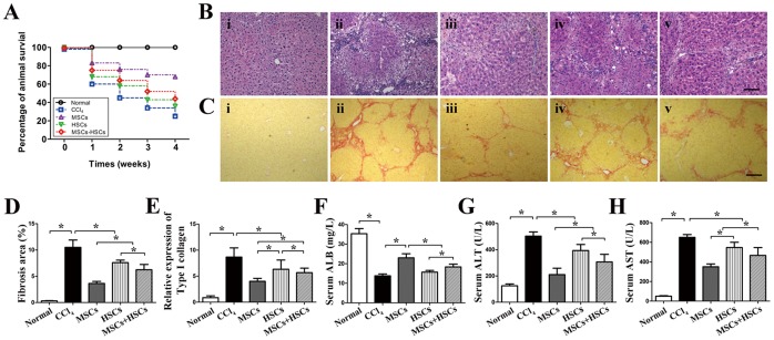
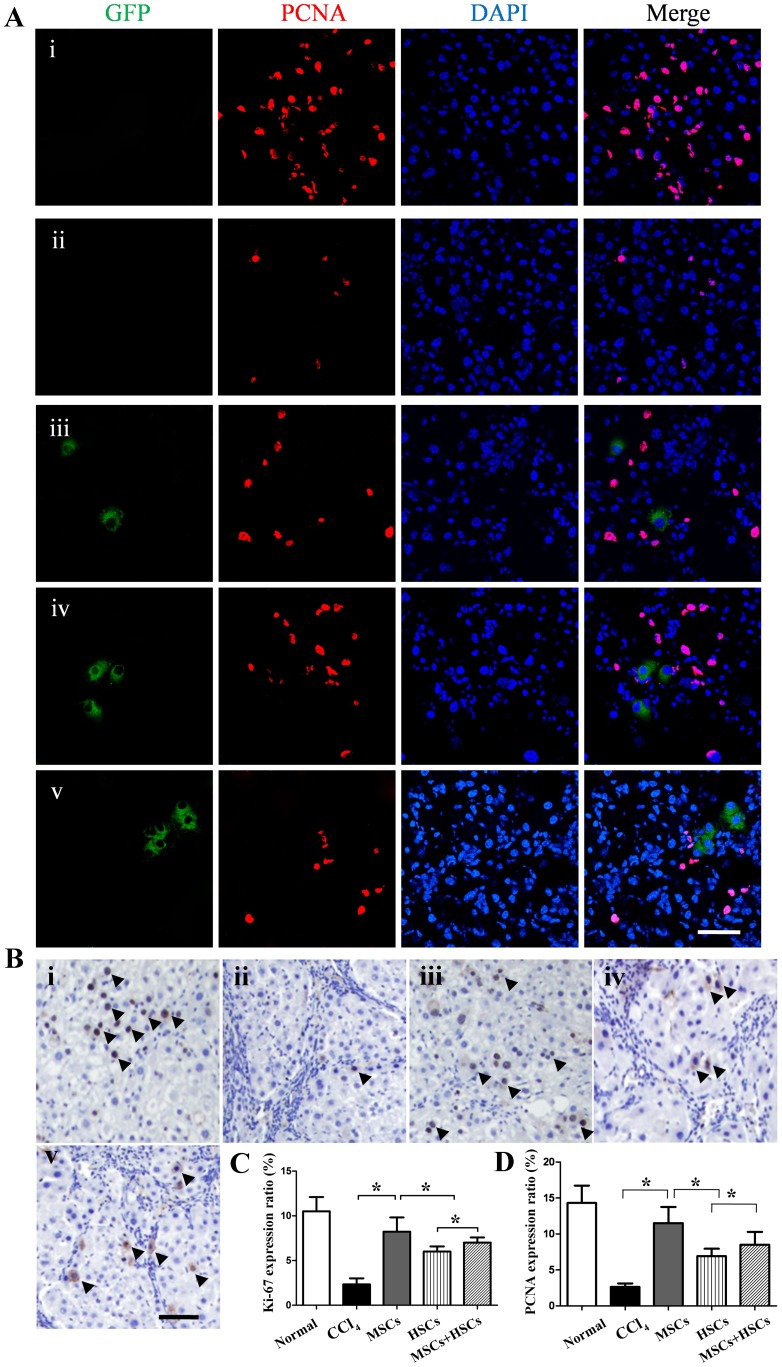
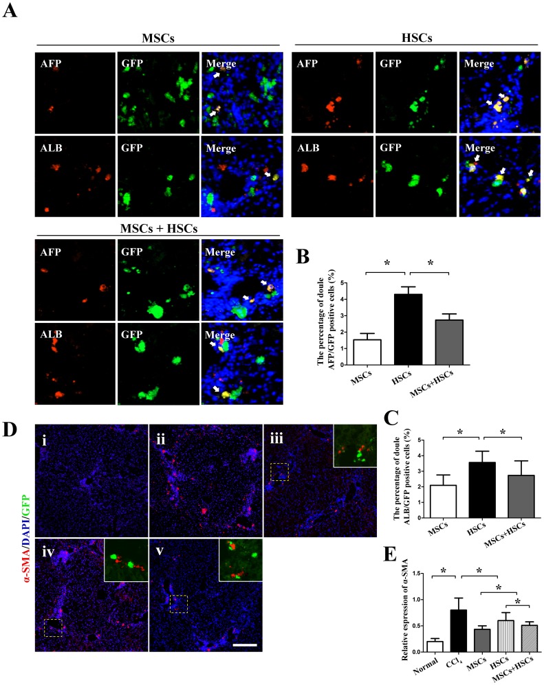
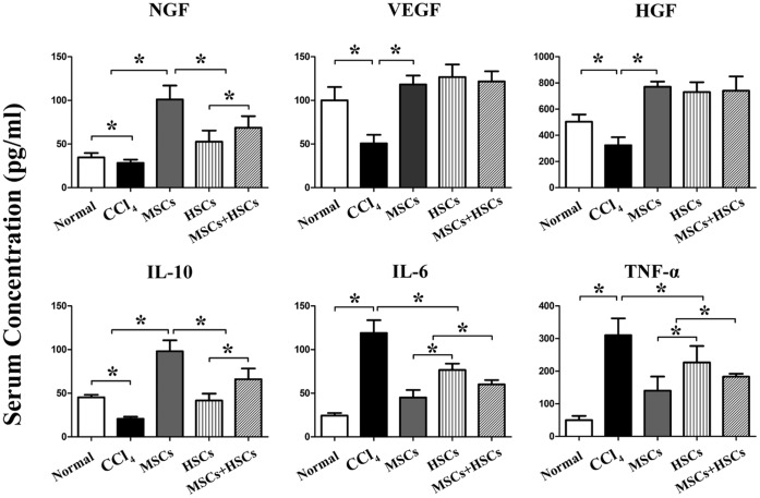
Methodology/principal findings: MSCs, HSCs and the combination of both cells were obtained from the bone marrow of male mice expressing enhanced green fluorescent protein(EGFP)and injected into the female mice with or without liver fibrosis. The distribution of the stem cells, survival rates, liver function, hepatocyte regeneration, growth factors and cytokines of the recipient mice were analyzed. We found that the liver content of the EGFP-donor cells was significantly higher in the MSCs group than in the HSCs or MSCs+HSCs group. The survival rate for the MSCs group was significantly higher than that of the HSCs or MSCs+HSCs group; all surpassed the control group. After MSC-transplantation, the injured livers were maximally restored, with less collagen than the controls. The fibrotic areas had decreased to a lesser extent in the mice transplanted with HSCs or MSCs+HSCs. Compared with mice in the HSCs group, the mice that received MSCs had better improved liver function. MSCs exhibited more remarkable paracrine effects and immunomodulatory properties on hepatic stellate cells and native hepatocytes in the treatment of the liver pathology. Synergistic actions of MSCs and HSCs were most likely not observed because the stem cells in liver were detected mostly as single cells, and single MSCs are insufficient to provide a beneficial niche for HSCs.

Conclusions/significance: MSCs exhibited a greater homing capability for the injured liver and modulated fibrosis and inflammation more effectively than did HSCs. Synergistic effects of MSCs and HSCs were not observed in liver injury.


Conflict of interest statement

Competing Interests: The authors have declared that no competing interests exist.

Figures

Figure 1
Figure 1. Characterization of MSCs derived from EGFP-transgenic mice.
(A) The morphologies of the MSCs in the first (i and ii) and third (iii and iv) passages observed under bright field and fluorescence microscopy, respectively. (B) Surface molecule characterization of the MSCs performed by FACS analyses after incubation with PE-conjugated antibodies (CD90, CD29, CD105, CD45, CD34 and CD80).
Figure 2
Figure 2. Detection of EGFP+ cells in recipient livers after transplantation.
EGFP+ cells from male donors were injected into liver-injured female mice. (A) After transplantation of stem cells, the EGFP fluorescence in the lung, liver, spleen and kidney was examined using bio- imaging system. A luminescent image from red (least intense) to yellow (most intense) represents the spatial distribution of the detected photons emitted from EGFP+ cells within the organs. The EGFP signal was not detected in the control mice. (B) Average radiance was quantified in the liver after stem cells transplantation. (C) PCR-based detection of donor-derived cells in the livers of different recipients. (D and E) Liver sections were stained with DAPI, and the distribution of EGFP+ cells in the portal lobe was quantified in the different groups at 4 weeks. White arrows show the stem cells in the sinusoids (i, MSCs group; ii, HSCs group; and iii, MSCs+HSCs group). (F and G) FACS analyses of CXCR4 expression on MSCs and HSCs. (H) mRNA levels of CXCR4 on MSCs and HSCs. P<0.05, n = 3.
Figure 3
Figure 3. Therapeutic effects of transplanted MSCs, HSCs and MSCs+HSCs on recovery in the CCl4-induced injury mouse model.
(A) A survival curve for the injured mice that underwent intravenous cell transplantation. Representative photomicrographs of H&E-stained (B) and Sirius red-stained (C) mouse livers from the different groups (i, normal mice; ii, CCl4 group; iii, MSCs group; iv, HSCs group; and v, MSCs+HSCs group).(D) Analyses of the fibrosis percentage using Image J software. (E) mRNA levels of type I collagen. (F, G and H) Serum ALB, ALT and AST levels in the experimental groups. P<0.05, n = 5, Scale bars: B = 150 µm, C = 200 µm.
Figure 4
Figure 4. Effect of MSC, HSC and MSC+HSC transplantation on hepatocyte regeneration.
(A) Immunohistochemistry analyses of PCNA expression in liver tissues (i, normal mice; ii, CCl4 group; iii, MSCs group; iv, HSCs group; and v, MSCs+HSCs group). (B) Immunohistochemistry analyses of Ki-67 expression in liver tissues (i, normal mice; ii, CCl4 group; iii, MSCs group; iv, HSCs group; and v, MSCs+HSCs group). (C) Quantitative image analyses of the percentage of Ki-67+ cells. (D) Quantitative image analyses of the percentage of PCNA+ cells. P<0.05, n = 5, Scale bar = 100 µm.
Figure 5
Figure 5. Differentiation of transplanted MSCs, HSCs and MSCs+HSCs in CCl4-induced injured livers at 4 weeks.
The liver sections were observed under fluorescence microscopy. (A, B and C) The liver sections were co-stained with either AFP or ALB to detect the differentiation of transplanted cells (white arrowhead) in the different groups. (D) The expression of α-SMA in the livers of CCl4-induced injured mice in the different groups (i, normal mice; ii, CCl4 group; iii, MSCs group; iv, HSCs group; and v, MSCs+HSCs group). (E) mRNA levels of α-SMA. P<0.05, n = 5, Scale bars: A = 50 µm, D = 150 µm.
Figure 6
Figure 6. The concentration of growth factors and cytokines in each group 4 w after transplantation of MSCs, HSCs or MSCs+HSCs.
P<0.05, n = 5. NGF, nerve growth factor; VEGF, vascular endothelial growth factor; HGF, hepatocyte growth factor; IL, interleukin; TNF-α, tumor necrosis factor-alpha.

Similar articles

Cited by

References

    1. Peng L, Xie DY, Lin BL, Liu J, Zhu HP, et al. (2011) Autologous bone marrow mesenchymal stem cell transplantation in liver failure patients caused by hepatitis B: short-term and long-term outcomes. Hepatology 54: 820–828. - PubMed
    1. Yannaki E, Anagnostopoulos A, Kapetanos D, Xagorari A, Iordanidis F, et al. (2006) Lasting amelioration in the clinical course of decompensated alcoholic cirrhosis with boost infusions of mobilized peripheral blood stem cells. Exp Hematol 34: 1583–1587. - PubMed
    1. am Esch JS 2nd, Knoefel WT, Klein M, Ghodsizad A, Fuerst G, et al (2005) Portal application of autologous CD133+ bone marrow cells to the liver: a novel concept to support hepatic regeneration. Stem Cells 23: 463–470. - PubMed
    1. Houlihan DD, Newsome PN (2008) Critical review of clinical trials of bone marrow stem cells in liver disease. Gastroenterology 135: 438–450. - PubMed
    1. Zhao Q, Ren H, Zhu D, Han Z (2009) Stem/progenitor cells in liver injury repair and regeneration. Biol Cell 101: 557–571. - PubMed

Publication types

Substances