

Potential of olfactory ensheathing cells from different sources for spinal cord repair
- PMID: 23638158
- PMCID: PMC3634744
- DOI: 10.1371/journal.pone.0062860
Potential of olfactory ensheathing cells from different sources for spinal cord repair
Abstract
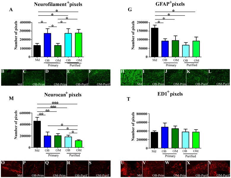
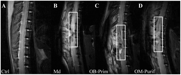
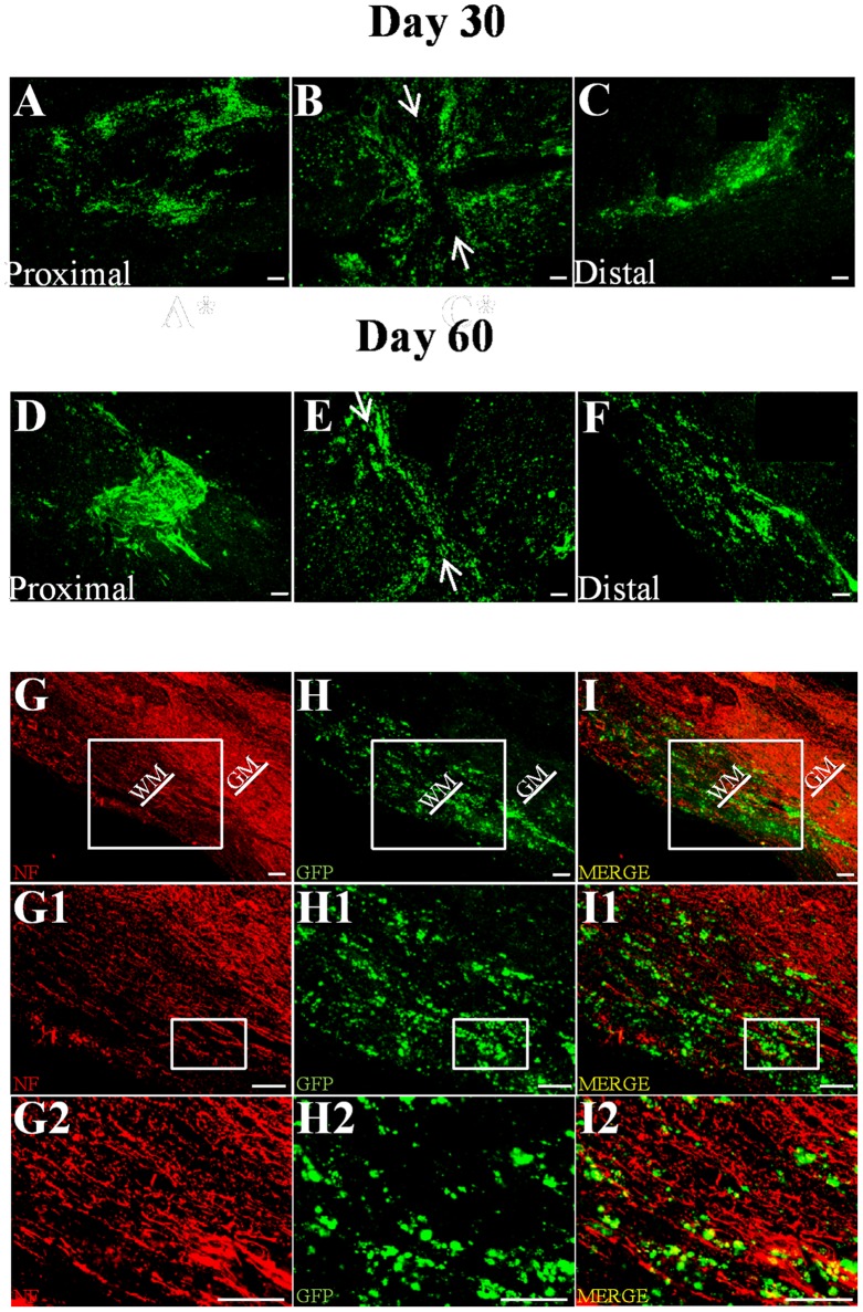
Spinal cord injury (SCI) induces a permanent disability in patients. To this day no curative treatment can be proposed to restore lost functions. Therefore, extensive experimental studies have been conducted to induce recovery after SCI. One of the most promising therapies is based on the use of olfactory ensheathing cells (OECs). OECs can be obtained from either the olfactory bulbs (OB-OECs) or from olfactory mucosa (OM-OECs), involving a less invasive approach for autotransplantation. However the vast majority of experimental transplantations have been focusing on OB-OECs although the OM represents a more accessible source of OECs. Importantly, the ability of OM-OECs in comparison to OB-OECs to induce spinal cord recovery in the same lesion paradigm has never been described. We here present data using a multiparametric approach, based on electrophysiological, behavioral, histological and magnetic resonance imaging experiments on the repair potential of OB-OECs and OM-OECs from either primary or purified cultures after a severe model of SCI. Our data demonstrate that transplantation of OECs obtained from OB or OM induces electrophysiological and functional recovery, reduces astrocyte reactivity and glial scar formation and improves axonal regrowth. We also show that the purification step is essential for OM-OECs while not required for OB-OECs. Altogether, our study strongly indicates that transplantation of OECs from OM represents the best benefit/risk ratio according to the safety of access of OM and the results induced by transplantations of OM-OECs. Indeed, purified OM-OECs in addition to induce recovery can integrate and survive up to 60 days into the spinal cord. Therefore, our results provide strong support for these cells as a viable therapy for SCI.
Conflict of interest statement
Figures
Similar articles
-
Transplantation of olfactory ensheathing cells to evaluate functional recovery after peripheral nerve injury.J Vis Exp. 2014 Feb 23;(84):e50590. doi: 10.3791/50590. J Vis Exp. 2014. PMID: 24637657 Free PMC article.
-
Effects of fibroblasts derived from the olfactory bulb and nasal olfactory mucosa on proliferation of olfactory ensheathing cells harvested from the olfactory bulb.J Vet Med Sci. 2011 Jan;73(1):133-7. doi: 10.1292/jvms.10-0344. J Vet Med Sci. 2011. PMID: 21293078
-
Co-transplantation of olfactory ensheathing cells from mucosa and bulb origin enhances functional recovery after peripheral nerve lesion.PLoS One. 2011;6(8):e22816. doi: 10.1371/journal.pone.0022816. Epub 2011 Aug 3. PLoS One. 2011. PMID: 21826209 Free PMC article.
-
Co-transplantation of autologous OM-MSCs and OM-OECs: a novel approach for spinal cord injury.Rev Neurosci. 2016 Apr 1;27(3):259-70. doi: 10.1515/revneuro-2015-0030. Rev Neurosci. 2016. PMID: 26574889 Review.
-
Cell therapy for spinal cord injury with olfactory ensheathing glia cells (OECs).Glia. 2018 Jul;66(7):1267-1301. doi: 10.1002/glia.23282. Epub 2018 Jan 13. Glia. 2018. PMID: 29330870 Review.
Cited by
-
A Novel Approach for Mucosal and Bulbar Olfactory Ensheathing Cells Isolation Based on the Non-adherent Subculture Technique.Basic Clin Neurosci. 2024 Mar-Apr;15(2):211-220. doi: 10.32598/bcn.2022.3579.1. Epub 2024 Mar 1. Basic Clin Neurosci. 2024. PMID: 39228451 Free PMC article.
-
ADAMTS proteoglycanases in the physiological and pathological central nervous system.J Neuroinflammation. 2013 Oct 31;10:133. doi: 10.1186/1742-2094-10-133. J Neuroinflammation. 2013. PMID: 24176075 Free PMC article. Review.
-
Comparison of the effects of two therapeutic strategies based on olfactory ensheathing cell transplantation and repetitive magnetic stimulation after spinal cord injury in female mice.J Neurosci Res. 2021 Jul;99(7):1835-1849. doi: 10.1002/jnr.24836. Epub 2021 May 7. J Neurosci Res. 2021. PMID: 33960512 Free PMC article.
-
Synergic effects of EPI-NCSCs and OECs on the donor cells migration, the expression of neurotrophic factors, and locomotor recovery of contused spinal cord of rats.J Mol Neurosci. 2015 Mar;55(3):760-9. doi: 10.1007/s12031-014-0416-2. Epub 2014 Sep 20. J Mol Neurosci. 2015. PMID: 25239519
-
Designing a Clinical Trial with Olfactory Ensheathing Cell Transplantation-Based Therapy for Spinal Cord Injury: A Position Paper.Biomedicines. 2022 Dec 6;10(12):3153. doi: 10.3390/biomedicines10123153. Biomedicines. 2022. PMID: 36551909 Free PMC article. Review.
References
Publication types
MeSH terms
LinkOut - more resources
Full Text Sources
Other Literature Sources
Medical
Miscellaneous
