. 2013 Apr 25;8(4):e62885.
doi: 10.1371/journal.pone.0062885. Print 2013.

Molecular characterization of the fecal microbiota in patients with nonalcoholic steatohepatitis--a longitudinal study

Affiliations

Molecular characterization of the fecal microbiota in patients with nonalcoholic steatohepatitis--a longitudinal study

Vincent Wai-Sun Wong et al. PLoS One. .

Abstract

Background: The human gut microbiota has profound influence on host metabolism and immunity. This study characterized the fecal microbiota in patients with nonalcoholic steatohepatitis (NASH). The relationship between microbiota changes and changes in hepatic steatosis was also studied.

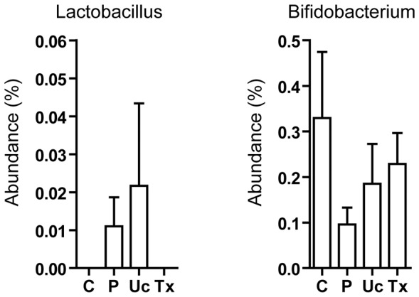
Methods: Fecal microbiota of histology-proven NASH patients and healthy controls was analyzed by 16S ribosomal RNA pyrosequencing. NASH patients were from a previously reported randomized trial on probiotic treatment. Proton-magnetic resonance spectroscopy was performed to monitor changes in intrahepatic triglyceride content (IHTG).

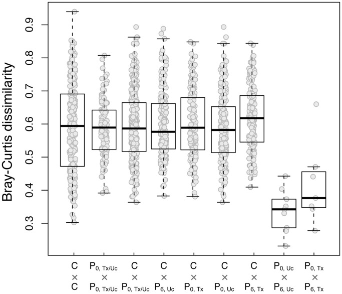
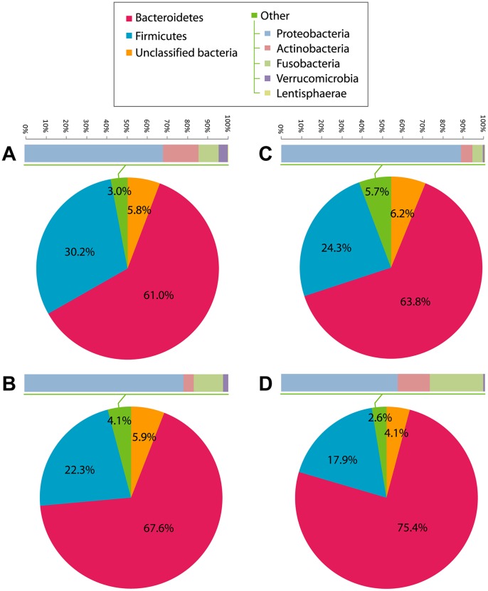
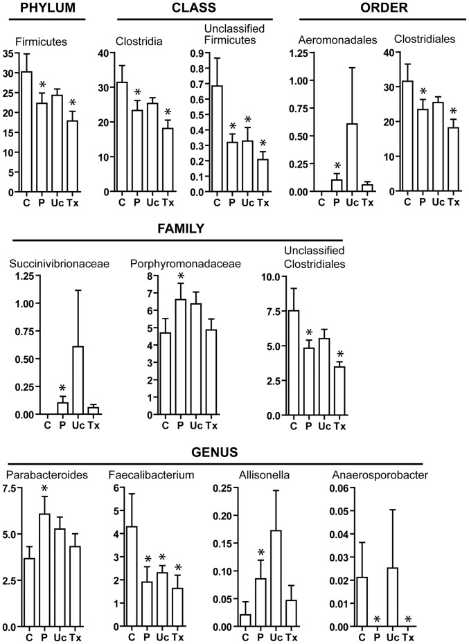
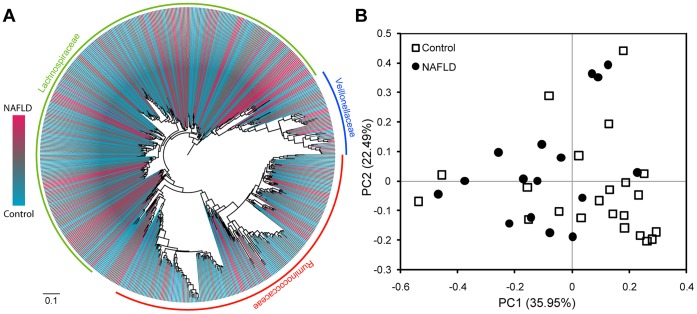
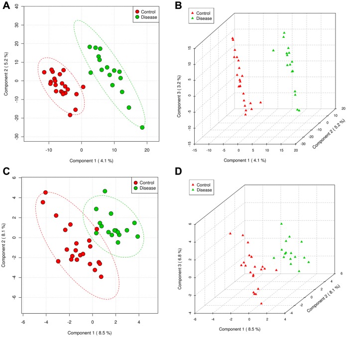
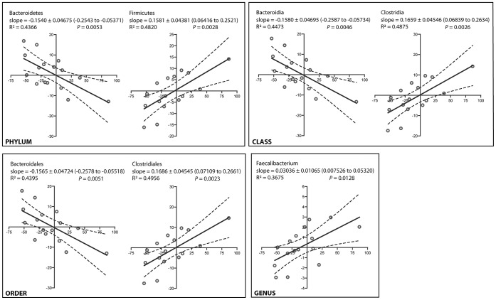
Results: A total of 420,344 16S sequences with acceptable quality were obtained from 16 NASH patients and 22 controls. NASH patients had lower fecal abundance of Faecalibacterium and Anaerosporobacter but higher abundance of Parabacteroides and Allisonella. Partial least-square discriminant analysis yielded a model of 10 genera that discriminated NASH patients from controls. At month 6, 6 of 7 patients in the probiotic group and 4 of 9 patients in the usual care group had improvement in IHTG (P=0.15). Improvement in IHTG was associated with a reduction in the abundance of Firmicutes (R(2)=0.4820, P=0.0028) and increase in Bacteroidetes (R(2)=0.4366, P=0.0053). This was accompanied by corresponding changes at the class, order and genus levels. In contrast, bacterial biodiversity did not differ between NASH patients and controls, and did not change with probiotic treatment.

Conclusions: NASH patients have fecal dysbiosis, and changes in microbiota correlate with improvement in hepatic steatosis. Further studies are required to investigate the mechanism underlying the interaction between gut microbes and the liver.


Conflict of interest statement

Competing Interests: Vincent Wai-Sun Wong has been an advisory board member of Roche, Novartis, Gilead and Otsuka, and received paid lecture fees from Roche, Novartis, Abbott Diagnostics and Echosens. Grace Lai-Hung Wong has been an advisory board member of Otsuka, and received paid lecture fees from Echosens and Otsuka. Henry Lik-Yuen Chan has been consultant for Abbott, Bristol-Myers Squibb, Merck, Novartis and Roche, and received paid lecture fees from Abbott, Bristol-Myers Squibb, Echosens, Gilead, Glaxo-Smith-Kline, Merck, Novartis and Roche.This does not alter the authors’ adherence to all the PLOS ONE policies on sharing data and materials.

Figures

Figure 1
Figure 1. Genus level Bray-Curtis dissimilarity between fecal samples.
Footnote: C, controls; P0, baseline samples of NASH patients; P6, month 6 samples of NASH patients; Uc, usual care group; Tx, probiotic treatment group.
Figure 2
Figure 2. Relative abundance of bacterial phyla.
(A) Controls (N = 22), (B) NASH patients at baseline (N = 16), (C) NASH patients at month 6 of usual care (N = 9), and (D) NASH patients at month 6 of probiotic treatment (N = 7).
Figure 3
Figure 3. Abundance of bacterial clades that differed between controls and NASH patients.
Footnote: Relative abundance is shown in percentage. C, controls; P, NASH patients; Uc, usual care group at month 6; Tx, probiotic treatment group at month 6. *<0.05 compared to controls.
Figure 4
Figure 4. Firmicutes phylogeny and principal component analysis (PCA) plot based on Unifrac distances between the Firmicutes sequences in control and NASH subjects.
(A) The Firmicutes phylogeny was reconstructed from the OTU representative sequences in the control and NASH samples, and their relative abundance was indicated by gradient color from red to blue. (B) PCA plot of controls and NASH patients. The percentage of variation explained by each principal component was indicated in the parenthesis.
Figure 5
Figure 5. Score plots of PLS-DA distinguishing between the microbial community data of controls and NASH patients.
(A and B) OTU level, (C and D) genus level.
Figure 6
Figure 6. Abundance of Lactobacillus and Bifidobacterium.
Lactobacillus and Bifidobacterium were the two bacterial genera contained in the probiotics used in this study. ‘C’, ‘P’, ‘Uc’ and ‘Tx’ refer to controls, NASH patients at baseline, NASH patient at 6 months after usual care and treatment of probiotic, respectively. There is no significant difference between each pair of study groups.
Figure 7
Figure 7. Correlation between the changes in intrahepatic triglyceride content and fecal bacterial abundance in 16 NASH patients over 6 months.
Footnote: The x-axis and y-axis represents changes of intrahepatic triglyceride content and abundance of the indicated bacterial groups in 6 months, respectively. Solid and dashed lines are linear regression fits and 95% confidence bands, respectively.

References

    1. Wong VW, Chu WC, Wong GL, Chan RS, Chim AM, et al. (2012) Prevalence of non-alcoholic fatty liver disease and advanced fibrosis in Hong Kong Chinese: a population study using proton-magnetic resonance spectroscopy and transient elastography. Gut 61: 409–415. - PubMed
    1. Xu C, Yu C, Xu L, Miao M, Li Y (2010) High serum uric acid increases the risk for nonalcoholic Fatty liver disease: a prospective observational study. PLoS One 5: e11578. - PMC - PubMed
    1. Ascha MS, Hanouneh IA, Lopez R, Tamimi TA, Feldstein AF, et al. (2010) The incidence and risk factors of hepatocellular carcinoma in patients with nonalcoholic steatohepatitis. Hepatology 51: 1972–1978. - PubMed
    1. Bhala N, Angulo P, van der Poorten D, Lee E, Hui JM, et al. (2011) The natural history of nonalcoholic fatty liver disease with advanced fibrosis or cirrhosis: an international collaborative study. Hepatology 54: 1208–1216. - PMC - PubMed
    1. Wong VW, Wong GL, Choi PC, Chan AW, Li MK, et al. (2010) Disease progression of non-alcoholic fatty liver disease: a prospective study with paired liver biopsies at 3 years. Gut 59: 969–974. - PubMed

Publication types

LinkOut - more resources