. 2013 Apr 30;8(4):e63213.
doi: 10.1371/journal.pone.0063213. Print 2013.

Small GTPase Rab40c associates with lipid droplets and modulates the biogenesis of lipid droplets

Affiliations

Small GTPase Rab40c associates with lipid droplets and modulates the biogenesis of lipid droplets

Ran Tan et al. PLoS One. .

Abstract

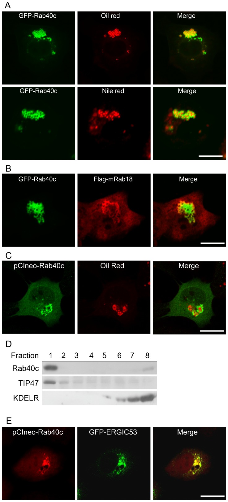
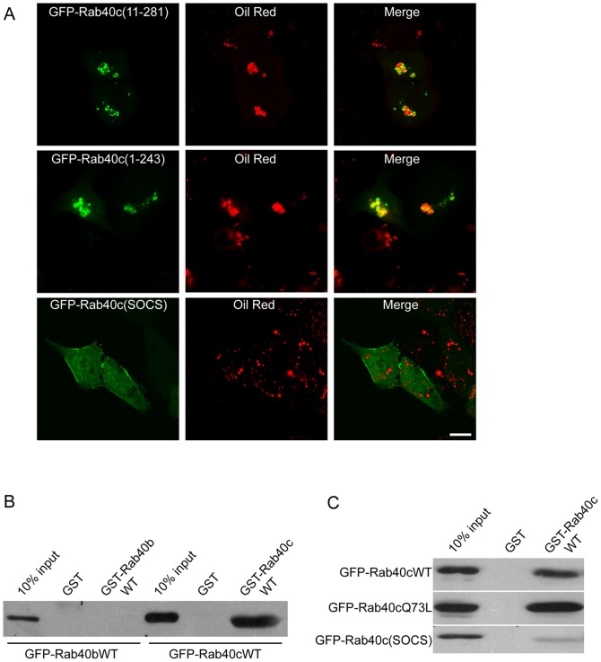
The subcellular location and cell biological function of small GTPase Rab40c in mammalian cells have not been investigated in detail. In this study, we demonstrated that the exogenously expressed GFP-Rab40c associates with lipid droplets marked by neutral lipid specific dye Oil red or Nile red, but not with the Golgi or endosomal markers. Further examination demonstrated that Rab40c is also associated with ERGIC-53 containing structures, especially under the serum starvation condition. Rab40c is increasingly recruited to the surface of lipid droplets during lipid droplets formation and maturation in HepG2 cells. Rab40c knockdown moderately decreases the size of lipid droplets, suggesting that Rab40c is involved in the biogenesis of lipid droplets. Stimulation for adipocyte differentiation increases the expression of Rab40c in 3T3-L1 cells. Rab40c interacts with TIP47, and is appositionally associated with TIP47-labeled lipid droplets. In addition, over-expression of Rab40c causes the clustering of lipid droplets independent of its GTPase activity, but completely dependent of the intact SOCS box domain of Rab40c. In addition, Rab40c displayed self-interaction as well as interaction with TIP47 and the SOCS box is essential for its ability to induce clustering of lipid droplets. Our results suggest that Rab40c is a novel Rab protein associated with lipid droplets, and is likely involved in modulating the biogenesis of lipid droplets.


Conflict of interest statement

Competing Interests: The authors have declared that no competing interests exist.

Figures

Figure 1
Figure 1. The expression of Rab40c.
A. sequence alignment showed that Rab40 family proteins possess conserve phosphate/Mg2+ (PM) binding motifs and SOCS box (the key residues for proper function of SOCS box are underlined), and hRab40c is likely the homolog of xRab40. B. PCR based examination of the transcript levelsof 3 Rab40 members. C. Rab40c is expressed in different tissues as examined by western-blot using Rab40c antibody. D. NRK cells were transfected with GFP-Rab40a, GFP-Rab40b or GFP-Rab40c, respectively. Immuno-fluorescence microscopy analysis showed different localization of Rab40 family proteins. Bar, 20 µm.
Figure 2
Figure 2. Rab40c is not associated with endocytic compartments and the Golgi apparatus.
A. NRK cells were transfected with GFP-Rab40c, and then immuno-labeled with the indicated antibodies. Immuno-fluorescence microscopy showed that GFP-Rab40c is not co-localized with Golgi marker GM130, late endosomal/lysomomal markers LBPA and Lamp2 in NRK cells. B. GFP-Rab40c is not co-localized with early endosomal marker EEA1 in Hela cells. Bar, 20 µm.
Figure 3
Figure 3. Rab40c is present in lipid droplets and structures marked by ERGIC-53.
A. NRK cells were transfected with GFP-Rab40c and labeled with Oil red or Nile red, then processed for immuno-fluorescence microscopy. The results showed that GFP-Rab40c is associated with LDs. B. NRK cells were co-transfected with GFP-Rab40c and Flag-Rab18, and processed for immuno-fluorescence microscopy, Rab40c is revealed by GFP and Rab18 is revealed by labeling with antibody against Flag tag. The results showed that GFP-Rab40c co-localizes partially with flag-Rab18. C. NRK cells were transfected with pCIneo-Rab40c and labeled with Oil red, and immuno-stained by antibody against Rab40c, showing that non-tagged Rab40c is also associated with LDs. D. Western blotting analysis of sucrose density-gradient fractions from HepG2. An equal volume from each fraction was loaded. Both TIP47 and Rab40c were enriched in the top floating LDs-containing fraction (lane 1). The ER-Golgi recycling protein KDEL receptor was detected in the bottom fractions. E. NRK cells were co-transfected with pCIneo-Rab40c and GFP-ERGIC-53, and immuno-stained by antibody against Rab40c. Rab40c is also associated with structures marked by ERGIC-53. Bar, 20 µm.
Figure 4
Figure 4. Rab40c is recruited to the LDs.
A. LDs formation was induced by Oleic acid in HepG2 transfected with GFP-Rab40c for 0, 12, 24 and 48 h, respectively, and LDs were revealed with Oil red. immuno-fluorescence microscopy showed that GFP-Rab40c is recruited to LDs upon stimulation in a time course dependent manner. B. HepG2 cells were co-transfected with pCIneo-Rab40c and GFP-ERGIC-53, then starved for 24 h (upper panels) or stimulated with Oleic acid for 24 h afterward(lower panels), the expressed Rab40c was revealed by immuno-staining with antibody against Rab40c. The data showed that most of the expressed Rab40c co-localized with ERGIC-53 under starvation condition. However, when stimulated with oleic acids, the expressed Rab40c segregated from ERGIC-53, and become associated with the enlarged vesicles characteristic of LDs (indicated by arrow heads). Bar, 20 µm.
Figure 5
Figure 5. The effects of Rab40c knockdown on lipid droplets formation.
A. HepG2 cells were transfected with shRNA-Rab40c or shRNA-control to achieve about 70% transfection efficiency. Western-blot showed that the protein level of Rab40c can be specifically knocked down by shRNA-Rab40c in HepG2 cells. Quantitative analysis was done by densitometry, and the data represent the mean value from 3 independent experiments. B. HepG2 cells were transfected with pSuper.GFP-shRNA-control, then stimulated with oleic acid for the indicated time, and LDs were revealed by Oil red, the data showed that shRNA-control has little effects on LDs. C. HepG2 cells transfected with pSuper.GFP-shRNA-Rab40c, and processed for immuno-fluorescence microscopy analysis as mentioned above, the results revealed that shRNA-Rab40c moderately decreased the size of LDs. D. Quantitative analysis of the mean size of LDs from 50 transfected cells, showing depletion of Rab40c significantly decreases the size of LD. *p<0.05. The error bars represent the deviation of three independent experiments. Bar, 20 µm.
Figure 6
Figure 6. Rab40c is involved in adipocyte differentiation and interacts with TIP47.
A. 3T3-L1 cells were induced to differentiate into adipocytes for the indicated times, then the cells were lysed and subjected for western-blot to detect β-tubulin, Rab40c, Perilipin, ADRP or TIP47. The results demonstrated that the level of Rab40c increased during adipocyte differentiation. B. 3-T3-L1 cell lysates were incubated with GST, GST-Rab40cWT or GST-Rab40c(SOCS) coupled to GST-Sepharose 4B resin. Proteins retained on the beads were processed for western-blot using antibodies against TIP47, Perilipin or ADRP. The data demonstrated that Rab40c can interact with TIP47 but not ADRP and Perilipin. Mutations of SOCS box in Rab40c disrupted the interaction between Rab40c and TIP47. C. Immuno-fluorescence microscopy revealed that GFP-Rab40c exhibites close apposition with TIP47 upon oleic acids stimulation in HepG2 cells. Bar, 20 µm.
Figure 7
Figure 7. Expression of Rab40c induces the clustering of LDs.
NRK cells were transfected with GFP-Rab40cWT (wild type), GFP-Rab40cG28N, GFP-Rab40cG28T or GFP-Rab40cQ73L, respectively. Then the cells were labeled with Oil red and processed for confocal microscopy analysis. The results demonstrated that GFP-Rab40cWT and all the mutants can significantly induce the clustering of LDs. Bar, 20 µm.
Figure 8
Figure 8. SOCS box in Rab40c mediates self-interaction and is essential for inducing the clustering of LDs.
A. NRK cells were transfected with the N-terminal truncated form GFP-Rab40c(11-281), C-terminal truncated form GFP-Rab40c(1-243) or GFP-Rab40c(SOCS) (mutant with mutations LPLP(212-215)AAAA in SOCS box), respectively. The cells were labeled with Oil red and processed for immuo-fulorescence microscopy. The results revealed that disruption of the SOCS box abolished the association of Rab40c with lipid droplets, and its ability to induce the clustering of LDs. B. MCF7 cell lysates expressing GFP-Rab40b or GFP-Rab40c were subjected to pull-down experiments using immobilized GST-Rab40b or GST-Rab40c. Western-blot experiments using GFP antibody demonstrated that GST-Rab40c can efficiently retain GFP-Rab40c, where as GST-Rab40b did not exhibit binding to GFP-Rab40b. C. MCF7 cell lysates expressing GFP-Rab40cWT, GFP-Rab40cQ73L or GFP-Rab40c(SOCS) were subjected to pull-down experiments using GST-Rab40c, showing that GST-Rab40c can bind to both GFP-Rab40c and GFP-Rab40cQ73L, but not GFP-Rab40c(SOCS) mutant. Bar, 20 µm.

Similar articles

Cited by

References

    1. Murphy DJ (2001) The biogenesis and functions of lipid bodies in animals, plants and microorganisms. Prog Lipid Res 40: 325–438. - PubMed
    1. Martin S, Parton RG (2006) Lipid droplets: a unified view of a dynamic organelle. Nat Rev Mol Cell Biol 7: 373–378. - PubMed
    1. Zehmer JK, Huang Y, Peng G, Pu J, Anderson RG, et al. (2009) A role for lipid droplets in inter-membrane lipid traffic. Proteomics 9: 914–921. - PMC - PubMed
    1. Soni KG, Mardones GA, Sougrat R, Smirnova E, Jackson CL, et al. (2009) Coatomer-dependent protein delivery to lipid droplets. J Cell Sci 122: 1834–1841. - PMC - PubMed
    1. Paar M, Jungst C, Steiner NA, Magnes C, Sinner F, et al. (2012) Remodeling of lipid droplets during lipolysis and growth in adipocytes. J Biol Chem 287: 11164–11173. - PMC - PubMed

Publication types

MeSH terms

LinkOut - more resources