. 2013 Apr 25;7(4):e2171.
doi: 10.1371/journal.pntd.0002171. Print 2013.

Transcriptional profiling of the circulating immune response to lassa virus in an aerosol model of exposure

Affiliations

Transcriptional profiling of the circulating immune response to lassa virus in an aerosol model of exposure

Shikha Malhotra et al. PLoS Negl Trop Dis. .

Abstract

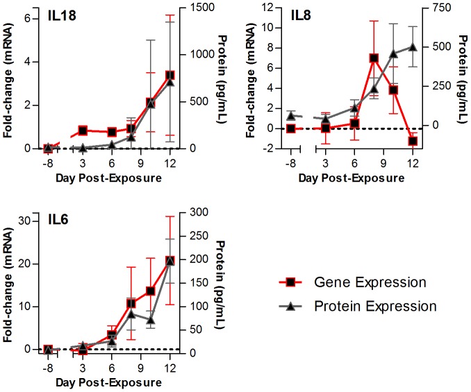
Lassa virus (LASV) is a significant human pathogen that is endemic to several countries in West Africa. Infection with LASV leads to the development of hemorrhagic fever in a significant number of cases, and it is estimated that thousands die each year from the disease. Little is known about the complex immune mechanisms governing the response to LASV or the genetic determinants of susceptibility and resistance to infection. In the study presented here, we have used a whole-genome, microarray-based approach to determine the temporal host response in the peripheral blood mononuclear cells (PBMCs) of non-human primates (NHP) following aerosol exposure to LASV. Sequential sampling over the entire disease course showed that there are strong transcriptional changes of the immune response to LASV exposure, including the early induction of interferon-responsive genes and Toll-like receptor signaling pathways. However, this increase in early innate responses was coupled with a lack of pro-inflammatory cytokine response in LASV exposed NHPs. There was a distinct lack of cytokines such as IL1β and IL23α, while immunosuppressive cytokines such as IL27 and IL6 were upregulated. Comparison of IRF/STAT1-stimulated gene expression with the viral load in LASV exposed NHPs suggests that mRNA expression significantly precedes viremia, and thus might be used for early diagnostics of the disease. Our results provide a transcriptomic survey of the circulating immune response to hemorrhagic LASV exposure and provide a foundation for biomarker identification to allow clinical diagnosis of LASV infection through analysis of the host response.


Conflict of interest statement

The authors have declared that no competing interests exist.

Figures

Figure 1
Figure 1. Confirmation of LASV aerosol model.
(A) Overview of the sequential sampling study samples. The table is organized by animal (column, represented with letter A–K) and day that the sample was collected (row). Samples from LASV-exposed NHP were sorted temporally by the day the sample was collected post-exposure, and further divided into three general categories (early, middle, and late induction) to ease interpretation. Pre-exposure samples were collected prior to LASV exposure and used to normalize data obtained from subsequent samples from the same animal. (B) Table describes the time of onset (early, middle, and late) of a given symptom (viremia, increased AST, anorexia onset, recumbency, and neurological signs/seizures) post-LASV exposure. Data is presented as the number and percentage of animals in which a clinical finding was detected, followed by the total number of animals that were examined at a given stage. (C) A conceptual diagram to explain the division of early, middle, and late stages of LASV in our sequential sampling NHP model. Early disease stage (0–5 dpe) is asymptomatic; middle disease stage (6–9 dpe) is early symptomatic with increase in viremia (dark blue line) and AST levels (light blue line); late disease stage (10–12 dpe) is depicting increasing signs of disease with increase in the onset of anorexia (dark green line) and neurological signs/seizures (light green line) in NHPs.
Figure 2
Figure 2. Strongly upregulated genes in LASV-exposed NHP.
(A) Data had been zero-transformed using the pre-exposure control sample from each individual monkey to normalize for animal-intrinsic signatures and establish a baseline. Data were then filtered to identify 361 genes that showed at least a 2.5 log2-fold differential expression, and then hierarchically clustered. Each row in the heatmaps represents data from an individual gene, and each column represents the individual PBMC sample taken at a specific time point. Samples from the dataset were grouped into early (days 2–3), middle (days 6–8) and late (days 10–12) disease categories based on the day the sample was collected post-viral challenge. Red and blue colors denote expression levels greater or less than baseline (white), respectively. Green boxes identify significant clusters of genes induced during early, middle, and late disease, and are labeled accordingly. An expanded view of these gene clusters is shown in (B), (C), and (D). The most significant functional groups (assigned by DAVID, p-value<0.001) found in the respective clusters are listed to the right of the heatmaps, along with the names of some representative genes.
Figure 3
Figure 3. A signaling networks of 65 genes which are strongly upregulated following LASV challenge of NHP.
Ingenuity Pathway Analysis software was used to analyze a list of 360 genes which were found to be strongly upregulated following LASV challenge. Of this list, 65 were found to be directly or indirectly connected according to Ingenuity's database of published interactions. Nodes represent individual gene products, with official gene symbols included. Edges without arrows indicate a direct interaction between two gene products, e.g. a protein-protein interaction. Edges with arrows represent a regulation of transcription, directed from the gene regulator to a regulated gene. Loops indicate self-regulation. Notable in the expression profiles is the upregulation of STAT1 and IRF7, MMP9 and ELANE signaling nodes.
Figure 4
Figure 4. Cytokine mRNA expression following LASV challenge.
Heatmaps show the gene expression levels of (A) previously analyzed cytokines in LASV disease: IL1β, IL6, IL8, IL10, IL12α, and IFNγ; (B) TNFα and TNF-responsive genes; (D) cytokines IL21, IL23α, IL24, and IL27; and (F) chemokines CCL23 and CCRL2 from the whole PBMC population; as well as (G) IL21 and IL27 from the CD4+ population (isolated from PBMCs). Line graphs show the expression levels (mRNA log2 fold change over time) of (C) IL1β, IL6, and TNFAIP6; and (E) IL21 and IL27 from the whole PBMC population; and (H) IL21 and IL27 in the CD4+ population.
Figure 5
Figure 5. Gene expression precedes the detection of viremia in LASV-exposed NHPs.
Comparison of observed mRNA expression level changes over the course of infection (red line) to the appearance of virus in the blood (grey line) in LASV-exposed NHPs. Shown here are probes for (A) the cytokine IL27; and (B) genes involved in the innate antiviral response: IRF7 (top left), STAT1 (top right), IFIT2 (bottom left), and CXCL12 (bottom right). Error bars represent S.E.
Figure 6
Figure 6. Comparison of the cytokine gene expression with the cytokine levels in the plasma of LASV-exposed NHPs.
Line graph to show the comparison of gene expression (red line) to the amount of cytokine detected (in pg/mL, grey line) in the plasma of LASV-exposed NHPs over the course of the disease. Shown here are probes for IL18 (top left panel), IL8 (top right panel), and IL6 (bottom left panel). Error bars represent S.E.

Similar articles

Cited by

References

    1. McCormick JB, Webb PA, Krebs JW, Johnson KM, Smith ES (1987) A prospective study of the epidemiology and ecology of Lassa fever. J Infect Dis 155: 437–444. - PubMed
    1. Fisher-Hoch SP, Tomori O, Nasidi A, Perez-Oronoz GI, Fakile Y, et al. (1995) Review of cases of nosocomial Lassa fever in Nigeria: the high price of poor medical practice. BMJ 311: 857–859. - PMC - PubMed
    1. Winn WC Jr, Walker DH (1975) The pathology of human Lassa fever. Bull World Health Organ 52: 535–545. - PMC - PubMed
    1. Hensley LE, Smith MA, Geisbert JB, Fritz EA, Daddario-DiCaprio KM, et al. (2011) Pathogenesis of Lassa fever in cynomolgus macaques. Virol J 8: 205. - PMC - PubMed
    1. Monath TP, Newhouse VF, Kemp GE, Setzer HW, Cacciapuoti A (1974) Lassa virus isolation from Mastomys natalensis rodents during an epidemic in Sierra Leone. Science 185: 263–265. - PubMed

Publication types

Substances