

Extranodal marginal zone lymphoma of mucosa-associated lymphoid tissue (MALT lymphoma) of the ileum in a 35-year-old Japanese woman
- PMID: 23638229
- PMCID: PMC3638108
Extranodal marginal zone lymphoma of mucosa-associated lymphoid tissue (MALT lymphoma) of the ileum in a 35-year-old Japanese woman
Abstract
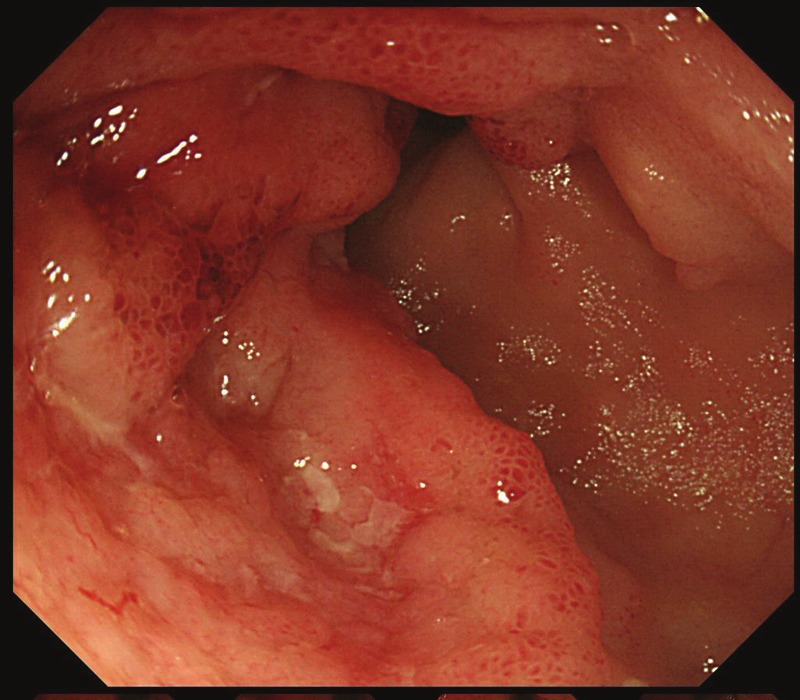
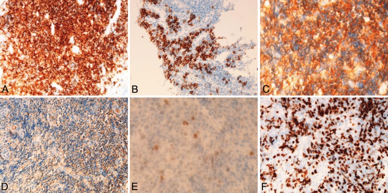
MALT lymphoma of the ileum is extremely rare: only several cases have been reported. A 34-year-old woman presented abdominal pain and melena. Colorectal and small intestinal endoscopes revealed multiple tumors and ulcers of the entire ileum. Biopsy was taken. Histologically, the biopsy consisted of 6 tissue specimens taken from the various sites of the ileum. All the tissue specimens showed infiltration of small atypical cells resembling centrocyte-like cells (CLC). Immunoblastic cells were scattered, though the number was scant. Monocytoid, plasma cell differentiation, and germinal centers were seen. Lymphoepithelial lesions (LEL) were scattered. Some small atypical lymphocyte were destructive the vessels and stromal tissues. Giemsa and Gram stains demonstrated no Helicobacter pylori and any bacteria. Immunohistochemically, the atypical small lymphocytes were positive for vimentin, but negative for various kinds of cytokeratins (CKs), EMA, CEA and CA19-9. The CK highlighted the LEL. They were positive for CD45, and B-cell markers (CD20, CD79a, CD10, CD23, bcl-2). CD138-positive plasma cells were seen in large number. CD68-positive macrophages were scattered. CD30- and CD15-positive immunoblastic cells were scattered. Most of the lymphoid cells were negative for T-cell markers (CD3, CD4, CD5, CD45RO, and CD43) and negative for NK cell markers (CD56 and CD57). The lymphoid cells were positive for κ-chain but negative for λ-chain; thus the light chain restriction was seen. TdT and cyclin D1 were negative. P53 was positive and Ki-67 labeling index was 67%. The lymphoid cells were negative for neuroendocrine markers (NCAM, NSE, chromogranin, and synaptophysin). The pathological diagnosis was MALT lymphoma of the ileum. Post-biopsy imaging techniques including CT, MRI, PET endoscope and gallium scintigraphy identified no tumors and no lymphadenopathy in the body except the ileum. The stomach was free from MALT lymphoma. She was treated by low dose chemotherapy and strictly followed up.
Keywords: Ileum; MALT lymphoma; histopathology; immunohistochemistry.
Figures

References
-
- Isaacson PG, Chott A, Nakamura S, Muller-Hermelink HK, Harris NL, Swerdlow SH. Extranodal marginal zone lymphoma of mucosa-associated lymphoid tissue (MALT lymphoma) In: Swerdlow SH, Campo E, Harris NL, Jaffe ES, Pileri SA, Stein H, Thiele J, Vardiman JW, editors. WHO classification of tumours of haematopoietic and lymphoid tissues. Lyon: IARC; 2008. pp. 214–217.
-
- Isaacson P, Wright DH. Malignant lymphoma of mucosa-associated lymphoid tissue: a distinctive type of B-cell lymphoma. Cancer. 1983;52:1410–1416. - PubMed
Publication types
MeSH terms
Substances
LinkOut - more resources
Full Text Sources
Medical
Research Materials
Miscellaneous