

Protective Effect of Safranal, a Constituent of Crocus sativus, on Quinolinic Acid-induced Oxidative Damage in Rat Hippocampus
- PMID: 23638295
- PMCID: PMC3637907
Protective Effect of Safranal, a Constituent of Crocus sativus, on Quinolinic Acid-induced Oxidative Damage in Rat Hippocampus
Abstract
Objective(s): Quinolinic acid (QA)-mediated excitotoxicity has been widely used as a model for studying neurodegenerative disorders. Recent studies suggested that saffron (Crocus sativus) or its active metabolite, i.e. safranal, exerts pharmacological actions on central nervous system including anxiolytic, anticonvulsant, and neuroprotective properties. The present study aimed to investigate the effect safranal pretreatment on QA-induced oxidative damage in rat hippocampus.
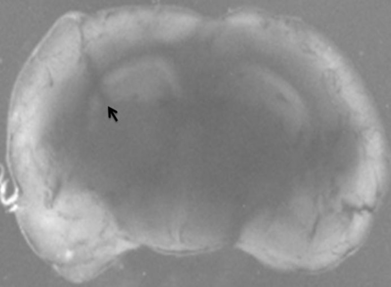
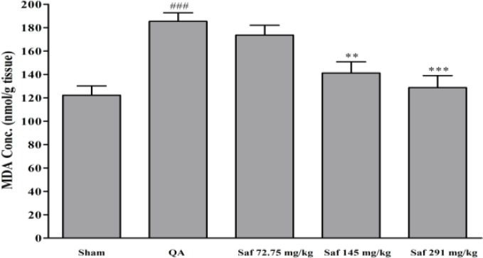
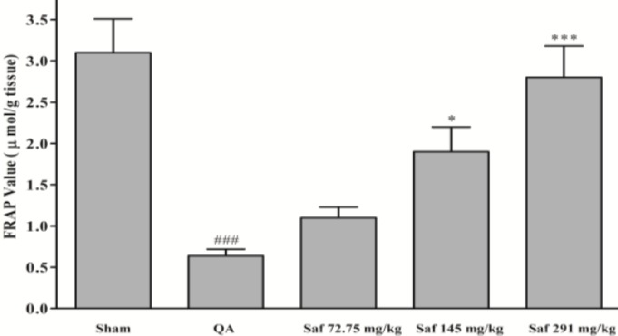
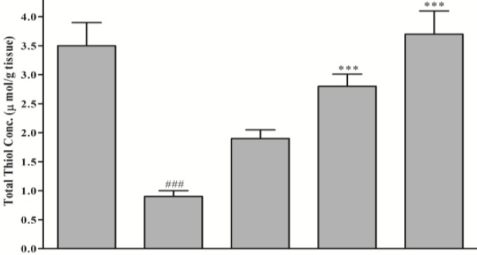
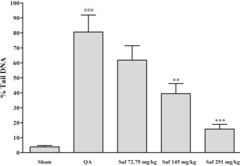
Materials and methods: Under anesthesia, a guide cannula was stereotaxically inserted into left ventral hippocampus of rats. The rats were then given either saline or safranal (72.75, 145.5, and 291 mg/kg, IP) 30 min before administration of QA (300 nmol, intrahippocampal injection). The markers of oxidative stress including thiobarbituric acid reactive substances (TBARS, as an index of lipid preoxidation), total sulfhydryl groups, antioxidant capacity of hippocampus (using FRAP assay), and oxidative DNA damage (%tail DNA, using comet assay) were measured in hippocampus.
Results: The QA induced a significant increase in TBARS levels and %tail DNA and remarkable decrease in antioxidant power (FRAP value) and total sulfhydryl content of hippocampus, in comparison with control animals. Systemic administration of safranal (291 mg/kg, IP), effectively and dose-dependently decreased the QA-induced lipid peroxidation (P<0.001) and oxidative DNA damage (P<0.001). Safranal also prevented the decrease of hippocampal thiol redox and antioxidant status (P<0.001) produced by QA.
Conclusion: Safranal have protective effects on different markers of oxidative damage in hippocampal tissue following QA administration. Our findings might raise a possibility of potential therapeutic application of safranal for preventing and treating neurodegenerative disorders such as Alzheimer's disease.
Keywords: Crocus sativus; Hippocampus; Neurodegenerative disorders; Oxidative stress; Quinolinic acid; Safranal.
Figures
Similar articles
-
Safranal, a constituent of Crocus sativus (saffron), attenuated cerebral ischemia induced oxidative damage in rat hippocampus.J Pharm Pharm Sci. 2005 Aug 22;8(3):394-9. J Pharm Pharm Sci. 2005. PMID: 16401389
-
Neuroprotective effect of safranal, an active ingredient of Crocus sativus , in a rat model of transient cerebral ischemia.Folia Neuropathol. 2017;55(3):206-213. doi: 10.5114/fn.2017.70485. Folia Neuropathol. 2017. PMID: 28984113
-
Safranal Attenuates Excitotoxin-Induced Oxidative OLN-93 Cells Injury.Drug Res (Stuttg). 2019 Jun;69(6):323-329. doi: 10.1055/a-0790-8200. Epub 2018 Nov 21. Drug Res (Stuttg). 2019. PMID: 30463091
-
Active constituents of saffron (Crocus sativus L.) and their prospects in treating neurodegenerative diseases (Review).Exp Ther Med. 2023 Apr 3;25(5):235. doi: 10.3892/etm.2023.11934. eCollection 2023 May. Exp Ther Med. 2023. PMID: 37114174 Free PMC article. Review.
-
Pharmacological effects of Safranal: An updated review.Iran J Basic Med Sci. 2023;26(10):1131-1143. doi: 10.22038/IJBMS.2023.69824.15197. Iran J Basic Med Sci. 2023. PMID: 37736506 Free PMC article. Review.
Cited by
-
1,3-Cyclohexadien-1-Als: Synthesis, Reactivity and Bioactivities.Molecules. 2021 Mar 22;26(6):1772. doi: 10.3390/molecules26061772. Molecules. 2021. PMID: 33809941 Free PMC article. Review.
-
Nigella sativa and thymoquinone attenuate oxidative stress and cognitive impairment following cerebral hypoperfusion in rats.Metab Brain Dis. 2019 Aug;34(4):1001-1010. doi: 10.1007/s11011-019-00394-4. Epub 2019 Apr 23. Metab Brain Dis. 2019. PMID: 31016464
-
The Extract of Crocus sativus and Its Constituent Safranal, Affect Serum Levels of Endothelin and Total Protein in Sensitized Guinea Pigs.Iran J Basic Med Sci. 2013 Sep;16(9):1022-6. Iran J Basic Med Sci. 2013. PMID: 24175050 Free PMC article.
-
The role of Safranal and saffron stigma extracts in oxidative stress, diseases and photoaging: A systematic review.Heliyon. 2021 Feb 10;7(2):e06117. doi: 10.1016/j.heliyon.2021.e06117. eCollection 2021 Feb. Heliyon. 2021. PMID: 33615006 Free PMC article. Review.
-
Synergistic effects of quercetin and regular exercise on the recovery of spatial memory and reduction of parameters of oxidative stress in animal model of Alzheimer's disease.EXCLI J. 2020 May 8;19:596-612. doi: 10.17179/excli2019-2082. eCollection 2020. EXCLI J. 2020. PMID: 32483406 Free PMC article.
References
-
- Gilgun-Sherki Y, Melamed E, Offen D. Oxidative stress induced-neurodegenerative diseases: the need for antioxidants that penetrate the blood brain barrier. Neuropharmacol. 2001;40:959–975. - PubMed
-
- Lau A, Tymianski M. Glutamate receptors, neurotoxicity and neurodegeneration. Eur J Physiol . 2010;460:525–542. - PubMed
-
- Heyes MP, Saito K, Crowley JS, Davis LE, Demitrack MA, Der M, et al. Quinolinic acid and kynurenine pathway metabolism in inflammatory and non-inflammatory neurological disease. Brain. 1992;115:1249–1273. - PubMed
-
- Myint AM. Kynurenines: from the perspective of major psychiatric disorders. FEBS J. 2012;279:1375–1385. - PubMed
-
- Stone TW. Neuropharmacology of quinolinic and kynurenic acids. Pharmacol Rev. 1993;45:309–379. - PubMed
LinkOut - more resources
Full Text Sources
Miscellaneous