. 2013 Mar 31;4(1):1-26.
Print 2013.

Relationship between the proteasomal system and autophagy

Affiliations

Relationship between the proteasomal system and autophagy

Alain Lilienbaum. Int J Biochem Mol Biol. .

Abstract

TWO MAJOR PATHWAYS DEGRADE MOST CELLULAR PROTEINS IN EUKARYOTIC CELLS: the ubiquitin-proteasome system (UPS), which usually degrades the majority of proteins, and autophagy, primarily responsible for the degradation of most long-lived or aggregated proteins and cellular organelles. Disruption of these processes can contribute to pathology of a variety of diseases. Further, both pathways are critical for the maintenance of several aspects of cellular homeostasis, but, until recently, were thought to be largely distinct. Recent advances in this field, however, now strongly suggest that their activities are carefully orchestrated through several interfacing elements that are presented and discussed in this review.

Keywords: Proteasome; autophagy; homeostasis; ubiquitin.


Figures
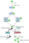
Figure 1
Figure 1
Schematic representation of the ubiquitination process that target proteins for degradation. A ubiquitin molecule (Ub) is first linked by its carboxy-terminal amino acid to an E1-activating enzyme (E1) through a high-energy bond on a cysteine residue, while consuming energy (ATP). Activated ubiquitin is then translocated to the E2-conjugating enzyme (E2). RING E3 ubiquitin ligases (E3 RING) catalyze the transfer of ubiquitin directly from E2 to the substrate, whereas HECT E3 enzymes (E3 HECT) accept activated ubiquitin from E2 before transferring it to the substrate. Following this first step, called monoubiquitination, the process may be repeated by some processive E2/E3 enzymes, or with the help of E4 enzymes and, finally, leads to polyubiquitination. Proteins targeted for degradation into amino acids can however be rescued by deubiquitining enzymes (DUB).
Figure 2
Figure 2
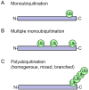
Different types of ubiquitination. Monoubiquitination represents modification of protein by a single ubiquitin (A), or by several single ubiquitins (B). Polyubiquitination (C) corresponds to the successive addition of several ubiquitins on one of their seven lysine residues. All types of ubiquitination involve isopeptide bonds anchoring ubiquitin to either the ε-NH2 group of a lysine in the target substrate, or to the α-NH2 group of its amino-terminal residue.
Figure 3
Figure 3
Assembly and constitution of the 26 S proteasome. The proteasome is constituted of subcomplexes of 20 S and 19 S core particles. The 20 S is a barrel-shaped structure, which consists of two inner β-rings with proteasic activities, each made up of seven subunits, and of two outer α-rings. The 19 S regulatory particle is composed of approximately 20 different proteins that form a lid and a base. Energy is required for the assembly of the complete 26 S complex (ATP), but also for the unfolding and translocation of the ubiquitinated (green spheres) protein to be degraded (black structure).
Figure 4
Figure 4
Autophagic pathways. Cytosolic proteins are degraded in the lysosomal lumen (yellow) through three different autophagic mechanisms. In microautophagy, the lysosomal membrane invaginates to engulf a small portion of cytosol with its contents. In chaperone-mediated autophagy (CMA), a targeting motif in the substrate proteins is recognized by a cytosolic chaperone (red sphere) that delivers it to a lysosomal receptor. This receptor multimerizes to form a translocation complex that mediates the translocation of the substrate protein into the lumen of the lysosome. In macroautophagy, a double membrane vesicle sequesters cargo proteins and a whole region of the cytosol, and then fuses with the lysosome for cargo delivering. Once in the lysosomal lumen, proteins as well as other macromolecules are rapidly degraded by multiple enzymes, including cathepsins.
Figure 5
Figure 5
Overview of the major components controlling the initiation step of mammalian autophagy. Several key molecular components participate in the initiation of autophagy. Autophagy inducers such as growth factor, glucose, or amino acid (a. a.) deprivation, DNA damage or hypoxia modulate the inhibitory interaction of mTORC1 with the ULK1/2 complex. Only some of the intermediate molecules are shown in the pathways from autophagy inducers to mTORC1 for the sake of clarity. Grey lines indicate connections between proteins that are not known to be direct. This upper part is condensed and adapted from [56]. The mTORC2 complex is shown only for comparison of its subunit composition with mTORC1, but its function is less well known. When activated by growth factors for example, the mTORC1 complex binds to the ULK1/2 complex and hyperphosphorylates its Atg13 subunit, resulting in the inactivation of the ULK1/2 complex. Starvation, resulting in glucose and amino acid deprivation, low level of growth factor, or stress factors such as DNA damage or hypoxia, results in the inhibition of the mTORC1 complex. As a consequence, the downregulated mTOC1 complex dissociates from the ULK1/2 complex. Subsequent dephosphorylation of the Atg13 subunit allows ULK1/2 activation, with its autophosphorylation, in addition of phosphorylation of Atg13 and FIP200. The activated ULK1/2 complex locates then at the initiating phagophore, where it stimulates the activity of Beclin 1 class III PI3K complex, through phosphorylation of Ambra1, and maybe through other putative interactions. The activated PI3 kinase Vps34 of the Beclin 1 complex produces phosphatidylinositol-3-phosphate (PI3P) at the phagophore membrane that binds to DFCP1 and WIPI 1 and 2 proteins. These proteins allow modifications at the phagophore membrane and subsequent recruitment of the Atg5-Atg12:Atg16 conjugation complex at the phagophore membrane, that in turn allows to anchor LC3, together with its homologous protein, GABARAP. These elongation steps allow subsequent autophagosome growth, closure, and maturation into autolysosomes.
Figure 6
Figure 6
Integrated view of mammalian autophagy. Autophagy is initiated (1) by the nucleation of the phagophore, also called isolation membrane. This membrane vesicle then elongates (2) and closes on itself as a double membraned vesicle, the autophagosome. It selectively engulfs proteins (black) or organelles such as mitochondria, as well as non-selectively a portion of the cytosol. The autophagosome usually fuses with endosomes coming from the endocytic pathway (3). The resulting amphisome then fuses with lysosomes to form autolysosomes, in which macromolecules and proteins are degraded in an acidic environment by lysosomal hydrolases. The Beclin1:hVps35:Atg14L complex controls the initiation process, and is regulated by several associated proteins, such as Ambra1, Bif-1, UVRAG, or Bcl-2. The kinase hVps34 allows the synthesis of phosphatidylinositol-3-phosphate (PtdIns3P), which is essential for the phagophore formation. In parallel, Atg9 and WIPI proteins contribute also to the nucleation and elongation of the phagophore. Below, LC3 proteins (Atg8 orthologs) are cleaved by Atg4, and linked to phosphatidylethanolamine (PE) by the Atg7, Atg3, and Atg12-Atg5:Atg16 conjugation complex, to be finally included into the autophagosomal membrane. It plays a crucial function in the elongation process, and in cargo anchoring to the autophagosome inner cavity, with the help of adaptors like p62. The second conjugation complex, Atg12-Atg5:Atg16, is bound to the inner and the outer membrane of the elongating phagophore, and is supposed to play a role in the elongation as well as the closure of the autophagosome.
Figure 7
Figure 7
Schematic representation of the structure of p62 and NBR1 adaptors, and of their binding partners. p62 and NBR1 bind preferentially to K63-ubiquitinated proteins with the UBA domain situated at their carboxy-terminus. LC3 molecules, which are situated at the inner membrane of the phagophore, and are involved in the selective autophagic process, recognize p62 through the LIR (LC3-interacting region)/LRS (LC3 recognition sequence). In addition, p62 can bind to the proteasome as a shuttling protein due to the presence of a ubiquitin-like (UBL) domain, situated at its amino-terminal end. Other sites in p62 allow binding of RIP proteins (ZnF: Zinc finger), Traf6 mediator in IL1 or NGF signaling (TB) and Keap1 (KIR). NBR1 posseses two LIR domains and two coiled-coil domains of oligomerization (CC1 and CC2). The common domain Phox and Bem 1 (PB1) can form self-oligomerization and hetero-oligomerization with other proteins containing the PB1 domain such as p62.
Figure 8
Figure 8
Schematic representation of factors situated at the interface between the ubiquitin-proteasome and the autophagy-lysosome degradative systems. A: Molecular interface between the UPS and autophagy. Misfolded proteins may be degraded preferentially by the UPS or by autophagy, or even by both degradative pathways. The fate of a given protein is dictated first by its altered conformation, and/or by molecular determinants such as exposed hydrophobic amino acids, specific signals (degrons), or N-terminal specific amino acids. These misfolded proteins are then recognized by the front-line detectors constituted of chaperones (Hsp70, Hsc70 or Hsp90) and co-chaperones (Bag1, Bag3, CHIP, HspBP1, HSJ1, etc.) that will attempt to refold it, or will make decisions (triage) about refolding or degrading the substrate protein. For simplicity, the case of Hsp90 has not be treated in this figure (see reference [156] for more details). Co-chaperones molecules can display a ubiquitin-ligase activity (CHIP), and recruit ubiquitin-conjugating enzymes such as Ubc4/5, or facilitate binding to the proteasome (Bag1). On the other hand, the co-chaperone Bag3 will favor K63- (Lysine 63) over K48-polyubiquitination (Lysine 48), and direct the substrate protein to autophagic degradation. The tagged protein is usually recognized then by several types of adaptor proteins that bring them to the proteasome or to autophagic vesicle. Certain adaptor proteins, such as p62, can play different roles for both degradative systems. There are proteins involved in the structure or in regulation of one degradative pathway, and are degraded by the second degradative system, that are thus contributing to the cross-talk between both systems (p53, LC3, or proteasome subunits, see text). At a higher level of integration, the degradative pathways are probably coordinated with more general homeostatic programs, such as response to stress factors (the ER stress response is one example of coordination of signaling pathways directing the activity of both degradative pathways). Another example of integrative program concerns the catabolic atrophy in muscles that involves the coordinating action of the FoxO transcription factors on both pathways. B: Energy interface as a possible balance element between UPS and autophagy activities. The UPS requires energy to assemble the proteasomal complex, activate ubiquitin, and unfold the substrate protein. Although the proteasome structure can be switched to an ATP-free one, and certain proteins can be degraded without needing ubiquitination, low levels of ATP may globally reduce the efficiency of this pathway. Conversely, low level of ATP, amino acids (a.a.), glucids or anabolic hormones (insulin, insulin-like growth ractor (IGF-1)) results in the activation of autophagy. Amino acids resulting from protein degradation may either be used for the synthesis of new proteins, sparing thus energy for their de novo synthesis (except for essential amino acids that have to be brought by nutrition), or may be totally degraded through a catabolic pathway to recover new energy.

Similar articles

Cited by

References

    1. Schoenheimer R, Ratner S, Rittenberg D. Studies in protein metabolism: VII. The metabolism of tyrosines. J Biol Chem. 1939;127:333–344.
    1. Rock KL, Gramm C, Rothstein L, Clarck K, Stein R, Dick L, Hwang D, Goldberg AL. Inhibitors of the proteasome block the degradation of most cell proteins and the generation of peptides presented on MHC class I molecules. Cell. 1994;78:761–771. - PubMed
    1. Hershko A, Ciechanover A. The ubiquitin system. Annu Rev Biochem. 1998;67:425–479. - PubMed
    1. Ciechanover A. Intracellular protein degradation: From a vague idea thru the lysosome and the ubiquitin-proteasome system and onto human diseases and drug targeting. Biochim Biophys Acta. 2012;1824:3–13. - PubMed
    1. Pickart CM, Eddins MJ. Ubiquitin: structures, functions, mechanisms. Biochim Biophys Acta. 2004;1695:55–72. - PubMed

LinkOut - more resources