

Multiple disseminated pyogenic granuloma after second degree scald burn: a rare two case
- PMID: 23638332
- PMCID: PMC3636660
Multiple disseminated pyogenic granuloma after second degree scald burn: a rare two case
Abstract
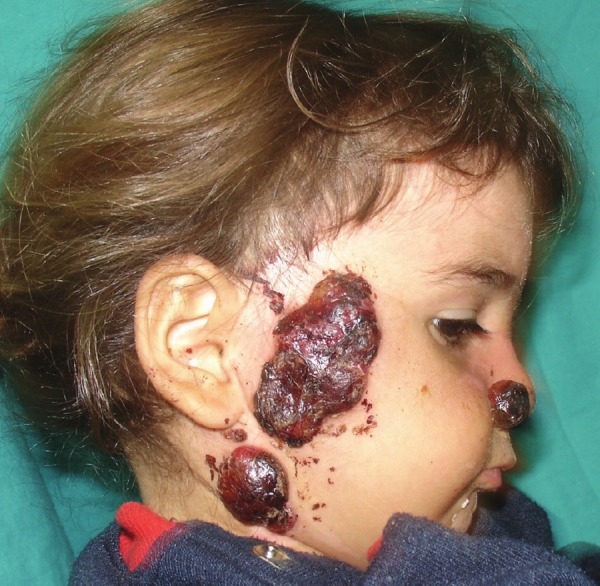
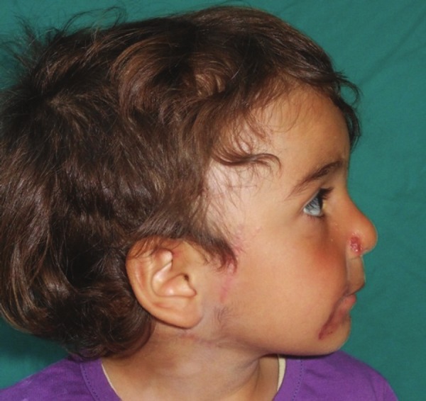
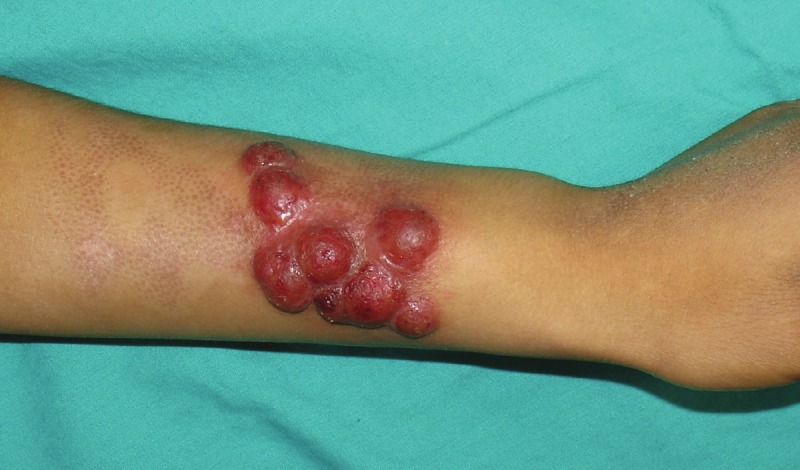
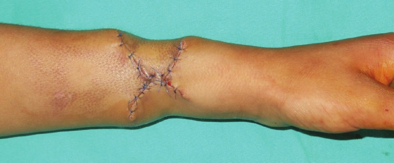
Pyogenic granuloma is a benign lesion and usually occurs after trauma. Disseminated pyogenic granuloma, is a rare form of pyogenic granuloma. There are 9 cases of disseminated pyogenic granuloma in literature and most of them are hot milk burns. First case describes an 18- month-old girl who developed disseminated pyogenic granulomas on her right cheek, neck, and right nasal ala. Lesions on her right cheek and neck were excised and sutured primarily. Lesion on right nasal ala was excised and repaired with full thickness skin graft. Second case describes a 7-years- old boy who developed pyogenic granulomas on his left forearm. These lesions were excised and sutured primarily. In both cases lesions were developed after scald burn. During 6-month follow-up, no recurrence was observed in both cases.
Keywords: Pyogenic granuloma; burn.
Figures
Similar articles
-
Infantile multiple large pyogenic granuloma on burned skin. Case report and review of literature.An Bras Dermatol. 2016 Apr;91(2):212-4. doi: 10.1590/abd1806-4841.20164060. An Bras Dermatol. 2016. PMID: 27192522 Free PMC article. Review.
-
Multiple giant disseminated pyogenic granuloma in a burn lesion.J Burn Care Res. 2006 Mar-Apr;27(2):247-9. doi: 10.1097/01.BCR.0000202642.08806.B7. J Burn Care Res. 2006. PMID: 16566577
-
Multiple giant disseminated pyogenic granuloma in three patients burned by boiling milk.Int J Dermatol. 1995 Oct;34(10):707-10. doi: 10.1111/j.1365-4362.1995.tb04658.x. Int J Dermatol. 1995. PMID: 8537159
-
Pyogenic granuloma with multiple dissemination in a burn lesion.Pediatr Dermatol. 1997 May-Jun;14(3):213-5. doi: 10.1111/j.1525-1470.1997.tb00240.x. Pediatr Dermatol. 1997. PMID: 9192415
-
Intravascular pyogenic granuloma arising in an acquired arteriovenous malformation: report of a case and review of the literature.Dermatol Surg. 2004 Jul;30(7):1050-3. doi: 10.1111/j.1524-4725.2004.30316.x. Dermatol Surg. 2004. PMID: 15209800 Review.
Cited by
-
Pyogenic granuloma after burns: a case report and review of the literature.Int J Burns Trauma. 2022 Jun 15;12(3):127-130. eCollection 2022. Int J Burns Trauma. 2022. PMID: 35891973 Free PMC article.
-
Oral propranolol and topical timolol in the treatment of post-burn pyogenic granuloma: Two cases and a review of the literature.Clin Case Rep. 2022 Nov 23;10(11):e6538. doi: 10.1002/ccr3.6538. eCollection 2022 Nov. Clin Case Rep. 2022. PMID: 36439384 Free PMC article.
-
Multiple disseminated pyogenic granuloma post-oil burning-Review literature.Clin Case Rep. 2020 Nov 6;9(1):169-172. doi: 10.1002/ccr3.3491. eCollection 2021 Jan. Clin Case Rep. 2020. PMID: 33489154 Free PMC article.
-
Multiple Disseminated Pyogenic Granuloma Post-Oil Burning: A Review of Literature.Indian J Dermatol. 2022 Nov-Dec;67(6):836. doi: 10.4103/ijd.IJD_371_18. Indian J Dermatol. 2022. PMID: 36998868 Free PMC article.
-
Infantile multiple large pyogenic granuloma on burned skin. Case report and review of literature.An Bras Dermatol. 2016 Apr;91(2):212-4. doi: 10.1590/abd1806-4841.20164060. An Bras Dermatol. 2016. PMID: 27192522 Free PMC article. Review.
References
-
- Netscher D, Spira M, Cohen V. Nonhemangiomatous vascular lesions. In: Achauer BM, editor. Plastic Surgery Indications, Operations and Outcomes. 1 edition. Missouri: Mosby; 2000. p. 310.
-
- Patrice SJ, Wiss K, Mulliken JB. Pyogenic granuloma (lobular capillary hemangioma): a clinicopathologic study of 178 cases. Pediatr Dermatol. 1991;8:267–276. - PubMed
-
- Ceyhan AM, Basak PY, Akkaya VB, Yildirim M, Kapucuoglu N. A case of multiple, eruptive pyogenic granuloma developed on a region of the burned skin: can erythromycin be a treatment option? J Burn Care Res. 2007;28:754–757. - PubMed
-
- Bozkurt M, Kulahci Y, Zor F, Askar I. Multiple giant disseminated pyogenic granuloma in a burn lesion. J Burn Care Res. 2006;27:247–249. - PubMed
-
- Aliagaoglu C, Bakan V, Atasoy M, Toker S. Pyogenic granuloma with multiple and satellite involvement after a burn in a 5-year-old child. J Dermatol. 2006;33:150–152. - PubMed
Publication types
LinkOut - more resources
Full Text Sources