

Bilateral peripheral neural activity observed in vivo following unilateral nerve injury
- PMID: 23638339
- PMCID: PMC3627524
Bilateral peripheral neural activity observed in vivo following unilateral nerve injury
Abstract
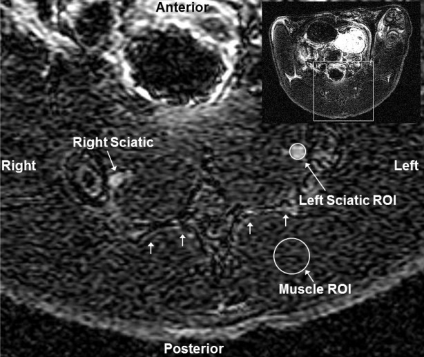
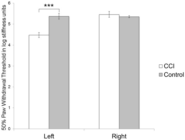
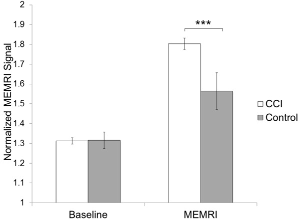
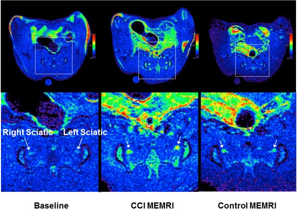
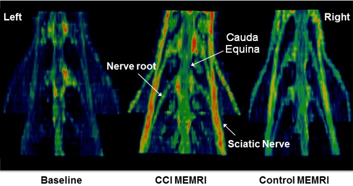
Manganese-enhanced magnetic resonance imaging (MRI) is a surrogate method to measure calcium content in nervous system since manganese physiologically follows calcium. Manganese is detectable in MRI and therefore visualizes structures and cell populations that actively regulate calcium. Since calcium is actively recruited for the transmission of action potentials, our purpose is to validate manganese-enhanced MRI for detection of changes in lumbar nerves related to nociception. A neuropathic pain model was created by chronic constrictive injury of the left sciatic nerve of Sprague-Dawley rats. Behavioral measurements, using von Frey's tests, confirmed the presence of significant allodynia in the left hind limb of animals in the injured group. T1-weighted fast spin echo images were obtained of the lumbar cord and plexus of animals with injured left sciatic nerve and uninjured animals (control) scanned in a 7 Tesla magnet after intraperitoneal manganese chloride administration four weeks after surgery. Lumbar nerve roots and sciatic nerves in the injured group show increased normalized manganese-enhanced MRI signal, representing manganese enhancement, compared to the control group. In conclusion, animals with neuropathic pain in the left hind limb show increased manganese uptake in not only the injured sciatic nerve but also in the contralateral uninjured sciatic nerve on manganese-enhanced MRI in vivo. Although poorly understood, this finding corroborates ex vivo finding of bilateral nociceptive-related molecular changes in the nervous system of unilateral pain models.
Keywords: Manganese-enhanced magnetic resonance imaging; animal model; chronic constrictive injury; neuropathic pain; sciatic nerve injury.
Figures
References
-
- Silva AC, Lee JH, Aoki I, Koretsky AP. Manganese-enhanced magnetic resonance imaging (MEMRI): methodological and practical considerations. NMR Biomed. 2004;17:532–543. - PubMed
-
- Aschner M, Aschner JL. Manganese neurotoxicity: cellular effects and blood-brain barrier transport. Neurosci Biobehav Rev. 1991;15:333–340. - PubMed
-
- Kita H, Narita K, Van der Kloot W. Tetanic stimulation increases the frequency of miniature end-plate potentials at the frog neuromuscular junction in Mn2+-, CO2+-, and Ni2+-saline solutions. Brain Res. 1981;205:111–121. - PubMed
-
- Narita K, Kawasaki F, Kita H. Mn and Mg influxes through Ca channels of motor nerve terminals are prevented by verapamil in frogs. Brain Res. 1990;510:289–295. - PubMed
LinkOut - more resources
Full Text Sources