. 2013 Mar 5:1:e51.
doi: 10.7717/peerj.51. Print 2013.

A critical role for suppressors of cytokine signaling 3 in regulating LPS-induced transcriptional activation of matrix metalloproteinase-13 in osteoblasts

Affiliations

A critical role for suppressors of cytokine signaling 3 in regulating LPS-induced transcriptional activation of matrix metalloproteinase-13 in osteoblasts

Anqi Gao et al. PeerJ. .

Erratum in

Abstract

Suppressor of cytokine signaling 3 (SOCS3) is a key regulator of cytokine signaling in macrophages and T cells. Although SOCS3 seems to contribute to the balance between the pro-inflammatory actions of IL-6 family of cytokines and anti-inflammatory signaling of IL-10 by negatively regulating gp130/Jak/Stat3 signal transduction, how and the molecular mechanisms whereby SOCS3 controls the downstream impact of TLR4 are largely unknown and current data are controversial. Furthermore, very little is known regarding SOCS3 function in cells other than myeloid cells and T cells. Our previous study demonstrates that SOCS3 is expressed in osteoblasts and functions as a critical inhibitor of LPS-induced IL-6 expression. However, the function of SOCS3 in osteoblasts remains largely unknown. In the current study, we report for the first time that LPS stimulation of osteoblasts induces the transcriptional activation of matrix metalloproteinase (MMP)-13, a central regulator of bone resorption. Importantly, we demonstrate that SOCS3 overexpression leads to a significant decrease of LPS-induced MMP-13 expression in both primary murine calvariae osteoblasts and a mouse osteoblast-like cell line, MC3T3-E1. Our findings implicate SOCS3 as an important regulatory mediator in bone inflammatory diseases by targeting MMP-13.

Keywords: Cytokine; Inflammation; Osteoblasts; Periodontitis.


Figures
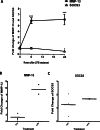
Figure 1
Figure 1. LPS induces expression of MMP-13 and SOCS3 in MC3T3-E1 cells and primary calvarial osteoblasts.
MC3T3-E1 cells (A) and primary calvarial osteoblasts (B and C) were incubated with 100 ng LPS/ml for indicated times. Total cellular RNA was isolated. Oligonucleotide primers used for real time PCR were MMP-13, SOCS3 and GAPDH. For panel A, data are presented as mean ± S. E. M., n = 4–5, and a representative experiment of a total of two similar experiments is shown. ∗ and ∗∗∗ indicate a statistically significant difference- p < 0.05 and p < 0.001, respectively.
Figure 2
Figure 2. Critical role of SOCS3 in regulating LPS-induced MMP-13 gene expression in MC3T3-E1 cells.
(A) MC3T3-E1 cells were infected with Adeno-SOCS3 at indicated MOI. 48 h after infection, the total protein extracts were subjected to Western blot using antibodies against SOCS3. (B) MC3T3-E1 cells were infected with Adeno-Ctrl or Adeno-SOCS3 at 100 X MOI. 48 h later, the cells were stimulated either with or without 100 ng LPS/ml for 6 h. Total cellular RNA was isolated. Oligonucleotide primers used for real time PCR were MMP-13 and GAPDH. Data are presented as mean ± S. E. M., n = 4–6. A representative experiment of a total of three similar experiments is shown. ∗∗∗ indicates a statistically significant difference-p < 0.001. (C) MC3T3-E1 cells were infected with Adeno-Ctrl or Adeno-sh-SOCS3 at a MOI of 100. 48 h after infection, the cells were incubated with 100 ng LPS/ml for 6 h. RNAs were isolated. Oligonucleotide primers used for real time PCR were SOCS3 and GAPDH. (D) MC3T3-E1 cells were infected with Adeno-Ctrl or Adeno-sh-SOCS3 at a MOI of 100. 48 h later, cells were incubated with or without 100 ng LPS/ml for for 6 h. RNAs were isolated. Oligonucleotide primers used for real time PCR were MMP-13 and GAPDH. Data are presented as mean ± S. E. M., n = 4–7. A representative experiment of a total of two similar experiments is shown. ∗∗ and ∗∗∗ indicate a statistically significant difference- p < 0.01 and p < 0.001, respectively.
Figure 3
Figure 3. Critical role of SOCS3 in regulating LPS-induced MMP-13 gene expression in primary calvarial osteoblasts.
Primary calvarial osteoblasts were infected with Adeno-Ctrl, Adeno-SOCS3, or Adeno-sh-SOCS3 at 100 X MOI. 48 h later, the cells were stimulated either with or without 100 ng LPS/ml for 6 h. Total cellular RNA was isolated. Oligonucleotide primers used for real time PCR were MMP-13 and GAPDH. Data are presented as mean ± S. E. M., n = 6. ∗, ∗∗, and ∗∗∗ indicate a statistically significant difference-p < 0.05, p < 0.01, and p < 0.001, respectively.
Figure 4
Figure 4. SOCS3 inhibits the activities of MMP-13 promoter-luciferase induced by LPS.
MC3T3-E1 cells were transiently transfected with 0.5 µg of DNA comprised of a mouse MMP-13 luciferase gene, thymidine kinase-luciferase gene, and either SOCS3 expression plasmid or control vector. 24 h after transfection, the cells were incubated with or without 100 ng LPS/ml for 4 h. Cell lysates were used to perform luciferase activity assay. Luminometer values were normalized for expression from a co-transfected thymidine kinase reporter gene. The data were expressed as means ± S. E. M., n = 3. A representative experiment of a total of two similar experiments is shown. ∗ and ∗∗∗ indicate a statistically significant difference- p < 0.05 and p < 0.001, respectively.
Figure 5
Figure 5. SOCS3 inhibits LPS-induced MMP-13 expression through p38 MAP Kinase pathway in MC3T3-E1 cells.
(A) MC3T3-E1 cells were infected with Adeno-Ctrl or Adeno-SOCS3 at 100 X MOI. 48 h later, the cells were stimulated either with or without 100 ng LPS/ml for the indicated time periods. Total proteins were subjected to Western blot by using rabbit anti-phospho-p38 (p-p38) antibody, rabbit anti-p38 antibody (p38), respectively. (B) Western blot were digitized and analyzed. p-p38 expression was normalized to p38 expression and the data are presented as a ratio. (C) MC3T3-E1 cells were infected with Adeno-Ctrl or Adeno-SOCS3 at 100 X MOI. 48 h later, the cells were stimulated either with or without 100 ng LPS/ml in the presence or absence of p38 MAP Kinase Inhibitor VII (10 µM) or vehicle control (DMSO) for 6 h. Total cellular RNA was isolated for real time PCR with primers for MMP-13 and GAPDH, respectively. Data are presented as mean ± S. E. M., n = 6. A representative experiment of a total of two similar experiments is shown. ∗∗ indicate a statistically significant difference-p < 0.01.

References

    1. Babon JJ, Nicola NA. The biology and mechanism of action of suppressor of cytokine signaling 3. Growth Factors. 2012;4:207–219. doi: 10.3109/08977194.2012.687375. - DOI - PMC - PubMed
    1. Barnes VM, Xu T, Shimizu E, Nakatani T, Jefcoat S, Vasilov A, Qin L, Partridge NC. Triclosan blocks Mmp 13 expression in hormone-stimulated osteoblasts. Journal of Periodontology. 2013 doi: 10.1902/jop.2013. Epub ahead of print Jan 31, 2013. - DOI - PMC - PubMed
    1. Cassatella MA, Gasperini S, Bovolenta C, Calzetti F, Vollebregt M, Scapini P, Marchi M, Suzuki R, Suzuki A, Yoshimura A. Interleukin-10 (IL-10) selectively enhances CIS3/SOCS3 mRNA expression in human neutrophils: evidence for an IL-10-induced pathway that is independent of STAT protein activation. Blood. 1999;94:2880–2889. - PubMed
    1. Chow JC, Young DW, Golenbock DT, Christ WJ, Gusovsky F. Toll-like receptor-4 mediates lipopolysaccharide-induced signal transduction. Journal of Biological Chemistry. 1999;274:10689–10692. doi: 10.1074/jbc.274.16.10689. - DOI - PubMed
    1. Croker BA, Kiu H, Pellegrini M, Toe J, Preston S, Metcalf D, O’Donnell JA, Cengia LH, McArthur K, Nicola NA, Alexander WS, Roberts AW. IL-6 promotes acute and chronic inflammatory disease in the absence of SOCS3. Immunology and Cell Biology. 2012;90:124–129. doi: 10.1038/icb.2011.29. - DOI - PMC - PubMed

LinkOut - more resources