. 2013 Mar 26:1:e60.
doi: 10.7717/peerj.60. Print 2013.

Pulmonary anatomy in the Nile crocodile and the evolution of unidirectional airflow in Archosauria

Affiliations

Pulmonary anatomy in the Nile crocodile and the evolution of unidirectional airflow in Archosauria

Emma R Schachner et al. PeerJ. .

Abstract

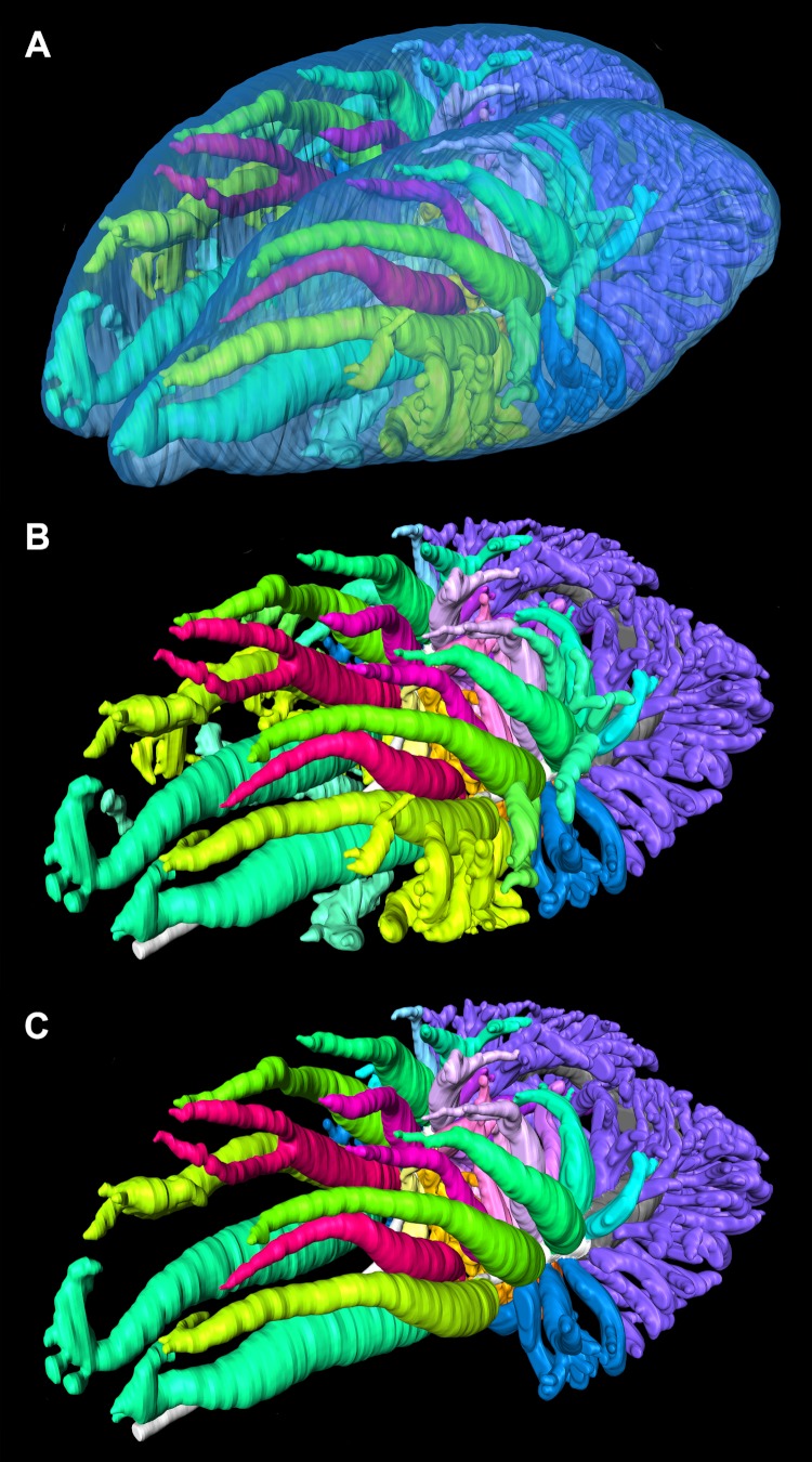
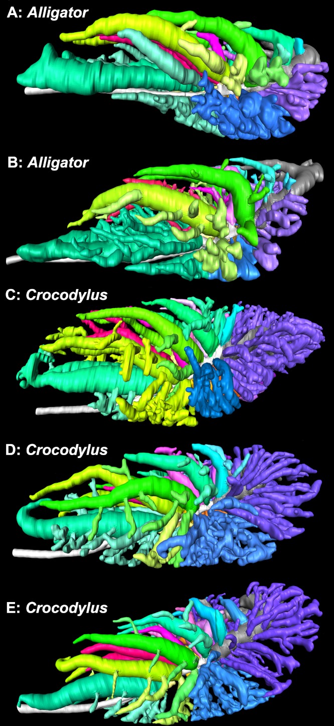
The lungs of birds have long been known to move air in only one direction during both inspiration and expiration through most of the tubular gas-exchanging bronchi (parabronchi). Recently a similar pattern of airflow has been observed in American alligators, a sister taxon to birds. The pattern of flow appears to be due to the arrangement of the primary and secondary bronchi, which, via their branching angles, generate inspiratory and expiratory aerodynamic valves. Both the anatomical similarity of the avian and alligator lung and the similarity in the patterns of airflow raise the possibility that these features are plesiomorphic for Archosauria and therefore did not evolve in response to selection for flapping flight or an endothermic metabolism, as has been generally assumed. To further test the hypothesis that unidirectional airflow is ancestral for Archosauria, we measured airflow in the lungs of the Nile crocodile (Crocodylus niloticus). As in birds and alligators, air flows cranially to caudally in the cervical ventral bronchus, and caudally to cranially in the dorsobronchi in the lungs of Nile crocodiles. We also visualized the gross anatomy of the primary, secondary and tertiary pulmonary bronchi of C. niloticus using computed tomography (CT) and microCT. The cervical ventral bronchus, cranial dorsobronchi and cranial medial bronchi display similar characteristics to their proposed homologues in the alligator, while there is considerable variation in the tertiary and caudal group bronchi. Our data indicate that the aspects of the crocodilian bronchial tree that maintain the aerodynamic valves and thus generate unidirectional airflow, are ancestral for Archosauria.

Keywords: Anatomy; Bronchi; Crocodylia; Endothermy; Evolution; Lung; Pneumaticity; Respiration.


Figures

Figure 1
Figure 1
Excised lungs from a 3.1 kg Nile crocodile (NNC1; Crocodylus niloticus) demonstrating probe placement, head is to the right. (A) Lungs in ventral view with the probe inserted in the right ventrobronchus (CVB); (B) the lungs in right dorsolateral view with the probe inserted in the second dorsobronchus (D2). Scale bars = 1 cm.
Figure 2
Figure 2
(A) Inflated lungs of a 1.01 kg Crocodylus niloticus (NNC3) in ventral view, head is to the right; (B) ventral view of the tracheal loop and heart (pericardium has been removed) of a 10.1 kg C. niloticus (FNC6), head is to the right. Scale bars = 1 cm. Arrows indicate tracheal loop; or lack thereof in the smaller individual (A).
Figure 3
Figure 3
3D segmented surface models of the bronchial trees of Crocodylus niloticus demonstrating the position of the caudal expansion of the caudal saccular regions of the primary bronchi within the lung, all in dorsal view. (A) The translucent lung surface and bronchial tree of NNC9; (B) the bronchial tree of NNC9; (C) the bronchial tree of NNC5; (D) the bronchial tree of NNC6. Abbreviations: CVB, cervical ventral bronchus; CSS, caudal sac-like structure; D2-D7, dorsobronchus 2-7; Ls, lung surface; Pb, primary bronchus. Bronchial trees are not to scale relative to one another.
Figure 4
Figure 4
Segmented airways and lung surface of a 0.5 kg specimen of Crocodylus niloticus (NNC9) generated from a µCT scan in left craniolateral view. The solid airways are visual representations of the negative spaces within the lung. (A) The primary, secondary, and tertiary bronchi positioned with respect to the lung surface (transparent blue); (B) the primary, secondary and tertiary bronchi; (C) the primary and secondary bronchi. For a detailed model of the anatomy see Figs. 5 and 6. Color scheme: translucent blue, lung surface; white, trachea and primary bronchi; mint green, cervical ventral bronchi (CVB); lime, D2; neon green, D3; aqua, D4; light aqua, D5; light blue, D6, periwinkle, D7; blue, laterobronchi; purple, caudal group bronchi (CGB); red, M1; neon pink, M2; medium pink, M3; light pink, M4; pale pink, M5; pale purple-deep pink-purples, M6-8; yellow-gold, cardiac lobes.
Figure 5
Figure 5
Primary bronchi, ventrobronchi (CVB), dorsobronchi (D), and medial bronchi (M) of a 0.5 kg Crocodylus niloticus (NNC9) generated from µCT. The ventrobronchus and dorsobronchi in (A) left craniolateral view; and (B) left lateral view. The ventrobronchus and medial bronchi in (C) right craniolateral view; and (D) left lateral view. The solid airways are visual representations of the negative spaces within the lung. Abbreviations: CVB, cervical ventral bronchus; D2-7, dorsobronchi 2-7; M1-8, medial bronchi 1-8; Pb, primary bronchus; R, right; Tr, trachea.
Figure 6
Figure 6
The primary bronchi, ventrobronchi, cardiac lobes, laterobronchi, and caudal group bronchi of a 0.5 kg Crocodylus niloticus (NNC9) generated from µCT. The lungs in (A) left craniolateral view; (B) dorsal view; (C) left lateral view; (D) ventral view. The solid airways are visual representations of the negative spaces within the lung. Abbreviations: C1-4, cardiac lobes 1-4; CGB, caudal group bronchi; CVB, cervicoventrobronchi; L, laterobronchi; Tr, trachea.
Figure 7
Figure 7
Lungs of a 0.5 kg specimen of Crocodylus niloticus (NNC9) injected with white latex, demonstrating the parabronchi (p) connecting the CVB and D2. (A) Lateral view of the right lung; (B) medial view of the sagittally-sectioned right lung stretched to expose the parabronchi indicated by the pink lines; (C) medial view of the sagittally-sectioned left lung. Pink arrows indicate the parabronchi. Scale bar in A and B = 1 cm; scale bar in C = 1.8 mm. Abbreviations: CVB, cervical ventral bronchus; D2-3, dorsobronchi 2-3; L, laterobronchi; P, parabronchi.
Figure 8
Figure 8
Airflow in the dorsobronchi and ventrobronchi measured in excised lungs with dual thermistor flow meters. A positive trace indicates that flow is caudal to cranial (black arrow); a negative trace shows airflow that is cranial to caudal (white arrow). (A) Direction of flow in D2 from NNC6; (B) direction of flow at the trachea while flow was recorded in D2 in NNC6; (C) direction of flow in D3 from NNC6; (D) direction of flow at the trachea while flow was recorded in D3 in NNC6; (E) direction of flow in D4 from NNC5; (F) direction of flow at the trachea while flow was recorded in NNC5 (G) direction of flow at the trachea while flow was recorded in the CVB in NNC5; (H) direction of flow at the trachea while flow was recorded in the CVB in NNC5.
Figure 9
Figure 9
3D segmented models of the bronchial tree of a 0.6 kg specimen of Crocodylus niloticus (NNC6) demonstrating the direction of airflow in the ventrobronchi and dorsobronchi in which airflow has been directly measured during both inspiration and expiration. (A) The primary, secondary, and tertiary bronchi in left lateral view; the color scheme is as in Figs. 2, 6 and 7. (B) The bronchial tree in left lateral view with the left ventrobronchus (CVB) and first three dorsobronchi highlighted to show direction of airflow. (C) The bronchial tree in dorsal view with the ventrobronchi and first three dorsobronchi highlighted to show direction of airflow. (D) The bronchial tree in dorsal view, with all of the secondary and tertiary bronchi removed except for the secondary bronchi in which airflow was directly measured (CVB, D2-D4). (E) The bronchial tree in left craniolateral view with all of the secondary and tertiary bronchi removed except for the secondary bronchi in which airflow was directly measured (CVB, D2-D4). Color scheme for B–E: blue, airflow is cranial to caudal during both phases of ventilation; green, airflow is caudal to cranial during both phases of ventilation; grey, primary bronchus.
Figure 10
Figure 10
Diagrammatic and highly simplified representation of airflow through the dorsobronchi and ventrobronchi during inspiration (A) and expiration (B) in the crocodilian lung, and inspiration (A) and expiration (D) in the avian lung. The avian model is a modification of the Hazelhoff loop (Hazelhoff, 1951). Arrows denote direction of airflow, white arrows show air flowing through the parabronchi, blue arrows show air entering the trachea, the red circled “X” demonstrates the location of the aerodynamic inspiratory valve (i.e., air does not flow through this location during inspiration). Colors represent hypothesized homologous regions of the lung in both groups. Abbreviations: d, dorsobronchi; P, parabronchi; Pb, primary bronchus; v, ventrobronchi.
Figure 11
Figure 11
3D segmented models of the bronchial tree of two live specimens of Alligator mississippiensis (in situ), and three specimens of Crocodylus niloticus generated from µCT and medical grade CT, all in dorsal view. (A) The primary, secondary, and tertiary bronchi of a 2.8 kg A. mississipiensis; (B) the primary, secondary, and tertiary bronchi of a 11 kg A. mississippiensis; (C) the primary, secondary, and tertiary bronchi of a 0.5 kg C. niloticus (NNC9); (D) the primary, secondary, and tertiary bronchi of a 0.8 kg C. niloticus (NNC6); (E) the primary, secondary, and tertiary bronchi of a 0.9 kg C. niloticus (NNC5). Images not to scale. Color scheme: white, trachea and primary bronchi; mint green, cervicoventrobronchi (CVB); lime, D2; neon green, D3; aqua, D4; light aqua, D5; light blue, D6, periwinkle, D7; blue, laterobronchi; purple, caudal group bronchi (CGB); red, M1; neon pink, M2; medium pink, M3; light pink, M4; pale pink, M5; pale purple-deep pink-purples, M6-8; yellow-gold, cardiac lobes.
Figure 12
Figure 12
3D segmented models of the bronchial tree of two live specimens of A. mississippiensis (in situ) and three cadaveric specimens of Crocodylus niloticus generated from µCT and medical grade CT, all in left lateral view. (A) The primary, secondary, and tertiary bronchi of a 2.8 kg A. mississippiensis; (B) the primary, secondary, and tertiary bronchi of a 11 kg A. mississippiensis; (C) the primary, secondary, and tertiary bronchi of a 0.5 kg C. niloticus (NNC9); (D) the primary, secondary, and tertiary bronchi of a 0.8 kg C. niloticus (NNC6); (E) the primary, secondary, and tertiary bronchi of a 0.9 kg C. niloticus (NNC5). Images not to scale. Color scheme: white, trachea and primary bronchi; mint green, cervicoventrobronchi (CVB); lime, D2; neon green, D3; aqua, D4; light aqua, D5; light blue, D6, periwinkle, D7; blue, laterobronchi; purple, caudal group bronchi (CGB); red, M1; neon pink, M2; medium pink, M3; light pink, M4; pale pink, M5; pale purple-deep pink-purples, M6-8; yellow-gold, cardiac lobes.
Figure 13
Figure 13
Diagrammatic representations of the crocodilian (A) and avian (B) lungs in left lateral view with colors identifying proposed homologous characters within the bronchial tree and air sac system of both groups. The image of the bird is modified from Duncker (1971). Abbreviations: AAS, abdominal air sac; CAS, cervical air sac; CRTS, cranial thoracic air sac; CSS, caudal sac-like structure; CTS, caudal thoracic air sac; d, dorsobronchi; GL, gas-exchanging lung; HS, horizontal septum; IAS, interclavicular air sac; L, laterobronchi; NGL, non-gas-exchanging lung; ObS, oblique septum; P, parabronchi; Pb, primary bronchus; Tr, trachea; v, ventrobronchi.

References

    1. Benson RBJ, Butler RJ, Carrano MT, O’Connor PM. Air-filled postcranial bones in theropod dinosaurs: physiological implications and the ‘reptile’-bird transition. Biological Reviews. 2011;87:168–193. doi: 10.1111/j.1469-185X.2011.00190.x. - DOI - PubMed
    1. Berner RA, VandenBrooks JM, Ward PD. Oxygen and evolution. Science. 2007;316:557–558. doi: 10.1126/science.1140273. - DOI - PubMed
    1. Bickler PE, Spragg RG, Hartman MT, White FN. Distribution of ventilation in American alligator, Alligator mississippiensis. American Journal of Physiology. 1985;249:477–481. - PubMed
    1. Boelert R. Sur la physiologie de la respirataion de l’ Alligator mississippiensis. Archives Internationales de Physiologie. 1942;52:57–72. doi: 10.3109/13813454209145577. - DOI
    1. Bonde N, Christiansen P. The detailed anatomy of Rhamphorhynchus: axial pneumaticity and its implications. London: Geological Society; 2003.