

Iris microhaemangioma: a management strategy
- PMID: 23638431
- PMCID: PMC3633769
- DOI: 10.3980/j.issn.2222-3959.2013.02.26
Iris microhaemangioma: a management strategy
Abstract
Aim: To analyse previous literature and to formulate a management strategy for iris microhaemangiomas (IMH).
Methods: A review of the literature in English language articles on IMH.
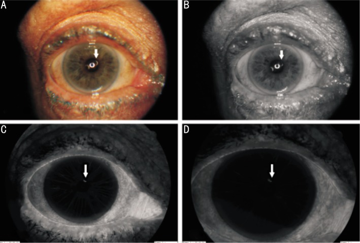
Results: Thirty five English language articles fulfilled the criteria for inclusion to the study and based on the contents on these articles a management strategy was formulated. Age at presentation ranged from 42 to 80 years with no sex or racial predisposition. Most patients with IMH have no systemic disease but a higher incidence had been reported in patients with diabetes mellitus, myotonic dystrophy, chronic obstructive pulmonary disease (COPD) and several other systemic and ophthalmic co-morbidities. Most patients remained asymptomatic until they experienced a sudden blurring of vision due to a hyphaema. Some patients only develop a self-limiting single episode of hyphaema and therefore the laser or surgical photocoagulation of iris should be reserved for the cases complicated with recurrent hyphaema. In some patients, several laser photocoagulation sessions may be needed and the recurrent iris vascular tufts may require more aggressive treatment. Iris fluorescein angiography (IFA) is useful in identifying the true extent of the disease and helps to improve the precision of the laser treatment. Surgical excision (iridectomy) should only be considered in patients who fail to respond to repeated laser treatment. In some cases IMHs has been initially misdiagnosed as amaurosis fugax, iritis and Posner-Schlossman syndrome.
Conclusion: Owing to its scarcity, there is no good quality scientific evidence to support the management of IMH. The authors discuss the various treatment options and present a management strategy based on the previous literature for the management for this rare condition and its complications.
Keywords: Argon laser photocoagulation; Cobb's tufts; capillary haemangioma of iris; iris microhaemangioma; iris vascular tufts.
Figures
Similar articles
-
Cobb's Tufts: A Systematic Review.Cureus. 2021 Dec 4;13(12):e20151. doi: 10.7759/cureus.20151. eCollection 2021 Dec. Cureus. 2021. PMID: 35003982 Free PMC article. Review.
-
Management of prominent iris vascular tufts causing recurrent spontaneous hyphema.Cornea. 2005 Mar;24(2):224-6. doi: 10.1097/01.ico.0000141236.33719.04. Cornea. 2005. PMID: 15725893
-
Active iris vascular tufts bleeding successfully treated with argon laser photocoagulation.Eur J Ophthalmol. 2018 Mar;28(2):241-242. doi: 10.5301/ejo.5001021. Eur J Ophthalmol. 2018. PMID: 29108395
-
Vascular tufts of pupillary margin of iris.Am J Ophthalmol. 1977 Jun;83(6):881-3. doi: 10.1016/0002-9394(77)90919-9. Am J Ophthalmol. 1977. PMID: 868991
-
Uveitis-Glaucoma-Hyphaema Syndrome. General review.Rom J Ophthalmol. 2017 Jan-Mar;61(1):11-17. doi: 10.22336/rjo.2017.3. Rom J Ophthalmol. 2017. PMID: 29450365 Free PMC article. Review.
Cited by
-
Cobb's Tufts: A Systematic Review.Cureus. 2021 Dec 4;13(12):e20151. doi: 10.7759/cureus.20151. eCollection 2021 Dec. Cureus. 2021. PMID: 35003982 Free PMC article. Review.
-
Oral tranexamic acid for acute management of active bleeding from iris microhemangiomatosis: A case report.Am J Ophthalmol Case Rep. 2024 Jan 19;33:102000. doi: 10.1016/j.ajoc.2024.102000. eCollection 2024 Mar. Am J Ophthalmol Case Rep. 2024. PMID: 38318444 Free PMC article.
-
Spontaneous hyphaemas requiring a closer look.BMJ Case Rep. 2016 Feb 23;2016:bcr2015213172. doi: 10.1136/bcr-2015-213172. BMJ Case Rep. 2016. PMID: 26907818 Free PMC article.
-
Spontaneous hyphaema secondary to bleeding from an iris vascular tuft in a patient with a supratherapeutic International normalised ratio: case report.BMC Ophthalmol. 2015 Jun 14;15:60. doi: 10.1186/s12886-015-0050-y. BMC Ophthalmol. 2015. PMID: 26071139 Free PMC article.
-
Acute iris vascular tuft hemorrhage treated successfully with intravitreal bevacizumab and pressure patching in a patient with branch retinal vein occlusion.Am J Ophthalmol Case Rep. 2022 Dec 13;29:101780. doi: 10.1016/j.ajoc.2022.101780. eCollection 2023 Mar. Am J Ophthalmol Case Rep. 2022. PMID: 36582845 Free PMC article.
References
-
- Blanksma LJ, Hooijmans JM. Vascular tufts of the pupillary border causing a spontaneous hyphaema. Ophthalmologica. 1979;178(6):297–302. - PubMed
-
- Ah-Fat FG, Canning CR. Recurrent visual loss secondary to an iris microhaemangioma (letter) Eye. 1994;8(Pt 3):357. - PubMed
-
- Akram I, Reck AC, Sheldrick J. Iris microhaemangioma presenting with total hyphema and elevated intraocular pressure. Eye. 2003;17(6):784–785. - PubMed
-
- Coleman SL, Green WR, Patz A. Vascular tufts of pupillary margin of iris. Am J Ophthalmol. 1977;83(6):881–883. - PubMed
-
- Elgohary MA, Sheldrick JH. Spontaneous hyphaema from pupillary vascular tufts in a patient with branch retinal vein occlusion. Eye (Lond) 2005;19(12):1336–1338. - PubMed
LinkOut - more resources
Full Text Sources