. 2013 Aug 9;425(15):2795-812.
doi: 10.1016/j.jmb.2013.04.019. Epub 2013 Apr 29.

E. coli ClpA catalyzed polypeptide translocation is allosterically controlled by the protease ClpP

Affiliations

E. coli ClpA catalyzed polypeptide translocation is allosterically controlled by the protease ClpP

Justin M Miller et al. J Mol Biol. .

Abstract

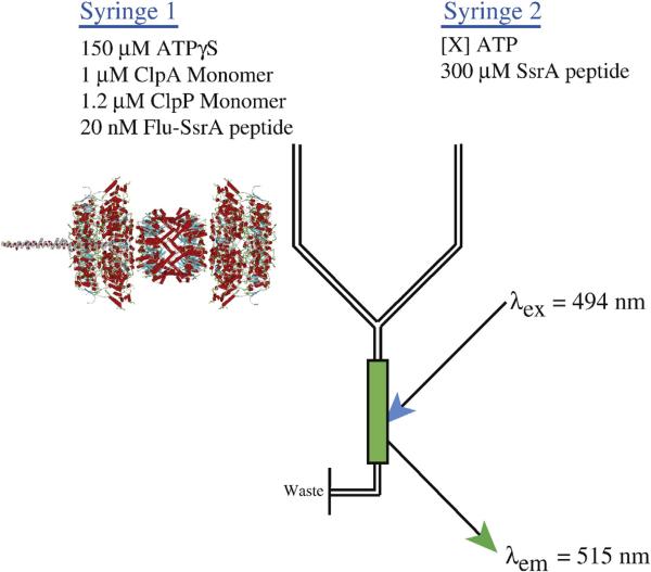
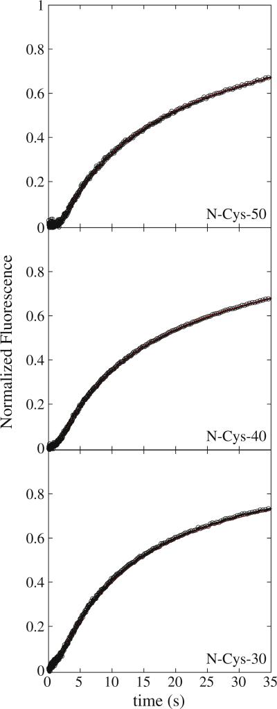
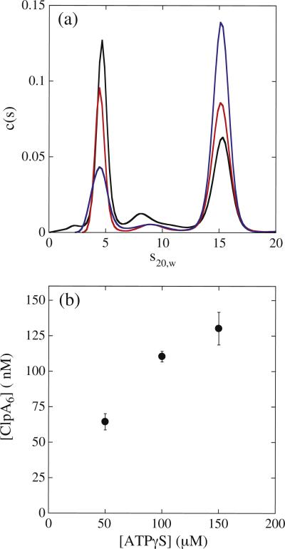
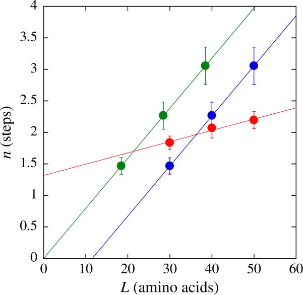
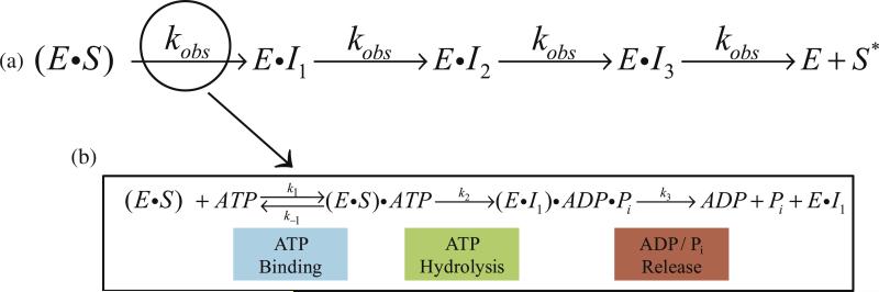
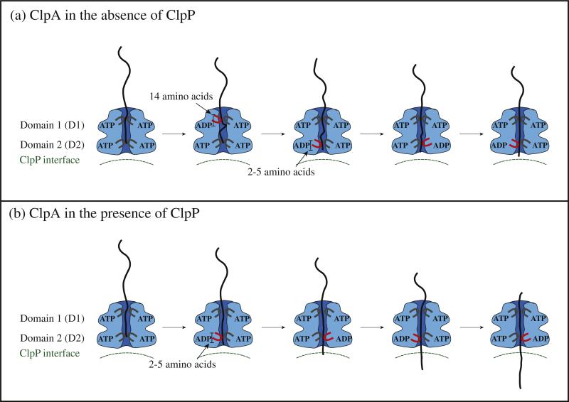
There are five known ATP-dependent proteases in Escherichia coli (Lon, ClpAP, ClpXP, HslUV, and the membrane-associated FtsH) that catalyze the removal of both misfolded and properly folded proteins in cellular protein quality control pathways. Hexameric ClpA rings associate with one or both faces of the cylindrically shaped tetradecameric ClpP protease. ClpA catalyzes unfolding and translocation of polypeptide substrates into the proteolytic core of ClpP for degradation through repeated cycles of ATP binding and hydrolysis at two nucleotide binding domains on each ClpA monomer. We previously reported a molecular mechanism for ClpA catalyzed polypeptide translocation in the absence of ClpP, including elementary rate constants, overall rate, and the kinetic step size. However, the potential allosteric effect of ClpP on the mechanism of ClpA catalyzed translocation remains unclear. Using single-turnover fluorescence stopped-flow methods, here we report that ClpA, when associated with ClpP, translocates polypeptide with an overall rate of ~35 aa s(-1) and, on average, traverses ~5 aa between two rate-limiting steps with reduced cooperativity between ATP binding sites in the hexameric ring. This is in direct contrast to our previously reported observation that, in the absence of ClpP, ClpA translocates polypeptide substrates with a maximum translocation rate of ~20 aa s(-1) with cooperativity between ATPase sites. Our results demonstrate that ClpP allosterically impacts the polypeptide translocation activity of ClpA by reducing the cooperativity between ATP binding sites.

Keywords: AAA+ motor proteins; ATP-dependent proteases; ATPγS; EDTA; FRET; GFP; NLLS; adenosine 5′-(γ-thio)-triphosphate; ethylenediaminetetraacetic acid; fluorescence resonance energy transfer; green fluorescent protein; nonlinear least squares; pre-steady-state kinetics; protein unfoldases.


Figures

Fig. 1
Fig. 1
Schematic representation of single-turnover stopped-flow translocation experiments. Syringe 1 contains the indicated reagents, ClpA, ClpP, ATPγS, and fluorescein-labeled polypeptide. The structure shown illustrates the contents of syringe 1 with the formation of the ClpAP complex with a single polypeptide bound (illustration is a schematic created by superimposing model structures for ClpA, ClpP, and model polypeptide substrate). Syringe 2 contains ATP to fuel polypeptide translocation and 300 μM SsrA peptide to serve as a trap for unbound ClpAP or any ClpAP that dissociates from polypeptide during the course of the reaction. The two reactants are rapidly mixed in the green-colored chamber and fluorescein is excited at λex = 494 nm. Fluorescein emissions are observed above 515 nm with a 515-nm-long pass filter. Upon mixing, the concentrations are 2-fold lower than in the preincubation syringe.
Fig. 2
Fig. 2
Fluorescence time courses for ClpAP catalyzed polypeptide translocation. Time courses represent 1 μM ClpA, 1.2 μM ClpP, 150 μM ATPγS, and 20 nM fluorescein-labeled polypeptide substrate pre-assembled prior to rapid mixing with 600 μM ATP and 300 μM SsrA. Shown are time courses for ClpAP catalyzed translocation of N-Cys-50, NCys-40, and N-Cys-30 polypeptide substrates. The red continuous lines represent a global NLLS fit using Scheme 2 for time courses collected with substrates I–III in Table 1. The resultant parameters are kT = (4.69 ± 0.09) s–1, kC = (0.12 ± 0.01) s–1, kNP = (0.02 ± 0.002) s–1, m = (4.6 ± 0.3) aa step–1, and mkT = (21.5 ± 1.1) aa s–1. Each time course was analyzed under a given set of conditions by constraining the parameters kT, kC, kNP, and h to be global parameters, while Ax, xx, and nx were allowed to float for each polypeptide length, where the subscript “x” represents the polypeptide substrate length.
Fig. 3
Fig. 3
Sedimentation coefficient distribution, c(s), dependence on ATPγS concentration. (a) c(s) distributions for 1 μM ClpA in the presence of 50 μM (black), 100 μM (red), and 150 μM (blue) ATPγS from the analysis of sedimentation velocity experiments performed as described in Materials and Methods in buffer H. (b) The concentration of ClpA hexamers was determined from analysis of the c(s) distributions collected at the three [ATPγS] using a non-interacting discrete species model. For 1 μM ClpA and 150 μM ATPγS, the concentration of ClpA hexamers was determined to be [ClpA6] = (130 ± 11) nM, which represents the average and standard deviation of eight replicates.
Fig. 4
Fig. 4
The dependence on polypeptide substrate length of the observed number of steps, n, determined from the analysis of ClpAP polypeptide translocation time courses shown in Fig. 2. Each time course was analyzed by constraining the parameters kT, kC, kNP, and h to be global parameters, while Ax, xx, and nx were allowed to float for each time course. The red-filled circles represent the determination of the number of steps, n, required to describe each time course in Fig. 2 using Scheme 1 [Eqs. (1) and (3) with h = 0]. The red continuous line represents a linear-least-squares fit with a slope of 0.018 and y-intercept of 1.32. The blue-filled circles represent the analysis of each time course in Fig. 2 using Scheme 2 [Eqs. (1) and (3) with h = 1]. The blue continuous line represents a linear-least-squares fit with a slope of 0.08 and y-intercept of –0.92. The green-filled circles represent the analysis of each time course in Fig. 2 using Scheme 2, but with each polypeptide length lacking the 11-aa SsrA sequence in analysis. The green continuous line represents a linear-least-squares fit with a slope of 0.08 and y-intercept of 1.2 × 10⁻⁶.
Fig. 5
Fig. 5
(a) Dependence of kT on [ATP], where the continuous line is the result of an NLLS fit to Eq. (4) with the Hill coefficient constrained to equal one for kT,max = (7.9 ± 0.2) s–1 and Ka = (4.8 ± 0.5) × 103 M–1. (b) Dependence of mkT on [ATP], where the continuous line is the result of a NLLS fit to Eq. (6) with the Hill coefficient constrained to equal one for mkT,max = 36.1 ± 0.7 aa s–1 and Ka = (4.8 ± 0.5) × 103 M–1. (c) Dependence of kC (filled circles) and kNP (filled squares) on [ATP], where the continuous line is the result of a NLLS fit to Eq. (4) with the Hill coefficient constrained to equal one. For kC and kNP, the equilibrium constant, Ka, is (2.8 ± 0.1) × 103 M–1 and (2.5 ± 0.1) × 103 M–1, respectively. The analysis of kC and kNP also yielded estimates of the maximum microscopic and macroscopic rates of translocation as 0.26 ± 0.003 s–1 and 0.045 ± 0.001 s–1, respectively.
Fig. 6
Fig. 6
Schematic representation of polypeptide translocation. In the n-step sequential model of polypeptide translocation, enzyme that is prebound to polypeptide substrate translocates polypeptide in discrete steps until reaching the end of the substrate and dissociating (a). Polypeptide translocation must occur through repeating cycles of ATP binding, hydrolysis, mechanical movement, various conformational changes, and ADP and Pi release, among other potentially significant kinetic steps (b). The observed rate constant, kobs, in the single-turnover experiments presented here represents the slowest step within this repeating cycle.
Fig. 7
Fig. 7
Proposed model of polypeptide translocation. In the absence of ClpP, ClpA translocates polypeptide with a mechanism that includes contributions from both ATP binding domains on each ClpA monomer. The D1 domain translocates polypeptide into the central cavity of ClpA by ~14 aa. In the time before D1 takes another translocation step, D2 must take three translocation steps of ~5 aa step–1. Upon association of ClpP, the D1 domain of ClpA undergoes a conformational change such that repeated cycles of ATP binding and hydrolysis no longer limit translocation and the rate of polypeptide translocation is limited by ATP hydrolysis and/or conformational changes taking place at D2 with each translocation step.
Scheme 1
Scheme 1
Simplest sequential n-step model. (ClpAP•S)L and (ClpAP•S)NP represent ClpAP bound to polypeptide substrate in the productive and nonproductive forms, respectively, and S is the unbound polypeptide substrate. kT is the translocation rate constant, kd is the dissociation rate constant, L is the polypeptide length, m is the average distance translocated between two steps with rate constant kT, and ‘i’ in I(L-im) represents i number of translocation steps.
Scheme 2
Scheme 2
Sequential n-step model with slow step relative to kT. All parameters are the same as in Scheme 1, with the exception of kc , which represents a step slower than translocation.

References

    1. Alberts B. The cell as a collection of protein machines: preparing the next generation of molecular biologists. Cell. 1998;92:291–294. - PubMed
    1. Neuwald AF, Aravind L, Spouge JL, Koonin EV. AAA+: A class of chaperone-like ATPases associated with the assembly, operation, and disassembly of protein complexes. Genome Res. 1999;9:27–43. - PubMed
    1. Gottesman S. Proteases and their targets in Escherichia coli. Annu. Rev. Genet. 1996;30:465–506. - PubMed
    1. Sauer RT, Baker TA. AAA+ proteases: ATP-fueled machines of protein destruction. Annu. Rev. Biochem. 2011;80:587–612. - PubMed
    1. Maglica Z, Kolygo K, Weber-Ban E. Optimal efficiency of ClpAP and ClpXP chaperone-proteases is achieved by architectural symmetry. Structure. 2009;17:508–516. - PubMed

Publication types

LinkOut - more resources