Review
. 2013 Jul;53(1):144-53.
doi: 10.1093/icb/ict020. Epub 2013 May 2.

What the clock tells the eye: lessons from an ancient arthropod

Affiliations
Review

What the clock tells the eye: lessons from an ancient arthropod

B-A Battelle. Integr Comp Biol. 2013 Jul.

Abstract

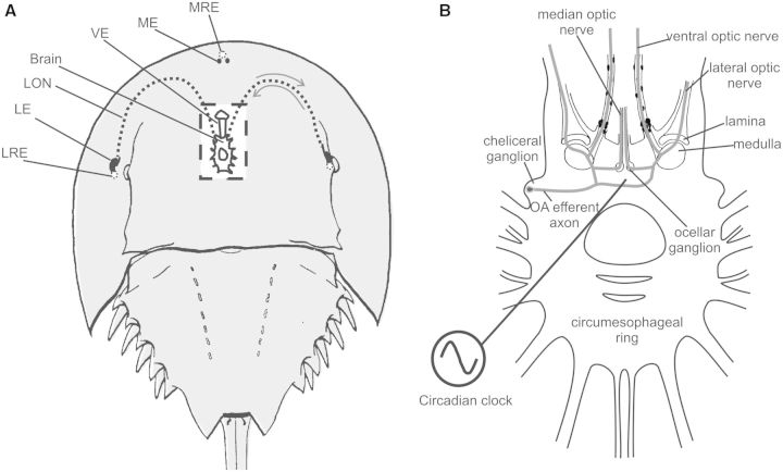
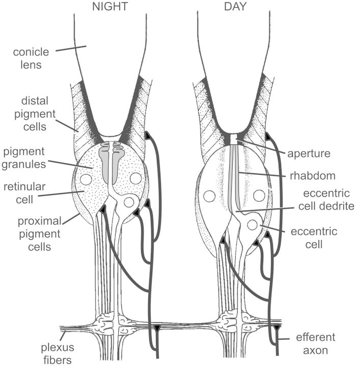
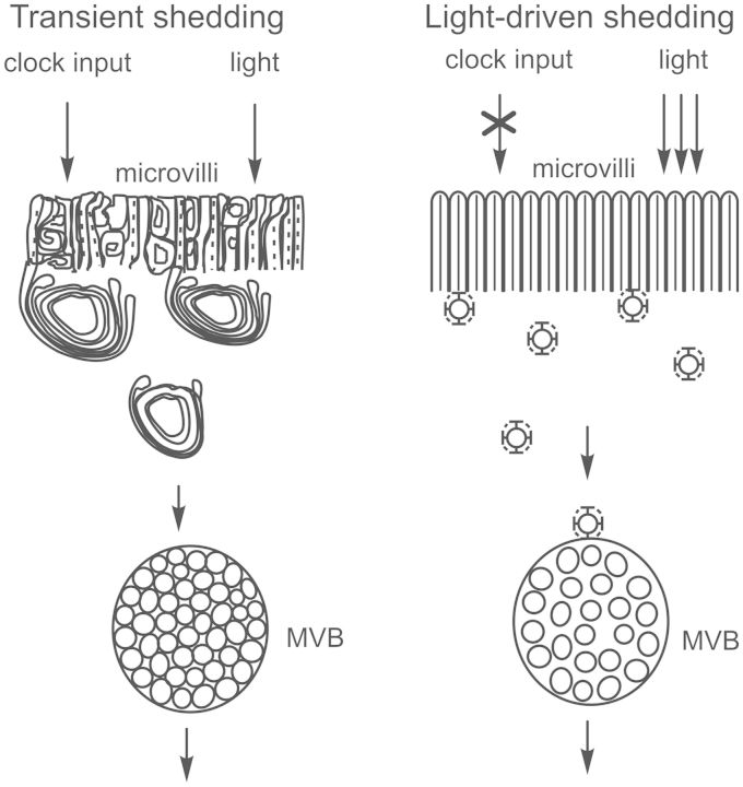
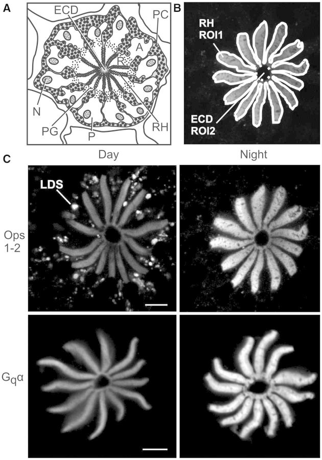
Circadian changes in visual sensitivity have been observed in a wide range of species, vertebrates, and invertebrates, but the processes impacted and the underlying mechanisms largely are unexplored. Among arthropods, effects of circadian signals on vision have been examined in most detail in the lateral compound eye (LE) of the American horseshoe crab, Limulus polyphemus, a chelicerate arthropod. As a consequence of processes influenced by a central circadian clock, Limulus can see at night nearly as well as they do during the day. The effects of the clock on horseshoe crab LE retinas are diverse and include changes in structure, gene expression, and rhabdom biochemistry. An examination of the known effects of circadian rhythms on LEs shows that the effects have three important outcomes: an increase in visual sensitivity at night, a rapid decrease in visual sensitivity at dawn, and maintenance of eyes in a relatively low state of sensitivity during the day, even in the dark. All three outcomes may be critically important for species' survival. Specific effects of circadian rhythms on vision will certainly vary with species and according to life styles. Studies of the circadian regulation of Limulus vision have revealed that these effects can be extremely diverse and profound and suggest that circadian clocks can play a critical role in the ability of animals to adapt to the dramatic daily changes in ambient illumination.


Figures

Fig. 1
Fig. 1
(A) Dorsal view of Limulus showing the locations of its eyes. Rectangle in center: cut-away to show the locations of the brain and the ventral eyes lying just under the ventral cuticle. The dashed lines show the projections of the lateral optic nerves. The arrows indicate that information in the optic nerve travels in two directions, from the eye to the brain and from the brain to the eye. LE, lateral eye; LON, lateral optic nerve; LRE, lateral rudimentary eye; ME, median eye; MRE, median rudimentary eye; VE, ventral eye. (B) The brain and circumesophageal ring of Limulus. The circadian clock(s) regulating the eyes is(are) in the brain. The location of one clock-driven efferent neuron is diagramed along with its proposed projections. Bilateral clusters of these efferent neurons are in the cheliceral ganglia, and each is thought to project to all of the eyes through the optic nerves. Based on Calman and Battelle (1991).
Fig. 2
Fig. 2
Longitudinal sections through LE ommatidia in their daytime and nighttime states. Based on Barlow et al. (1980) and Chamberlain and Barlow (1987). See text for details. The schematic also shows projections of the clock-driven efferent neurons innervating all cell-types in the LE. Based on Fahrenbach (1981).
Fig. 3
Fig. 3
Schematic differentiating clock-primed, light-triggered transient rhabdom-shedding from light-driven shedding. Transient shedding is primed by clock input during the night and triggered by the dim light of dawn. It is characterized by a rapid, transient disorganization of microvilli in the rays of the rhabdom and a breakdown of the actin cytoskeleton, accompanied by the formation of large whorls of opsin-containing membranes that accumulate between the rays of the rhabdom as densely packed multivesicular bodies (MVBs). Light-driven shedding is a progressive process driven by brighter light that does not require clock input. It is characterized by the clathrin-mediated endocytosis of opsin-containing membranes from the base of the microvilli that then accumulates in loosely packed MVBs. Transient shedding begins at about sunrise and is largely complete by 1 h after sunrise; light-driven shedding continues throughout the remaining daylight hours. Based on Chamberlain and Barlow (1979), Sacunas et al. (2002), and Battelle (2013).
Fig. 4
Fig. 4
Immunoreactive intensities of Ops1-2 and Gqα over rhabdoms changes significantly from day to night. (A) Cross-section of a LE ommatidium. The following are labeled: A, arhabdomeral segment; ECD, eccentric cell dendrite; N, nucleus; PC pigment cells; PG, pigment granules in photoreceptors; R, rhabdomeral segment; and RH, rhabdom. (B) Confocal image of Ops1-2-ir in the R-segment and proximal A-segment of an ommatidium from a LE fixed during the night. Shown are the regions of interest (ROIs) drawn to quantify average immunoreactive intensities over rhabdoms. Total intensity of ROI1 minus ROI2 was divided by total area of ROI1 minus ROI2 to calculate the average intensity over rhabdoms/µm2. (C) Representative images of single confocal optical sections showing Ops1-2- and Gqα-ir in the R-segment and proximal A-segment of LEs fixed at midday (Day) and during the night (Night), between 4 and 6 h after sunset. Day-images and night-images of each antigen were immunostained and imaged together during the same confocal session, using identical confocal settings. Images were intensified in Photoshop to exactly the same extent and then assembled in CorelDraw. LDS, opsin-containing membranes shed during the day by light-driven shedding. Scale bar = 10 µm.
Fig. 5
Fig. 5
Clock input drives increases in concentrations of (A) RhOps1-2 and of (B) Rh Gqα. The average intensity of RhOps1-2- and RhGqα-ir per µm2 of rhabdom is plotted versus time of day in hours relative to sunset (SS) and sunrise (SR). All animals were exposed to natural illumination. The shaded area indicates when it was dark in the room. The figure summarizes three series of studies: (1) A Dusk Series that included LEs fixed between midday (D) and SS + 4. (2) A Dawn Series that included LEs fixed from SR-1 to SR + 3. Concentrations of RhOps1-2 and RhGqα did not change significantly between SS + 4 and SR-1; therefore, these series could be combined. The data are normalized relative to the maximum concentration. All eyes assayed in Series 1 and 2 had intact optic nerves and thus received normal clock input (+Clock) (filled circles and dotted line). The asterisk in the dusk series indicates when concentrations of rhabdomeral protein were significantly higher than at midday (D). In the dawn series, the asterisk indicates when RhOps1-2 concentrations were significantly higher than at SR + 1 and RhGqα concentrations were significantly higher than at SR + 2 (P < 0.05). (3) In series 3, direct comparisons were made between RhOps1-2 or RhGqα concentrations in LEs from the same animal, one with, and the other without, clock input. The open circles and dashed line shows the average concentrations of RhOps1-2 or RhGqα in LEs lacking clock input (− Clock) relative to that in LEs with clock input (+ Clock). The asterisks (*) associated with these points indicate when there was a significant difference between RhOps1-2 or RhGqα concentrations in LEs with, and without, clock input. Means are plotted ± SEM. The number at each time-point indicates the number of animals assayed. The fall in concentration of RhOps1-2 during the morning is attributed to transient rhabdom shedding (TRS), and light-driven shedding (LDS), as indicated. For details, see Battelle (2013).
Fig. 6
Fig. 6
Octopamine and forskolin increase concentrations of RhOps1-2 and RhGqα during daytime dark-adaptation in vitro. LEs were dissected from animals at midday. Each LE was cut in half to yield four slices from each animal. One slice from each animal was fixed immediately in the light. The other three slices were incubated 4 h at room temperature in the dark in an organ-culture medium (Katti et al. 2010) +0.08% dimethyl sulfoxide and one of the following: Dk, no further additions; OA, octopamine (40 µM)+ IBMX (1 mM); FSK, forskolin (10 µM) + IBMX (1 mM). RhOps1-2 and RhGqα were quantified in each slice as described in Fig. 5. Data are expressed as mean immunoreactive intensities/µm2 of rhabdom ± SEM. Rhabdomeral protein concentrations in each dark-adapted slice were normalized to those in the light-adapted slice of the same animal. The light-adapted concentrations are expressed as 1. The number of animals assayed is shown in the first bar of each dataset. The significances of differences among the dark-adapted treatments are as follows: *P < 0.05; **P < 0.01. From Battelle et al. (2013).
Fig. 7
Fig. 7
The relative level of Ops1-2 and Ops5 in rhabdoms changes from day to night. Shown for each time-point are confocal images of sequential scans of single optical sections and their merged images (Ops1-2-ir, green; Ops5-ir, red). Sections were immunostained at the same time and imaged during a single confocal session using identical settings. All images were intensified in Photoshop to exactly the same extent. During the night, the intensity of RhOps1-2-ir is considerably higher than during the day, whereas the intensity of RhOps5-ir does not change significantly from day to night. Rhabdomeral membrane shed during the day by LDS contains both Ops1-2- and Ops5-ir, although Ops1-2-ir in debris is clearly more intense. Scale bar = 10 µm.

Similar articles

Cited by

References

    1. Arshavsky VY, Burns ME. Photoreceptor signaling: supporting vision across a wide range of sight sensitivities. J Biol Chem. 2012;287:1620–6. - PMC - PubMed
    1. Barlow RB, Chamberlain SC, Levinson J. Limulus brain modulates the structure and function of the lateral eyes. Science. 1980;210:1037–9. - PubMed
    1. Barlow RB. Circadian-rhythms in the Limulus visual-system. J Neurosci. 1983;3:856–70. - PMC - PubMed
    1. Barlow RB, Bolanowski SJ, Brachman ML. Efferent optic neve fibers mediate circadian rhythms in the Limulus eye. Science. 1977;197:86–9. - PubMed
    1. Barlow RB, Kaplan E, Renninger GH, Saito T. Circadian-rhythms in Limulus photoreceptors. 1. Intracellular studies. J Gen Physiol. 1987;89:353–78. - PMC - PubMed

Publication types

MeSH terms

Substances