. 2013 Jul 15;379(2):195-207.
doi: 10.1016/j.ydbio.2013.04.021. Epub 2013 Apr 29.

Myf5 expression during fetal myogenesis defines the developmental progenitors of adult satellite cells

Affiliations

Myf5 expression during fetal myogenesis defines the developmental progenitors of adult satellite cells

Stefano Biressi et al. Dev Biol. .

Abstract

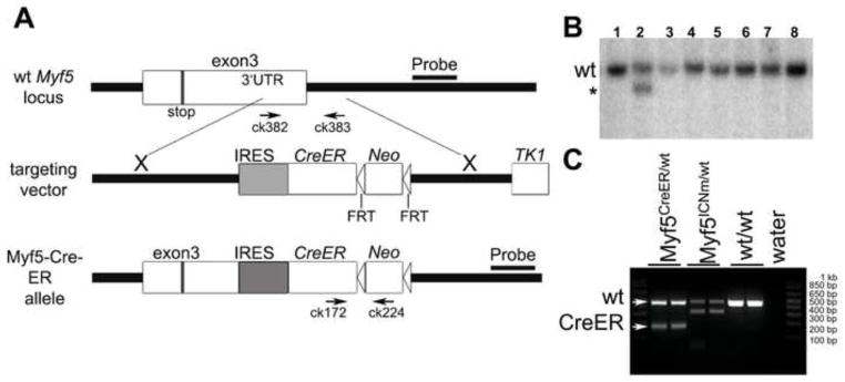
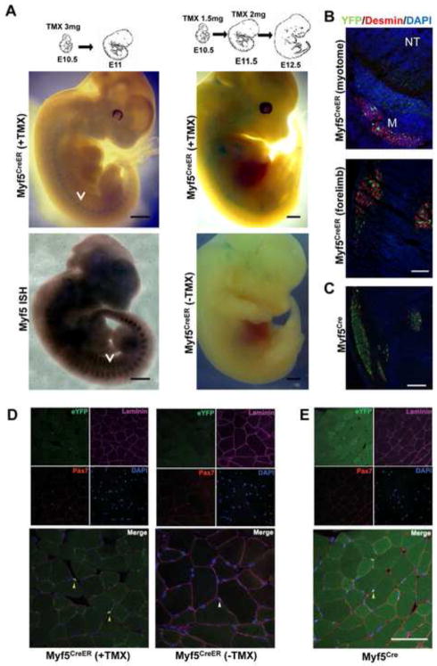
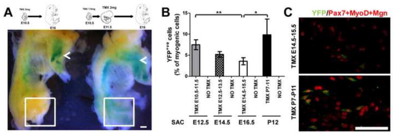
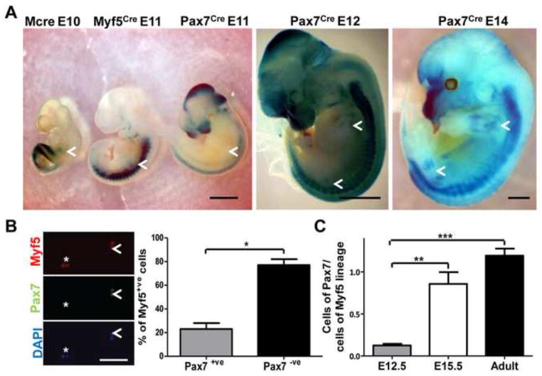
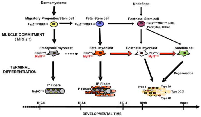
Myf5 is a member of the muscle-specific determination genes and plays a critical role in skeletal muscle development. Whereas the expression of Myf5 during embryonic and fetal myogenesis has been extensively studied, its expression in progenitors that will ultimately give rise to adult satellite cells, the stem cells responsible for muscle repair, is still largely unexplored. To investigate this aspect, we have generated a mouse strain carrying a CreER coding sequence in the Myf5 locus. In this strain, Tamoxifen-inducible Cre activity parallels endogenous Myf5 expression. Combining Myf5(CreER) and Cre reporter alleles, we were able to evaluate the contribution of cells expressing Myf5 at distinct developmental stages to the pool of satellite cells in adult hindlimb muscles. Although it was possible to trace back the origin of some rare satellite cells to a subpopulation of Myf5(+ve) progenitors in the limb buds at the late embryonic stage (∼E12), a significant number of satellite cells arise from cells which expressed Myf5 for the first time at the fetal stage (∼E15). These studies provide direct evidence that adult satellite cells derive from progenitors that first express the myogenic determination gene Myf5 during fetal stages of myogenesis.


Figures

Fig. 1
Fig. 1. Generation of Myf5CreER mice
(A) A targeting vector was designed for the insertion of a multifunctional cassette into the 3′ UTR of the Myf5 gene. At the AseI site in exon 3, an IRES-CreER construct was inserted to allow bi-cistronic expression of Myf5 and CreER. An FRT-Neo-FRT cassette was inserted 3′ to IRESCreER. The positions of PCR genotyping primers ck172, ck224, ck382 and ck383 are shown. Neo; neomycin resistance gene. Stop; stop codon. AseI; AseI restriction endonuclease site. (B) Southern blot analysis of ES cell clones using a 3′ external probe. One correctly targeted clone (line 2) shows a band (asterisk), specific to the mutant Myf5CreER allele, which is 4 kb smaller than the wild type band. (C) Genotyping PCR shows Myf5-CreER (182 bp), wild type (454 bp), and Myf5-ICNm (Haldar et al., 2007) (312 bp) alleles, respectively.
Fig. 2
Fig. 2. Characterization of Myf5CreER mice
(A) Whole-mount Xgal staining of Myf5CreER/wt; R26RLacZ/wt embryos. TMX administration protocols and stages as indicated. Myf5CreER/wt; R26RLacZ/wt embryos injected with corn oil were used as negative controls. Whole-mount in situ hybridization (ISH) of E11.5 embryos with Myf5-specific probes is shown to highlight the pattern of Myf5 expression. Arrows mark the myotomes. Bar, 1 mm. (B) Transverse sections of Myf5CreER/wt; R26RYFP/wt E12.5 embryos treated with TMX (5 mg) at E10.5 and E11.5 were stained with antibodies recognizing YFP and Desmin. DAPI was used to stain the nuclei. YFP+ve cells localize to the myotome (M; Desmin+ve) lateral to the neural tube (NT) and in the forming muscle masses in the forelimbs. Bar, 50 μm. (C) Sections of the limb of E12.5 Myf5Cre/wt; R26RYFP/wt are shown as term of comparison. Bar, 50 μm. (D) Immunofluorescence analysis of skeletal muscle from adult Myf5CreER/wt; R26RYFP/wt mice 2 weeks after TMX injections, or without TMX administration. Bar, 100 μm. (E) Immunofluorescence analysis of skeletal muscle from Myf5Cre; R26RYFP/wt mice. Frozen sections of tibialis anterior muscles were immunostained for YFP, Pax7, and Laminin. Nuclei were counterstained with DAPI. Yellow arrows point to YFP+ve satellite cells (D, E). A white arrow on the merged image shows a YFP−ve satellite cell localized beneath the basal membrane (D).
Fig. 3
Fig. 3. Temporal control and efficiency of TMX-induced labeling in developing Myf5CreER mice
(A) Whole-mount Xgal staining of Myf5CreER/wt; R26RLacZ/wt skinned E16 fetuses. Alternative TMX administration protocols are indicated. Depending on the protocol used, Cre-mediated recombination was induced in the hindlimb muscles (dashed box) in addition to forelimb muscles (arrows). Bar, 1.2 mm. (B) Cells are isolated from hindlimb muscles of Myf5CreER/wt; R26RYFP/wt mice treated with the indicated regimen of TMX and sacrificed (Sac) at the indicated stages, and are stained with antibodies recognizing YFP and the myogenic markers Pax7, MyoD and Myogenin (Mgn). The labeling efficiency is quantified by counting the number of myogenic cells (positive for Pax7, MyoD or Mgn) that are YFP+ve. Results are expressed as mean ± standard deviation. The labeling efficacy detected at E16.5 is lower compared to that obtained postnatally and at earlier embryonic stages (*P<0.05; **P<0.01). (C) Representative images of cells obtained from E16.5 fetuses and P12 pups treated with TMX at the indicated times are shown. Bar, 50 μm.
Fig. 4
Fig. 4. Adult satellite cells derive from progenitors expressing Myf5 during prenatal development
(A) An example of a YFP+ve satellite cell (arrow) associated with an EDL fiber of an adult Myf5CreER/wt; R26RYFP/wt mouse treated with TMX at E10.5–E11.5. Staining with antibodies recognizing YFP and the satellite cell markers Pax7 and Syn4 is presented. Bar, 20 μm. (B) Xgal staining of an EDL fiber from an adult Myf5CreER/wt; R26RLacZ/wt mouse treated with TMX at E10.5–E11.5. A β-gal+ve satellite cell is shown (arrow and insets). Bar, 30 μm. (C) An example of a satellite cell expressing the reporter gene YFP (arrowhead) after administration of TMX to E14.5–E15.5 is shown. Note that due to the relatively low efficiency of recombination, most of the satellite cells are YFP−ve (asterisk). DAPI was used to stain the nuclei. Bar, 40 μm. (D) The fraction of the VCAM+ve/CD45−ve/CD31−ve/Sca1−ve satellite cells expressing the lineage marker YFP (FITC) was quantified by FACS on cells obtained from adult Myf5CreER/wt; R26RYFP/wt mice after TMX administration at different developmental stages, as indicated. Satellite cells from Pax7CreER/wt; R26RYFP/wt mice injected with TMX at adult stage were used as positive controls. Note that the analysis of the satellite cells obtained from Myf5CreER/wt; R26RYFP/wt not treated with TMX reveals a virtual absence of recombination. (E) Cells were isolated by enzymatic digestion from hindlimb muscles of Myf5CreER/wt; R26RYFP/wt mice treated with TMX at the indicated stages and allowed to adhere overnight in vitro. Cells were then stained with an antibody to YFP and with a cocktail of antibodies to Pax7, MyoD, and Mgn to reveal the total pool of myogenic cells. Bar, 25 μm. (F) Data from replicate studies shown in panel E were quantified to determine the percentage of myogenic cells that were marked by YFP. Results are expressed as mean ± standard deviation. The recombination obtained after TMX administration at E14.5–E15.5 or P7–P11 is significantly higher compared that detected at earlier stages (** P<0.01, * P<0.05). (G) Data from replicate studies shown in panel E were quantified to determine the percentage of myogenic cells that were marked by YFP and were normalized for the average efficiency of recombination observed after TMX treatment at different stages (see Fig. 3B). Each square represents the normalized value obtained in a single replicate (n=3 for each condition). Note that a minority of satellite cells traces its origin back to progenitors expressing Myf5 at the embryonic (E10.5–E11.5) or early fetal (E12.5–E13.5) stage, whereas >75% of the satellite cells derive from progenitors expressing Myf5 at the mid-fetal (E14.5–15.5) or neonatal (P7–11) stage.
Fig. 5
Fig. 5. Enrichment in cells of the Pax7-lineage in the limb muscles at the fetal stage
(A) Whole-mount Xgal staining of MCre+/; R26RLacZ/wt, Myf5Cre/wt; R26RLacZ/wt and Pax7Cre/wt; R26RLacZ/wt mice at the indicated developmental stages. Limbs are indicated with arrows. Bar, 3 mm. (B) DsRED+ve cells were isolated by FACS from the hindlimb muscles of Myf5Cre/wt; Z/RED+/ E12.5 embryos, cultured for 4 hours in vitro in proliferating conditions in presence of the cytokinesis blocker cytochalasin, and stained with antibodies recognizing Pax7 and Myf5. The percentage of Myf5+ve doublets (proliferating myoblasts) positive (arrowhead) or negative (asterisk) for Pax7 is quantified. Bar, 25 μm. (C) DsRED+ve cells obtained from the hindlimb muscles of Myf5Cre/wt; Z/RED+/ and Pax7Cre/wt; Z/RED+/ mice at E12.5, E15.5 and adult stages were quantified by FACS. Data from the Pax7 line are expressed as percentage of DsRED+ve cells from the Myf5 line at the same stage.
Fig. 6
Fig. 6. Proposed lineage scheme for hindlimb muscles
Pax3+ve cells delaminate from the somitic dermomyotome and migrate into the limb buds, where they generate embryonic myoblasts which terminally differentiate into primary fibers. As development proceeds into the fetal stage, muscle stem cells resident in the limb activate the expression of Pax7. Pax7+ve/MRF−ve progenitors remain throughout fetal myogenesis and after birth as source of myoblasts (fetal and postnatal) and possibly satellite cells. During secondary myogenesis they generate fetal myoblasts by expressing Myf5 and other MRFs. Fetal Myf5+ve cells alternatively terminally differentiate during secondary myogenesis or remain as committed, but undifferentiated cells until successive phases of muscle development. After birth they contribute to the pool myoblasts responsible for post-natal growth and give rise to the majority of the adult satellite cells. A minority of the satellite cells resident in the adult limb muscles is derived from pericytes present in the neonatal muscles and possibly from other postnatal stem cells. The dashed arrow indicates the still poorly defined lineage relationship between embryonic and fetal myoblasts. The red color and the weight of the arrows highlight the predominance of the lineage described in this study.

Similar articles

Cited by

References

    1. Armand O, Boutineau AM, Mauger A, Pautou MP, Kieny M. Origin of satellite cells in avian skeletal muscles. Arch Anat Microsc Morphol Exp. 1983;72:163–181. - PubMed
    1. Asakura A, Seale P, Girgis-Gabardo A, Rudnicki MA. Myogenic specification of side population cells in skeletal muscle. J Cell Biol. 2002;159:123–134. - PMC - PubMed
    1. Atit R, Sgaier SK, Mohamed OA, Taketo MM, Dufort D, Joyner AL, Niswander L, Conlon RA. Beta-catenin activation is necessary and sufficient to specify the dorsal dermal fate in the mouse. Dev Biol. 2006;296:164–176. - PubMed
    1. Beauchamp JR, Heslop L, Yu DS, Tajbakhsh S, Kelly RG, Wernig A, Buckingham ME, Partridge TA, Zammit PS. Expression of CD34 and Myf5 defines the majority of quiescent adult skeletal muscle satellite cells. J Cell Biol. 2000;151:1221–1234. - PMC - PubMed
    1. Biressi S, Messina G, Collombat P, Tagliafico E, Monteverde S, Benedetti L, Cusella De Angelis MG, Mansouri A, Ferrari S, Tajbakhsh S, Broccoli V, Cossu G. The homeobox gene Arx is a novel positive regulator of embryonic myogenesis. Cell Death Differ. 2008;15:94–104. - PubMed

Publication types