. 2013 Jul 1;305(1):G84-94.
doi: 10.1152/ajpgi.00430.2012. Epub 2013 May 2.

Hepatocytes produce TNF-α following hypoxia-reoxygenation and liver ischemia-reperfusion in a NADPH oxidase- and c-Src-dependent manner

Affiliations

Hepatocytes produce TNF-α following hypoxia-reoxygenation and liver ischemia-reperfusion in a NADPH oxidase- and c-Src-dependent manner

Netanya Y Spencer et al. Am J Physiol Gastrointest Liver Physiol. .

Abstract

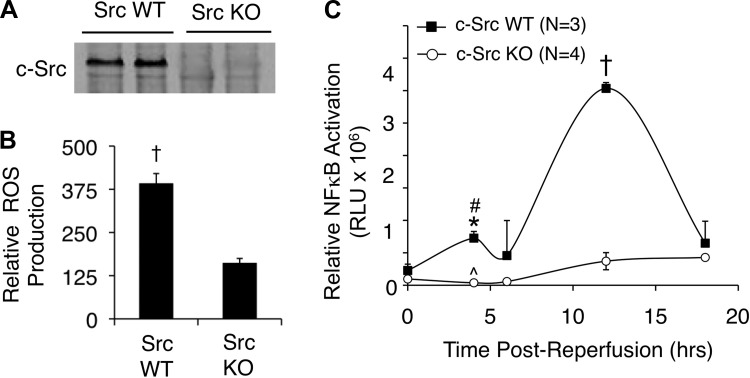
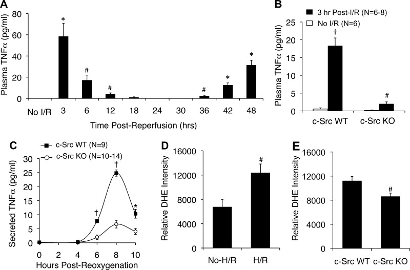
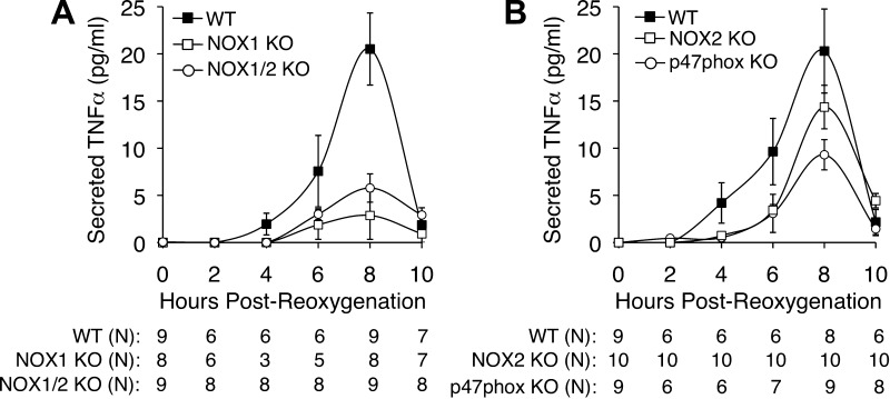
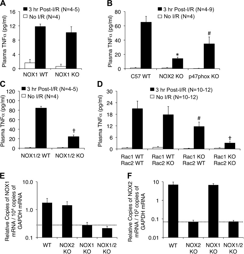
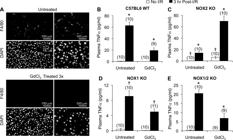
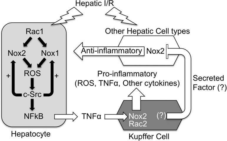
Cell line studies have previously demonstrated that hypoxia-reoxygenation (H/R) leads to the production of NADPH oxidase 1 and 2 (NOX1 and NOX2)-dependent reactive oxygen species (ROS) required for the activation of c-Src and NF-κB. We now extend these studies into mouse models to evaluate the contribution of hepatocytes to the NOX- and c-Src-dependent TNF-α production that follows H/R in primary hepatocytes and liver ischemia-reperfusion (I/R). In vitro, c-Src-deficient primary hepatocytes produced less ROS and TNF-α following H/R compared with controls. In vivo, c-Src-KO mice also had impaired TNF-α and NF-κB responses following partial lobar liver I/R. Studies in NOX1 and p47phox knockout primary hepatocytes demonstrated that both NOX1 and p47phox are partially required for H/R-mediated TNF-α production. To further investigate the involvement of NADPH oxidases in the production of TNF-α following liver I/R, we performed additional in vivo experiments in knockout mice deficient for NOX1, NOX2, p47phox, Rac1, and/or Rac2. Cumulatively, these results demonstrate that NOX2 and its activator subunits (p47phox and Rac) control the secretion of TNF-α by the liver following I/R. Interestingly, in the absence of Kupffer cells and NOX2, NOX1 played a dominant role in TNF-α production following hepatic I/R. However, NOX1 deletion alone had little effect on I/R-induced TNF-α. Thus Kupffer cell-derived factors and NOX2 act to suppress hepatic NOX1-dependent TNF-α production. We conclude that c-Src and NADPH oxidase components are necessary for redox-mediated production of TNF-α following liver I/R and that hepatocytes play an important role in this process.

Keywords: NADPH oxidase; c-Src; hepatocyte; redox; reperfusion injury.


Figures

Fig. 1.
Fig. 1.
c-Src deficiency reduces hepatic reactive oxygen species (ROS) production and NF-κB activation following hepatic ischemia-reperfusion (I/R). A: immunoprecipitation and Western blot for c-Src in liver lysates from wild-type (WT) or c-Src knockout (KO) mice. B: NADPH-dependent ROS production by hepatic endomembranes by lucigenin detection following liver I/R in WT and c-Src KO mice (45 min ischemia and 30 min reperfusion, N = 3 mice, ±SE). A statistically significant difference in ROS production between genotypes was observed by use of the Student's t-test (†P < 0.002). C: animals were preinfected with a recombinant adenoviral NF-κB-luciferase reporter vector 72 h prior to partial lobar hepatic I/R. Then 45 min of partial lobar ischemia was performed and NF-κB-luciferase reporter activity was assessed by biophotonic imaging on an IVIS imaging system. The zero time point represents pre-I/R luciferase activity and was not significantly different between genotypes (P = 0.1447). In all panels results depict means ± SE. The N independent animals are given. Statistically significant differences between genotypes were determined by a 2-tailed Student's t-test (*P < 0.001; †P < 0.0001). Statistically significant differences between the same genotypes comparing pre-I/R to post-I/R at the 4-h time point were determined by a 2-tailed paired Student's t-test (#P < 0.026; ^P = 0.1615).
Fig. 2.
Fig. 2.
c-Src deficiency inhibits hepatic and hepatocyte secretion of TNF-α following I/R and hypoxia-reoxygenation (H/R), respectively. A: kinetics of plasma TNF-α levels in C57BL6 mice following 60 min of partial lobar ischemia followed by the various time points of reperfusion. Blood was collected by terminal cardiac bleed (N = 3 independent animals for each data point). The first point represents nonischemic controls (labeled No I/R). B: plasma TNF-α levels prior to and following 45 min of partial lobar ischemia and 3 h of reperfusion. C: primary hepatocytes were generated from c-Src KO and c-Src WT littermates. TNF-α secretion into the media of these cultures was then evaluated following 5-h hypoxia and the indicated times of oxygenation. In all panels results depict means ± SE. The N independent animals or replicates are given in B and C. D and E: dihydroethidium (DHE) determination of ROS production in ICR (D) and c-Src WT and KO (E) cultured primary hepatocytes following 5 h of hypoxia and 20 min of reoxygenation. No H/R controls were also treated with DHE for 20 min. Fluorescent photomicrographs were quantified by use of Metamorph software; graphs depict the relative DHE intensity per cell (±SE) from N = 3–5 independent primary cultures. Statistically significant differences were determined by a 2-tailed Student's t-test (#P < 0.05, *P < 0.005; †P < 0.0001). Comparisons in A are between the preischemia and various postreperfusion time points. Comparisons in B are as follows: #c-Src KO preischemia vs. c-Src KO postreperfusion; †c-Src KO postreperfusion vs. c-Src WT postreperfusion, and c-Src WT preischemia vs. c-Src WT postreperfusion. Comparisons in C are between genotypes at each specific time point.
Fig. 3.
Fig. 3.
Nox1 and p47phox deficiency inhibits hepatocellular secretion of TNF-α following H/R. A: primary hepatocytes were generated from NOX1 WT, NOX1 KO, and NOX1/2 double-KO littermates. TNF-α secretion into the media was then studied following 5 h hypoxia and the indicated times of oxygenation. B: primary hepatocytes were generated from C57BL6 inbred p47phox KO mice, NOX2 KO mice, or WT C57BL6 mice. TNF-α secretion into the media was then measured following 5 h hypoxia and the indicated times of reoxygenation. In both A and B, results depict means ± SE. The N independent replicates (number of plates of hepatocytes treated with H/R) are given below each graph. Statistically significant differences between genotypes at the 8-h time point were determined by a 2-tailed Student's t-test (WT vs. NOX1 KO, P < 0.0020; WT vs. NOX1/2 KO, P < 0.0025; WT vs. NOX2 KO, P = 0.2270; WT vs. p47phox KO, P < 0.0278).
Fig. 4.
Fig. 4.
Nox2, Rac1/2, and p47phox deficiency inhibits hepatic secretion of TNF-α following partial lobar liver I/R. The indicated genetic strains were subjected to 60 min of partial lobar ischemia followed by 3 h of reperfusion. Blood was then harvested by cardiac bleeds and plasma was assessed for TNF-α levels. A: comparison of plasma TNF-α levels between Nox1 KO and Nox1 WT littermates. B: comparison of plasma TNF-α levels between C57 inbred mice with WT, NOX2 KO, or p47phox KO genotypes. C: comparison of plasma TNF-α levels between NOX1/2 WT and NOX1/2 double-KO mice. D: comparison of plasma TNF-α levels between Rac1flx/flx/Rac2-WT (Rac1-WT/Rac2-WT), Rac1flx/flx/Rac2-KO (Rac1-WT/Rac2-KO), AlbCRE-Rac1flx/flx/Rac2-WT (Rac1-KO/Rac2-WT), and AlbCRE-Rac1flx/flx/Rac2-KO (Rac1-KO/Rac2-KO) mice. In all panels results depict means ± SE. The N independent animals are given in each graph. Statistically significant differences in AD were determined by a 2-tailed Student's t-test (#P < 0.05, *P < 0.005; †P < 0.0005). Marked comparisons are between the WT and KO postreperfusion time points. E and F: quantitative RT-PCR for NOX1 (E) and NOX2 (F) mRNA from total liver RNA generated from WT, NOX2 KO, NOX1 KO, and NOX1/2 double-KO mice. GAPDH was used as internal control using ΔCt calculations for relative abundance. Values show means (± SE, N = 5 independent animals) relative abundance of NOX transcripts normalized to GAPDH transcripts as 2−(Nox:Ct − GAPDH:Ct) values. Dotted lines indicate the background levels of detection.
Fig. 5.
Fig. 5.
Kupffer cell depletion differentially affects hepatic secretion of TNF-α in a Nox1- and Nox2-dependent fashion following partial lobar liver I/R. The indicated genetic strains were treated with or without GdCl3 and subjected to 60 min of partial lobar ischemia followed by 3 h of reperfusion. Blood was then harvested by cardiac bleeds and plasma was assessed for TNF-α levels. A: macrophage marker (F4/80) staining of livers from animals untreated or treated with GdCl3 demonstrating the depletion of Kupffer cells. BE: comparison of plasma TNF-α levels prior to and following I/R for untreated and GdCl3-treated C57BL6 WT mice (B), NOX2 KO mice (C), NOX1 KO mice (D), or NOX1/2 double-KO mice (E). In all panels results depict means ± SE. The N independent animals are given in brackets above each data point. Statistically significant differences were determined by a 2-tailed Student's t-test for the indicated marked comparisons (*P < 0.001, †P < 0.02).
Fig. 6.
Fig. 6.
Working model for the NOX dependence of TNF-α production by the liver following I/R injury. We propose that the hepatocyte is one of the first cell types to respond to I/R injury by secreting low levels of NOX1/2-dependent TNF-α, priming Kupffer cells to mount an increased inflammatory response including high levels of TNF-α production. In this context, both hepatocyte Rac1/Nox1 and/or Rac1/Nox2 can contribute to ROS production by the hepatocyte, which activates c-Src and NF-κB pathways to induce TNF-α production by the hepatocyte. As in other cell types (28), c-Src in the hepatocyte appears to potentiate H/R-induced NOX-mediated ROS production in the hepatocyte. On the basis of our Kupffer cell-depletion experiments on the various NOX KO backgrounds, we propose that, in vivo, Kupffer cell-derived factors repress a partially NOX2-dependent pathway (labeled “Anti-inflammatory”) that otherwise would inhibit NOX1 activity in the liver. In vivo, this anti-inflammatory pathway may limit NOX1 activity in hepatocytes. In cultured primary hepatocytes (i.e., ex vivo), where this extrinsic factor is absent, NOX1 predominates as the pathway to activate TNF-α following H/R in the absence of other hepatic cell types. In NOX1 KO mice, NOX2 in the hepatocyte compensates for the loss of NOX1, and I/R responses are similar to WT animals. In NOX2 KO mice, NOX1 can still lead to low levels of acute-phase TNF-α production following I/R; however, the amplification of proinflammatory responses by the Kupffer cells is absent since NOX2 is the predominant NOX in this cell type. In Nox1/2 double-KO mice, ROS-dependent signaling that activates much of the TNF-α response following I/R is absent. In WT mice lacking Kupffer cells, a NOX2-dependent anti-inflammatory state is induced by the lack of Kupffer cell-secreted proinflammatory factors, and I/R-mediated TNF-α responses are attenuated. In NOX1 KO animals lacking Kupffer cells, the NOX2-dependent anti-inflammatory state is maintained, whereas Kupffer cell proinflammatory contributions are eliminated, leading to diminished I/R-mediated plasma TNF-α levels. In NOX2 KO Kupffer cell depleted animals, the NOX2-dependent anti-inflammatory state is no longer present and this transitions to a proinflammatory state raising both the baseline and I/R-induced TNF-α production by hepatocytes and potentially other non-Kupffer hepatic cell types. In this case (the absence of NOX2 and Kupffer cells), NOX1 activity may increase in the hepatocyte and/or other cell types, leading to enhanced NOX1-dependent production of TNF-α. In NOX1/2 double-KO animals depleted for Kupffer cells, the proinflammatory state caused by absence of both NOX2 and Kupffer cells is attenuated since NOX1 is not present to induce TNF-α production.

Similar articles

Cited by

References

    1. Abe J, Takahashi M, Ishida M, Lee JD, Berk BC. c-Src is required for oxidative stress-mediated activation of big mitogen-activated protein kinase 1. J Biol Chem 272: 20389– 20394, 1997 - PubMed
    1. Bhogal RH, Curbishley SM, Weston CJ, Adams DH, Afford SC. Reactive oxygen species mediate human hepatocyte injury during hypoxia/reoxygenation. Liver Transpl 16: 1303– 1313, 2010 - PubMed
    1. Boros P, Bromberg JS. New cellular and molecular immune pathways in ischemia-reperfusion injury. Am J Transplant 6: 652– 658, 2006 - PubMed
    1. Chen YX, Sato M, Kawachi K, Abe Y. Neutrophil-mediated liver injury during hepatic ischemia-reperfusion in rats. Hepatobiliary Pancreat Dis Int 5: 436– 442, 2006 - PubMed
    1. Diaz-Cruz A, Vilchis-Landeros MM, Guinzberg R, Villalobos-Molina R, Pina E. NOX2 activated by α1-adrenoceptors modulates hepatic metabolic routes stimulated by β-adrenoceptors. Free Radic Res 45: 1366– 1378, 2011 - PubMed

Publication types

MeSH terms