. 2013 Jul 15;305(2):G163-71.
doi: 10.1152/ajpgi.00003.2013. Epub 2013 May 2.

Cicletanine stimulates eNOS phosphorylation and NO production via Akt and MAP kinase/Erk signaling in sinusoidal endothelial cells

Affiliations

Cicletanine stimulates eNOS phosphorylation and NO production via Akt and MAP kinase/Erk signaling in sinusoidal endothelial cells

Songling Liu et al. Am J Physiol Gastrointest Liver Physiol. .

Erratum in

  • Corrigendum for Liu et al., volume 305, 2013, p. G163-G171.
    [No authors listed] [No authors listed] Am J Physiol Gastrointest Liver Physiol. 2022 Apr 1;322(4):G457-G458. doi: 10.1152/ajpgi.00003.2013_COR. Am J Physiol Gastrointest Liver Physiol. 2022. PMID: 35315727 Free PMC article. No abstract available.

Abstract

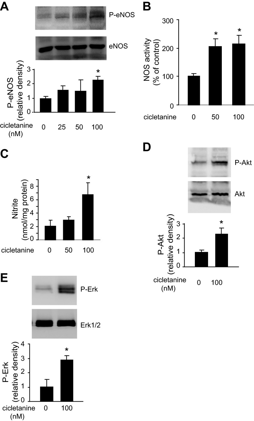
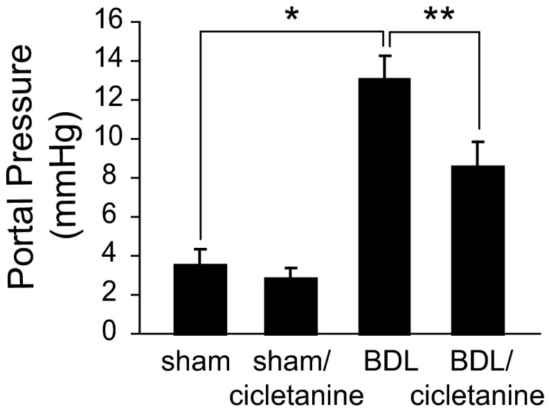
The function of the endothelial isoform of nitric oxide synthase (eNOS) and production of nitric oxide (NO) is altered in a number of disease states. Pharmacological approaches to enhancing NO synthesis and thus perhaps endothelial function could have substantial benefits in patients. We analyzed the effect of cicletanine, a synthetic pyridine with potent vasodilatory characteristics, on eNOS function and NO production in normal (liver) and injured rat sinusoidal endothelial cells, and we studied the effect of cicletanine-induced NO on stellate cell contraction and portal pressure in an in vivo model of liver injury. Sinusoidal endothelial cells were isolated from normal and injured rat livers. After exposure to cicletanine, eNOS phosphorylation, NO synthesis, and the signaling pathway regulating eNOS activation were measured. Cicletanine led to an increase in eNOS (Ser¹¹⁷⁷) phosphorylation, cytochrome c reductase activity, L-arginine conversion to L-citrulline, as well as NO production. The mechanism of the effect of cicletanine appeared to be via the protein kinase B (Akt) and MAP kinase/Erk signaling pathways. Additionally, cicletanine improved NO synthesis in injured sinusoidal endothelial cells. NO production induced by cicletanine in sinusoidal endothelial cells increased protein kinase G (PKG) activity as well as relaxation of stellate cells. Finally, administration of cicletanine to mice with portal hypertension induced by bile duct ligation led to reduction of portal pressure. The data indicate that cicletanine might improve eNOS activity in injured sinusoidal endothelial cells and likely activates hepatic stellate cell NO/PKG signaling. It raises the possibility that cicletanine could improve intrahepatic vascular function in portal hypertensive patients.

Keywords: endothelial isoform of nitric oxide synthase; extracellular/signal-regulated kinase; mitogen-activated protein kinase; portal hypertension; protein kinase B; signaling; stellate cell.


Figures

Fig. 1.
Fig. 1.
Effect of cicletanine on the function of endothelial isoform of nitric oxide synthase (eNOS) in sinusoidal endothelial cells. A: sinusoidal endothelial cells were isolated from rat livers and exposed to cicletanine (100 nM) for 2 h, cell lysates were harvested, and phosphor (P)-eNOS (Ser1177), total eNOS, and β-actin were detected by immunoblotting. In the graph shown on the bottom, bands corresponding to phospho-eNOS were quantified (n = 4, *P < 0.01 vs. control). B: the activity of nitric oxide synthase (NOS) in normal sinusoidal endothelial cells was examined in the same cells. NOS activity without exposure to cicletanine was arbitrarily set to 100; n = 3, *P < 0.01 vs. control (no cicletanine), **P < 0.01 vs. cicletanine. l-NAME, NG-nitro-l-arginine methyl ester. C: cells were treated with cicletanine, S-cicletanine, and R-cicletanine as indicated for 2 h, conditioned medium was harvested, and nitrite levels were measured as in materials and methods (n = 3, *P < 0.001 and **P < 0.05 vs. control).
Fig. 2.
Fig. 2.
Cicletanine induces eNOS activity in a time- and dose-response manner. A: sinusoidal endothelial cells were isolated from rat livers and exposed to cicletanine (100 nM) from 1 to 4 h, and cell lysates were subjected to immunoblotting to detect phosphor- or total eNOS. Blots shown are representative of 3 others. In the lower panel, nitrite levels were measured in conditioned medium form the same cells, and quantitative data are shown in the graph below. Medium from cells without treatment served as a control (n = 3, *P < 0.05 and **P < 0.01 vs. time “0”). B: sinusoidal endothelial cells were exposed to 50 or 100 nM cicletanine for 2 h, and cell lysates were subjected to immunoblotting to detect phosphor- or total eNOS. Blots shown are representative of 3 others. In the lower panel, nitrite levels were measured in conditioned medium form the same cells, and quantitative data are shown in the graph below; medium from cells without treatment served as a control (n = 3, *P < 0.01 vs. control). C: normal sinusoidal endothelial cells were exposed to indicated concentrations of cicletanine, and nitrite was measured in conditioned medium at the indicated times; medium from cells without treatment served as controls (n = 3, *P < 0.01 and **P < 0.05 vs. control). D: normal sinusoidal endothelial cells were preexposed to l-NAME (1 μM) or not for 4 h and then exposed to the cicletanine (at the indicated concentrations) for a further 1 or 2 h. In the upper panel is shown a representative immunoblot of cell lysates. Conditioned medium was collected, and nitrite levels were measured as in materials and methods (n = 3, *P < 0.05 vs. 1-h control, ** P < 0.01 vs. 2-h control, #P < 0.01 indicates significant difference between indicated groups). E: sinusoidal endothelial cells were exposed to cicletanine (100 nM) for either 2 or 6 h, cells were harvested, and NOS activity was measured in cell lysates, which was normalized to the level observed at the 2-h time point without cicletanine (n = 3, *P < 0.05 vs. 2-h exposure control, **P < 0.001 vs. 6-h exposure controls).
Fig. 3.
Fig. 3.
Cicletanine potentiates eNOS cytochrome c reductase activity. Sinusoidal endothelial cells were isolated from rat livers and exposed to cicletanine (100 nM) for 2 h, cell lysates were harvested, and NADPH cytochrome c reductase activity was measured as in materials and methods. Cells without treatment served as a control. The rate of basal cytochrome c reductase activity and the cytochrome c reductase activity in the presence of calmodulin (CaM) are shown graphically (n = 3, *P < 0.01 vs. control).
Fig. 4.
Fig. 4.
Cicletanine-induced eNOS activity is protein kinase B (Akt) dependent. A: sinusoidal endothelial cells were isolated from rat livers and were exposed to 50 or 100 nM cicletanine for 2 h, and cell lysates were harvested and subjected to immunoblotting to detect phospho (Ser473)- and total Akt. In the graph shown on bottom, bands corresponding to phospho-Akt were quantified (n = 3, *P < 0.01 vs. control). B: sinusoidal endothelial cells were exposed to 50 or 100 nM cicletanine for 1 or 2 h, and cell lysates were harvested and subjected to immunoblotting to detect phospho-eNOS, total eNOS, phospho-Akt (Ser473), total Akt, and β-actin. Blots representative of 3 others are shown. C: normal sinusoidal endothelial cells were infected with adenovirus (Ad, multiplicity of infection 250) encoding constitutively active Akt (Ad-myrAkt), dominant-negative Akt (Ad-dnAkt), or an empty vector (Ad-EV) for 24 h before exposure to cicletanine (100 nM) for an additional 2 h. Conditioned medium was harvested, and nitrite levels were measured and presented in the graph; medium from cells treated with Ad-EV alone served as a control (n = 3, *P < 0.05 and **P < 0.01 vs. control).
Fig. 5.
Fig. 5.
Cicletanine-induced eNOS activity is Erk dependent. A: sinusoidal endothelial cells were isolated from rat livers and were exposed to 50 or 100 nM cicletanine for 2 h, and cell lysates were subjected to immunoblotting to detect phospho-eNOS, total eNOS, phospho-Erk, total Erk1/2, and β-actin. In the graphs shown on bottom, bands corresponding to phospho-Erk were quantified (n = 3, *P < 0.05 vs. control). B: sinusoidal endothelial cells as in A were pretreated with the MAP kinase inhibitor PD-98059 (10 μM) for 30 min and then stimulated with cicletanine (100 nM) for an additional 2 h; eNOS and Erk phosphorylation were detected by immunoblotting. In the graph shown on bottom, bands corresponding to phospho-Erk were quantified (n = 3, *P < 0.01 vs. no cicletanine control, **P < 0.005 vs. cicletanine control). C: conditioned supernatants from cells as in B were collected, and nitrite levels were measured (n = 3, *P < 0.05 vs. no cicletanine control, **P < 0.01 vs. cicletanine control).
Fig. 6.
Fig. 6.
Effects of cicletanine in injured sinusoidal endothelial cells. A: sinusoidal endothelial cells were isolated from rat livers 10 days after bile duct ligation (BDL), allowed to adhere overnight, and then exposed to the indicated concentrations of cicletanine for 2 h; cells were harvested, and cell lysates were subjected to immunoblotting to detect phospho- and total eNOS. In the graph shown on bottom, bands corresponding to phospho-eNOS were quantified (n = 3, *P < 0.01 vs. control). B: sinusoidal endothelial cells as in A were harvested, and NOS activity was measured in cell lysates; NOS activity was normalized to that of control cells and is presented graphically (n = 3, *P < 0.005 vs. control). C: conditioned medium as in A was harvested, and nitrite levels were measured as in materials and methods (n = 3, *P < 0.05 vs. control). D and E: sinusoidal endothelial cells as in A were harvested, and cell lysates were immunoblotted to detect phospho- and total Akt and phospho- and total Erk1/2, respectively. Bands corresponding to phospho-Akt and phospho-Erk were quantitated, normalized, and shown in the lower graphs (D: n = 3, *P < 0.01 vs. control; E: n = 3, *P < 0.005 vs. control).
Fig. 7.
Fig. 7.
Cicletanine-induced nitric oxide (NO) production in sinusoidal endothelial cells causes protein kinase G (PKG) activation and stellate cell relaxation. A: hepatic stellate cells and sinusoidal endothelial cells were isolated separately from rat livers and cocultured (each at a density of 100,000 cells/lattice) on collagen lattices for 5 days as in materials and methods. Cells were then serum starved overnight, exposed to 50 or 100 nM cicletanine for 2 h, and then stimulated with endothelin-1 (ET-1, 10 nM). Collagen lattices were released from their substrata, and gel contraction was measured over time. A representative experiment with images of changes in gel area at the indicated time points is shown on left. Gel areas were measured, quantified, and depicted quantitatively in the graph on the right (n = 4, *P < 0.05 and **P < 0.001 vs. control). B: hepatic stellate cells (HSC) and sinusoidal endothelial cells (EC) were isolated, and HSC were cultured alone or cocultured (HSC + EC) as in materials and methods. Cells were harvested, and cell lysates were immunoblotted to detect phospho-vasodilator-stimulated phosphoprotein (VASP) for PKG activity. HSC or HSC + EC without cicletanine served as controls. Bands corresponding to phospho-VASP were quantitated, normalized, and shown in the lower graph (n = 3, *P < 0.01 and **P < 0.001 vs. HSC + EC control). C: cells (HSC + EC) as in B were preexposed to l-NAME (1 μM) for 4 h and then exposed to cicletanine (100 nM) for an additional 1–2 h. Phospho-VASP, PKG, and β-actin were detected in cell lysates by immunoblotting, and representative images are shown.
Fig. 8.
Fig. 8.
Cicletanine reduces portal pressure. Portal hypertension was induced by performing BDL in BALB/c mice; sham operations were also performed as described in materials and methods. Cicletanine (5 mg·kg−1·day−1) or vehicle was given by gavage in a single daily dose for 10 days before portal pressure measurement (which was performed at day 28) as described in materials and methods and presented graphically (n = 5 for each group; *P < 0.001 and **P < 0.005 for differences between indicated groups).
Fig. 9.
Fig. 9.
A proposed model of cicletanine effects on sinusoidal endothelial cells. Cicletanine, a furopyridine derivative, stimulated eNOS phosphorylation in sinusoidal endothelial cells (SECs). The data suggest a dual role, including at the level of Akt-eNOS and Erk-eNOS signaling. Cicletanine-mediated NO generation via enhanced eNOS activity appears to stimulate soluble guanylate cyclase in stellate cells, which activates cGMP/PKG and (downstream) VASP, leading to stellate cell relaxation.

Similar articles

Cited by

References

    1. Akamatsu N, Sawada S, Komatsu S, Tamagaki T, Hiranuma O, Kawahara T, Tsuda Y, Kono Y, Higaki T, Tada Y, Yamasaki S, Imamura H, Sato T, Tsuji H, Nakagawa M. Effect of cicletanine on the nitric oxide pathway in human umbilical vein endothelial cells. J Cardiovasc Pharmacol 38: 174–182, 2001 - PubMed
    1. Bernier SG, Haldar S, Michel T. Bradykinin-regulated interactions of the mitogen-activated protein kinase pathway with the endothelial nitric-oxide synthase. J Biol Chem 275: 30707–30715, 2000 - PubMed
    1. Bukoski RD, Bo J, Xue H, Bian K. Antiproliferative and endothelium-dependent vasodilator properties of 1,3-dihydro-3-p-chlorophenyl-7-hydroxy-6-methyl-furo-(3,4c) pyridine hydrochloride (cicletanine). J Pharmacol Exp Ther 265: 30–35, 1993 - PubMed
    1. Busconi L, Michel T. Endothelial nitric oxide synthase. N-terminal myristoylation determines subcellular localization. J Biol Chem 268: 8410–8413, 1993 - PubMed
    1. Calder JA, Schachter M, Sever PS. Interaction between cicletanine and the eicosanoid system in human subcutaneous resistance arteries. J Pharm Pharmacol 44: 574–578, 1992 - PubMed

Publication types

MeSH terms

Substances

LinkOut - more resources